Why Immunopeptidome Profiling?
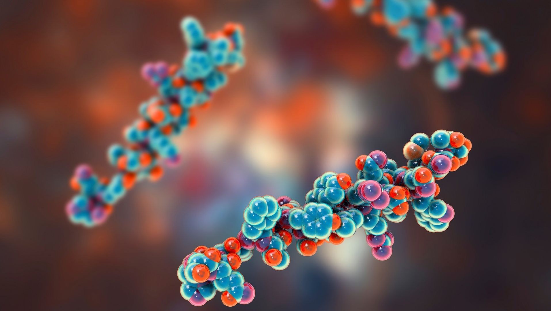
The immunopeptidome—comprising peptides naturally presented by HLA molecules—serves as a real-time molecular signature of cellular state. It reveals changes driven by mutations, infections, or immune dysregulation long before phenotypic symptoms appear.
By profiling these HLA-bound peptides, researchers gain an actionable readout of the immune system’s interaction with tumor cells, pathogens, or self-antigens.
What Makes Immunopeptidomics Powerful?
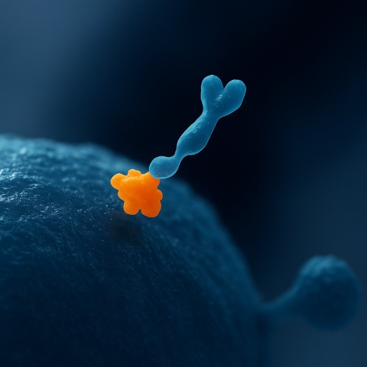
Unlike transcriptomics or proteomics, immunopeptidomics directly captures what the immune system can "see"—the actual surface-displayed peptides recognized by T cells. It bridges the gap between expression and immune recognition.
Key Application Areas
When paired with genomics, transcriptomics, and proteomics, immunopeptidomics adds a functional layer that reveals how molecular changes translate into immune-detectable differences—crucial for drug discovery, biomarker validation, and precision immunotherapy.

Cancer Immunotherapy
Identify tumor-specific and mutated peptides (neoantigens) for designing personalized vaccines, TCR therapies, and immune checkpoint strategies.

Infectious Disease
Reveal pathogen-derived peptides actively presented on infected cells to inform epitope-based vaccine development and antiviral drug targeting.

Autoimmune Research
Characterize self-peptides implicated in T cell misrecognition, enabling the study of molecular mimicry and autoimmune triggers.
What We Offer: Comprehensive Immunopeptidome Profiling and Beyond
Creative Proteomics provides an end-to-end immunopeptidomics service for the in-depth characterization of naturally presented HLA-bound peptides. By integrating advanced mass spectrometry platforms, custom enrichment workflows, and sophisticated bioinformatics pipelines, we empower researchers to identify, quantify, and interpret the dynamic antigen landscape in both physiological and disease states.
What We Can Analyze

HLA Class I Peptidome
Comprehensive coverage of intracellular and nuclear-derived peptides (8–12mers), ideal for CD8⁺ T cell epitope studies.

HLA Class II Peptidome
Profiling of extracellular, endocytic, and autophagy-derived peptides (13–25mers) relevant to CD4⁺ T cell immunity.

Post-Translationally Modified Peptides
Identification of phosphorylated, glycosylated, citrullinated, and other modified epitopes without prior enrichment.

Non-Canonical / Cryptic Peptides
Detection of peptides derived from lncRNAs, introns, upstream ORFs, repeat elements, or alternative splicing.

Treatment-Responsive Peptidome
Assess the immunopeptidomic changes under cytokine stimulation, drug exposure, gene editing, or environmental perturbations.

Neoantigen Candidate Discovery
Extraction and prioritization of mutation-derived HLA ligands for vaccine design or T cell immunotherapy development.
Specialized Add-On Modules (Optional)

Immunopeptide Affinity Screening
High-throughput screening of peptides against target antigens using ELISA, surface plasmon resonance (SPR), or molecular docking. Ranking based on affinity and specificity.

Custom Immune Peptide Library Construction
Design and synthesis of diverse peptide libraries optimized for immunogenicity, PTM coverage, or structure-based selection.

Multiplexed MS Quantification (TMT/iTRAQ)
Isobaric labeling for comparative peptidomics across treatment arms or patient cohorts.

HLA Binding Motif Prediction & Prioritization
Includes motif clustering, allele assignment, and anchor residue analysis.
Platform Advantages
Low Sample Input Compatibility
Requires as little as 1×10⁶ cells or ~5 mg of tissue; suitable for frozen, fresh, or limited biopsy samples.
Dual-Class HLA Analysis
Simultaneous profiling of HLA-I and HLA-II peptides from the same sample.
HLA Binding Motif Characterization
Motif deconvolution and editing pattern analysis for tapasin-, ERAP1/2-, HLA-DM/DO-dependent regulation.
Quantification-Ready Workflows
Supports DDA, DIA, and PRM/SureQuant for both relative and absolute quantification.
HLA Deconvolution & Allele Assignment
Compatible with monoallelic, multiallelic, and unknown donor samples; supports allele-specific peptide clustering.
Comprehensive Bioinformatics Outputs
Includes peptide ID lists, motif logos, binding affinities, source protein mapping, and pathway enrichment (upon request).
Step-by-Step Immunopeptidome Profiling Workflow
Sample Reception & QC
Visual check, integrity, sample logging
HLA Complex Enrichment
Antibody-based pulldown (Class I & II)
Acidic Peptide Elution
Native peptide release from HLA
LC-MS/MS Analysis
Orbitrap Astral or Exploris 480 platform
Data Processing
FragPipe, MaxQuant, Spectronaut workflows
Report Output & Visualization
Peptide ID lists, motifs, PTMs, HLA binding
1
Sample Reception & Quality Control
Visual inspection, sample integrity check, and pre-enrichment validation. We accept fresh, frozen cells, tissues, or clinical materials.
2
HLA Complex Immunoaffinity Enrichment
Selective isolation of HLA class I and/or II complexes using allele-specific or pan-HLA antibodies.
3
Peptide Elution (Acid-Based)
Native peptides are gently released from the HLA groove using acid treatment while preserving biological structure.
4
LC-MS/MS Analysis
High-resolution mass spectrometry is performed using Orbitrap Astral™ or Exploris 480™ for deep and accurate peptide detection.
5
Bioinformatics & Data Analysis
Peptides are identified, quantified, and annotated using FragPipe, Spectronaut, or MaxQuant, with optional PTM and motif profiling.
6
Report Generation & Delivery
Comprehensive data reports include peptide sequences, source protein mapping, binding motifs, PTM status, and HLA restriction predictions.
Deep and Accurate Peptidome Discovery
At Creative Proteomics, our immunopeptidomics workflow is powered by next-generation Orbitrap mass spectrometry platforms—Orbitrap Astral™ and Orbitrap Exploris™ 480—ensuring exceptional sensitivity, resolution, and throughput for comprehensive HLA ligand profiling.
Whether you're working with rare cell types or large cohorts, our platform delivers reproducible, deep-coverage datasets that accelerate epitope discovery and immunological insight.
Technical Highlights
- >90% MS/MS Coverage
High-resolution tandem MS enables identification of thousands of HLA-bound peptides with superior confidence and spectral clarity. - 1% FDR Stringent Filtering
Strict false discovery rate (FDR) control ensures reliable peptide identification and reproducibility across biological replicates. - PRM & SureQuant™-Enabled Quantification
Supports both relative and absolute quantification, with enhanced sensitivity for low-abundance immune peptides. - Broad Input Range: 1M–100M Cells
Optimized for scarce biospecimens or bulk materials—our platform scales with your project’s needs.
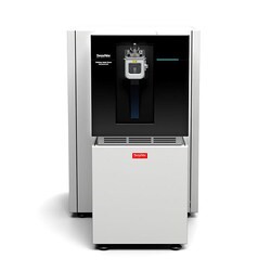 Orbitrap Astral™
(Fig from Thermo Scientific)
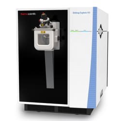 Orbitrap Exploris™ 480
(Fig from Thermo Scientific)
Orbitrap Astral™ vs. Orbitrap Exploris™ 480 — Technical Comparison
| Feature | Orbitrap Astral™ | Orbitrap Exploris™ 480 |
|---|---|---|
| MS/MS Coverage | >90% with high-speed, deep proteome coverage | >90% with robust, reproducible coverage |
| FDR Filtering | 1% FDR stringent filtering | 1% FDR stringent filtering |
| Quantification Support | PRM / SureQuant™ for absolute quantification | PRM / SureQuant™ for absolute quantification |
| Sample Input Range | Deep coverage from 100K–100M cell input | Deep coverage from 1M–100M cell input |
| Scan Speed | Up to 200 Hz (ultra-high speed) | ~40 Hz (high speed) |
| Sensitivity | Ultra-high sensitivity (single-cell compatible) | High sensitivity |
| Mass Accuracy | Sub-ppm mass accuracy (Orbitrap-based) | Sub-ppm mass accuracy (Orbitrap-based) |
Sample Requirements for Immunopeptidomes
| Sample Type | Minimum Amount | Preservation | Shipping Condition | Notes |
|---|---|---|---|---|
| Cell Pellet | ≥1×10⁶ cells | Snap-frozen (dry ice) | Dry ice, overnight courier | Avoid freeze-thaw; PBS wash preferred before freezing |
| Adherent Cells | ≥80% confluent in 6-well plate × 2 | Harvested & frozen as pellet | Dry ice, avoid liquid medium | Include cell count and viability if possible |
| Tissue (Fresh/Frozen) | ≥5 mg | Snap-frozen preferred | Dry ice | No fixatives or embedding media (e.g., paraffin) |
| PBMCs | ≥1×10⁶ viable cells | Cryopreserved in DMSO or fresh | Dry ice or room temp for fresh overnight | Prefer Ficoll-purified human PBMCs |
| Tumor Tissue | ≥10 mg | Snap-frozen or OCT embedded | Dry ice | Provide pathology report if available |
| FFPE Tissue (upon request) | ≥2 unstained 10 μm sections | FFPE block or slides | Ambient (slides) / Dry ice (block) | Please consult us prior to submission; yield may vary |
| Whole Blood | ≥500 μL (for PBMC prep) | EDTA tube, processed ≤2 h | Cold pack, same-day delivery | For downstream PBMC isolation; not ideal for direct MS |
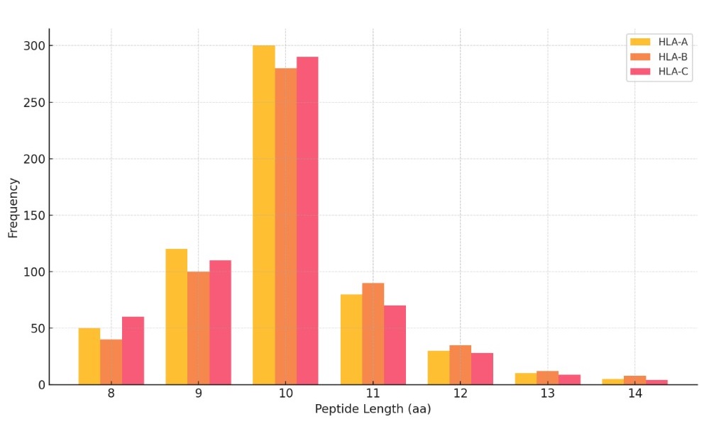
Demo Results
Peptide Length Distribution by HLA Type
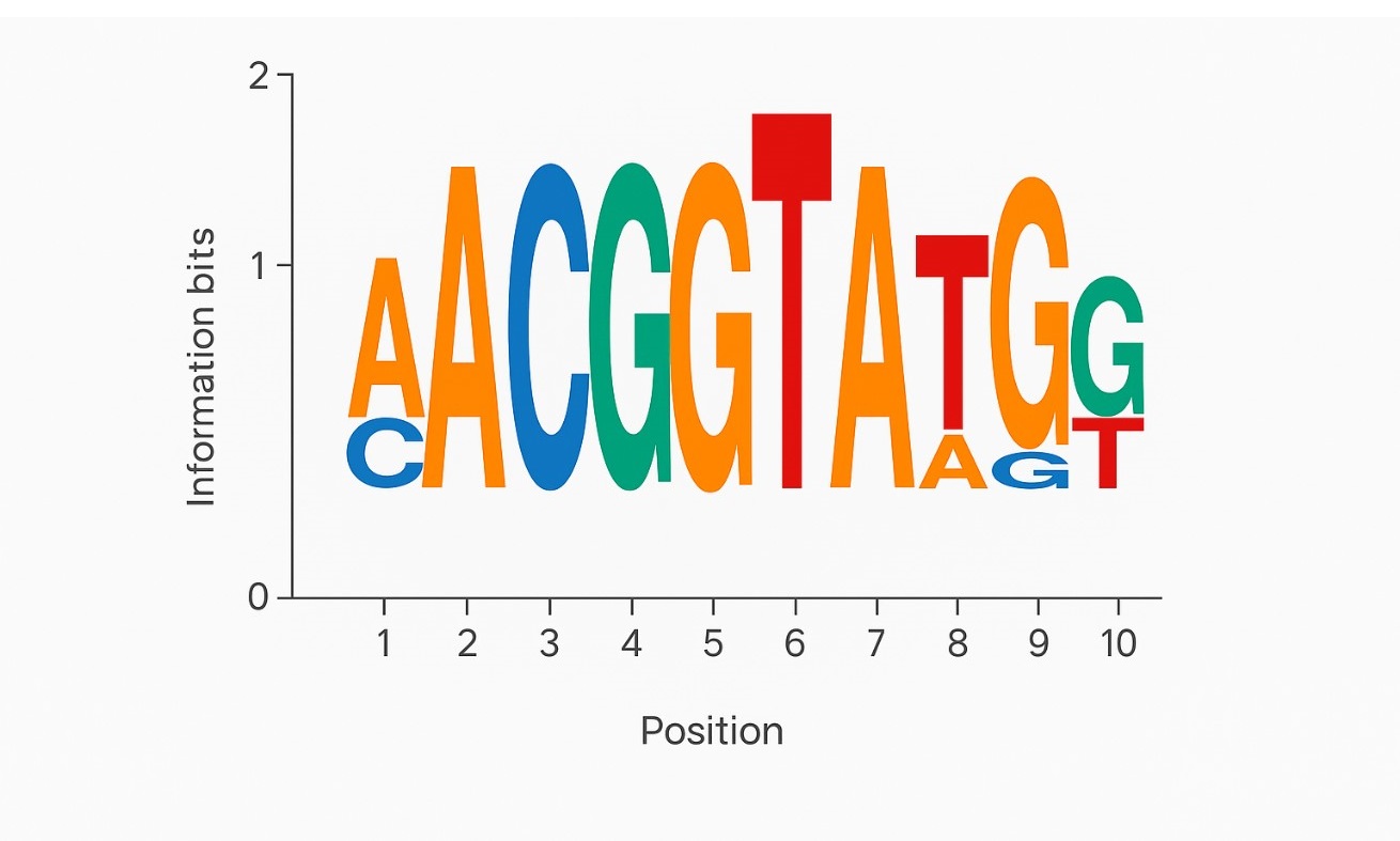
Motif Logo Plot
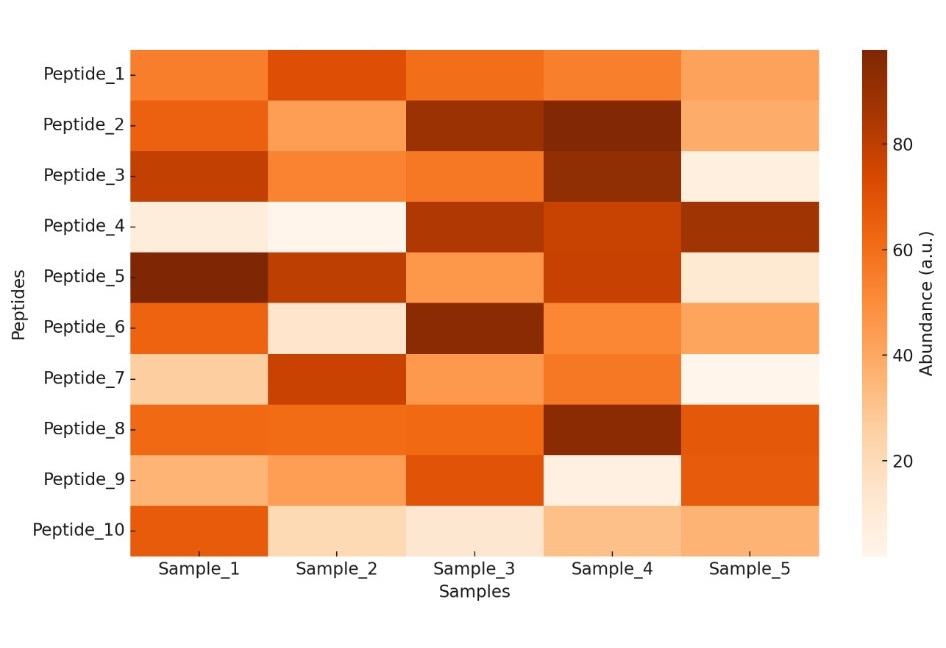
Heatmap of Peptide Abundance Across Samples
Volcano Plot of Differential Peptide Abundance

Deliverables | What You Will Receive
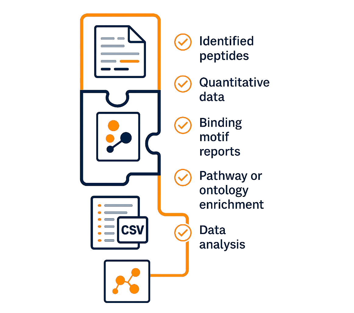
- Identified Peptides (CSV Format)
List of all HLA-bound peptides with sequences, lengths, charge states, and spectral quality scores. - Quantitative Data (Normalized)
Relative peptide abundance (intensity or spectral count) across sample groups, ready for statistical analysis or visualization. - HLA Binding Motif Reports
Sequence logos and binding motif tables derived from deconvoluted peptide sets, categorized by HLA alleles. - PTM and Variant Annotations (if applicable)
Detected phosphorylation, glycosylation, or citrullination sites, and candidate neoantigen peptides with mutation context. - Pathway & Ontology Enrichment (Optional)
Functional insights into peptide source proteins, including GO terms, KEGG pathways, and immune-related processes. - Graphical Summary PDF
A visual executive summary with peptide length distribution, volcano plots, and key motif logos.
FAQ for Immunopeptidome Analysis
How many peptides can I expect to identify per sample? +
Depending on sample type and quality, we typically detect 5,000–12,000 peptides per HLA class. Dual-class enrichment (Class I + II) may increase total yield.
Are post-translational modifications (PTMs) analyzed by default? +
Only common PTMs (e.g., oxidation, deamidation) are included in baseline searches. For targeted PTM analysis (e.g., phosphorylation, citrullination), please request this in advance.
Do you offer replicates or technical reproducibility testing? +
Yes, technical replicates and enrichment replicates can be included upon request to evaluate reproducibility or inter-sample variability.
Is it possible to analyze secreted or extracellular vesicle-derived immunopeptides? +
In most cases, no. HLA-bound peptides are cell surface-associated. Secretome/exosome profiling requires alternative proteomics workflows, which we can advise on separately.
Can you distinguish monoallelic vs. multiallelic HLA-derived peptides? +
Yes. When HLA typing is provided, we can assign peptides to specific alleles. In multi-allelic samples, we apply deconvolution algorithms to cluster peptides by binding motifs and anchor preferences, though exact allele attribution may have ambiguity without monoallelic controls.
Do you support rare or non-classical HLA alleles (e.g., HLA-E, -F, -G)? +
We focus primarily on classical HLA-A, -B, -C, -DR, -DQ, and -DP. Non-classical alleles can be supported only with custom antibodies and motif definitions, which must be validated separately.
What is the false discovery rate (FDR) applied to peptide identification? +
All datasets are filtered at a strict 1% peptide-level FDR, calculated by decoy database search. Optionally, protein-level and motif-level FDR thresholds can also be applied depending on use case.
Can neoantigen candidates be ranked by predicted immunogenicity? +
Yes. We can integrate NetMHCpan or IEDB prediction pipelines to score candidate peptides for HLA binding affinity, source mutation context, and optionally T-cell epitope prediction models, allowing prioritization for vaccine or TCR development.
Do you analyze source protein bias or protein turnover influence? +
Upon request, we can perform integration with matched proteome or transcriptome datasets to assess presentation bias, protein abundance correlation, and DRiP vs. stable protein presentation ratios.
How are shared vs. private peptides determined across samples? +
We use sequence alignment and motif clustering to define shared immunopeptides. Differential expression and allele-specific restrictions are considered to identify truly "shared" vs. patient-specific epitopes.
Can immunopeptidomics be used to monitor treatment-induced antigen shifts? +
Absolutely. We offer comparative profiling pre- and post-treatment (e.g., IFN-γ, checkpoint inhibitors, small molecules) to capture antigenic remodeling, upregulated ligands, and loss-of-presentation signatures.
Is differential HLA ligand presentation quantifiable across conditions? +
Yes. With PRM or DIA-based workflows, we can quantify ligand abundance changes with statistical modeling, enabling identification of differentially presented peptides with fold-change and p-value.
Can I submit pooled or bulked clinical samples? +
Yes, but this may obscure sample-specific HLA restriction and reduce discovery of private epitopes. If pooling is necessary, we recommend maintaining consistent HLA background across the pool.
What limitations should I expect in low-abundance antigen detection? +
Detection of low-abundance peptides is influenced by sample input, peptide stability, enrichment efficiency, and instrument sensitivity. For low-input samples (<1M cells), PRM or SureQuant is recommended for targeted detection.
Large-Scale Immunopeptidome Analysis Reveals Recurrent Posttranslational Splicing of Cancer- and Immune-Associated Genes
Journal: Molecular & Cellular Proteomics
Published: Volume 22, Issue 4, 2023
Summary
Using advanced mass spectrometry and a novel spliced peptide prediction pipeline, researchers analyzed immunopeptidomic data from cancer patients and cell lines. They identified over 150 recurrent posttranslational spliced peptides presented by HLA class-I and class-II molecules, several of which derived from cancer-related genes like DAPK1 and HLA-E. Importantly, two of these peptides were validated using synthetic standards and shown to
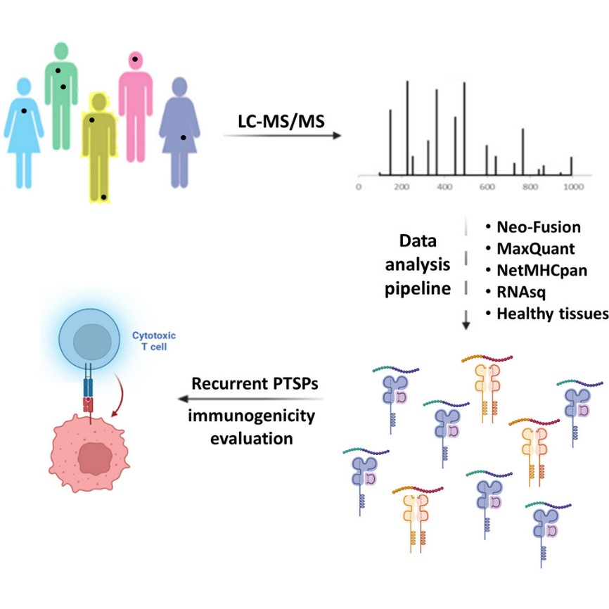
Methods
This study presents a novel computational pipeline combining Neo-Fusion and MaxQuant to stringently identify and validate recurrent PTSPs across multiple cancer types using large-scale MS-based immunopeptidomics data.
Key Technical Features:
- Sample types: Patient-derived melanoma lines, breast cancer (MCF7), HNSCC (SCC47), and mouse melanoma clones
- Peptide identification: Neo-Fusion detects spliced sequences; MaxQuant performs spectrum matching
- Stringent filtration: Peptides were filtered for binding affinity (NetMHCpan), MS/MS coverage, retention time, transcriptomic origin (RNA-seq), and presence in normal or microbial proteomes
- Validation: Light and heavy synthetic peptides were used to confirm MS identifications
- Immunogenicity tests: Conducted using TILs and PBMC-derived T cells
- Recurrent peptides: Defined by presence across multiple independent patients or metastases
Creative Proteomics can offer advanced immunopeptidomics services that mirror and support this research pipeline, including:
- HLA peptidomics profiling using high-resolution LC-MS/MS
- Neoantigen discovery and PTSP identification
- HLA class-I and class-II ligandome mapping
- Quantitative and comparative immunopeptidome analysis across samples or treatment conditions
- Synthetic peptide validation and custom bioinformatics pipelines for spliced peptide filtering
These services empower cancer immunotherapy research, vaccine target screening, and immune evasion mechanism studies.
Results
PTSP Frequency Is Low but Significant
- PTSPs constitute <3.1% of HLA class-I and <0.5% of HLA class-II immunopeptidomes.
- Despite the low proportion, PTSPs outnumber neoantigens derived from somatic mutations, highlighting their potential clinical relevance.
Recurrent PTSPs Identified Across Patients
- 143 HLA class-I and 10 HLA class-II recurrent PTSPs were discovered.
- These peptides are associated with cancer-related genes such as MITF, DAPK1, IL17RE, and HLA-E.
Immunogenicity Validated
- Two PTSPs (from DAPK1 and HLA-E) elicited significant CD8+ T cell responses in autologous TILs and PBMC-derived T cells.
- Flow cytometry showed upregulation of activation markers (e.g., 4-1BB, IFN-γ, TNF-α).
- Dextramer staining confirmed peptide-specific CD8+ T cell populations in healthy donors.
Spliced Peptides Recur in Single-Cell Clones
- Spliced peptides were consistently detected across 5–15 single-cell clones derived from the same mouse tumor, demonstrating biological reproducibility.
Pipeline Validation and Stringency
- Most spurious hits were removed by Delta score and Leu/Ile ambiguity filters.
- Leu/Ile replacement filter alone provided a 30–40× discrimination between WT and spliced peptide false-positives.
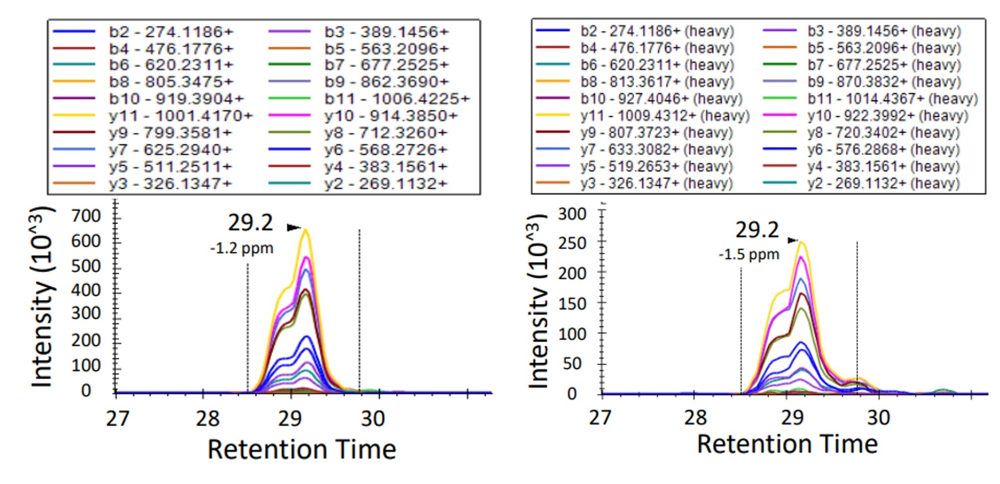 Overlay of endogenous and isotopically labeled HLA-E–derived peptide (WSDSSGGKGGSY) chromatograms in targeted immunopeptidomics of 83T melanoma cells, visualized via Skyline.
Reference
- Levy, Ronen, et al. "Large-scale immunopeptidome analysis reveals recurrent posttranslational splicing of cancer-and immune-associated genes." Molecular & cellular proteomics 22.4 (2023). https://doi.org/10.1016/j.mcpro.2023.100519
Combined Omics Approaches Reveal Resistance Mechanisms to Beet Curly Top Virus in Sugar Beet Genotypes
Journal: International Journal of Molecular Sciences
Published: 2023
https://doi.org/10.3390/ijms241915013BRCA1 Antibodies Matter: Benchmarking Reagent Specificity in Cancer Research
Journal: International Journal of Biological Sciences
Published: 2021
https://doi.org/10.7150/ijbs.63115In Vitro Identification of Phosphorylation Sites on TcPolβ by Protein Kinases TcCK1, TcCK2, TcAUK1, and TcPKC1 and Effect of Phorbol Ester on Activation by TcPKC of TcPolβ in Trypanosoma cruzi Epimastigotes
Trypanosoma cruzi DNA Polymerase β Is Phosphorylated In Vivo and In Vitro by Protein Kinase C (PKC) and Casein Kinase 2 (CK2)
The Interplay Between GSK3β and Tau Ser262 Phosphorylation During the Progression of Tau Pathology
Journal: International Journal of Molecular Sciences
Published: 2022
https://doi.org/10.3390/ijms231911610Inactivation of Myeloperoxidase by Benzoic Acid Hydrazide
Journal: Archives of Biochemistry and Biophysics
Published: 2015
https://doi.org/10.1016/j.abb.2015.01.028