Why Single-Cell Neuropeptidomics Matters in Heterogeneous Tissues
The central nervous system is highly complex, and traditional bulk tissue analysis often masks the critical signals of rare, functionally distinct neuron populations. Unlocking this complexity requires single-neuron peptide profiling.
While single-cell RNA sequencing (scRNA-seq) has revolutionized cell typing, transcript levels rarely correlate perfectly with bioactive peptide secretion. Transcript ≠ peptide ≠ active signaling. Prohormones undergo complex, cell-type specific enzymatic cleavages and post-translational modifications (PTMs) that RNA data simply cannot predict. To truly map neuro-circuit biology and unravel disease mechanisms, researchers must perform neuropeptide heterogeneity analysis to observe the functional signaling molecules exactly where they are produced.
When to Use Single-Cell Neuropeptidomics
Our specialized single-cell neuropeptidomics service is designed to overcome critical bottlenecks in advanced neuroscience and drug discovery. It is the definitive solution when:
| Scenario / Challenge | Why Single-Cell Neuropeptidomics is Required |
|---|---|
| Bulk/tissue-level proteomics fails to resolve signal | High-abundance proteins and surrounding glial cells drown out the low-abundance neuropeptides of target neurons. |
| scRNA-seq completed but lacks functional validation | You need to prove that the identified mRNA actually translates into functional, mature neuropeptides in specific clusters. |
| Reviewer requires cell-type specific peptide evidence | High-impact journals demand direct proteomic validation of spatial or transcriptomic claims. |
| Studying rare neurons or specific brain regions | Your target biology involves discrete cell populations (e.g., within the hypothalamus or amygdala) driving specific behaviors. |
| Drug response heterogeneity at the cellular level | You need to understand why only a subset of cells responds to pharmacological intervention. |
Service Scope: What We Deliver in Single-Cell Neuropeptidomics
We provide an end-to-end analytical engine tailored for high-resolution discovery:
Single-Cell Neuropeptidomics Workflow and Technical Capabilities
Capturing attomole-level peptides requires flawless execution. Our workflow integrates proprietary stabilization with next-generation instrumentation:
Single-Cell Isolation & Handling
Native Peptide Stabilization
Peptidome Enrichment
High-Sensitivity MS Acquisition
Data Processing
1
Single-Cell Isolation & Handling
We utilize high-precision microdissection, cell picking, and microfluidic technologies with picoliter-scale control to isolate intact cells without inducing stress-related artifactual processing.
2
Native Peptide Stabilization
Neuropeptides degrade in seconds. We employ rapid thermal inactivation and immediate protease quenching to freeze the native peptidome in its true physiological state.
3
Low-Molecular-Weight Peptidome Enrichment
Using advanced MWCO (Molecular Weight Cut-Off) and SPE (Solid-Phase Extraction) enrichment, we actively isolate the short, non-tryptic peptide fraction from larger structural proteins.
4
High-Sensitivity MS Acquisition
Leveraging the unprecedented sensitivity of the Orbitrap Astral™ and timsTOF Pro platforms, we perform PTM-aware DIA/DDA acquisition optimized specifically for low-mass endogenous fragments.
5
Single-Cell Data Processing
Our bioinformatic engine conducts 1% FDR-controlled peptide identification, mapping truncated sequences back to their prohormone precursors to reconstruct the processing map.
Project Phasing & Estimated Turnaround Time
To manage project risk and ensure data quality, we structure single-cell projects into clear milestones:
- Phase 1 (1–2 Weeks): Cell isolation, micro-extraction, and peptide stabilization QC. (Go/No-Go Checkpoint)
- Phase 2 (3–4 Weeks): Ultra-high sensitivity LC-MS/MS acquisition across single-cell cohorts.
- Phase 3 (2–3 Weeks): Bioinformatics processing, single-cell clustering, and final report generation.
Technical Benchmarks: Sensitivity, Depth, and Reproducibility
When conducting single-cell peptidomics for CNS research, data integrity is paramount. Achieving single-cell resolution requires moving beyond standard proteomics hardware. By coupling picoliter-scale nanodroplet sample preparation (such as nanoPOTS methodologies) with the latest generation of ion mobility and high-resolution mass spectrometers, we set the industry standard for analytical rigor:
- Detection Depth: >50–100 unique endogenous peptides successfully identified per single cell.
- Sensitivity: Reliable detection of neuropeptides at the attomole (10-18 mole) level.
- Reproducibility: Excellent quantitative precision with a CV <15% across biological single-cell replicates.
- Resolution: Accurate sequencing of ultra-short signaling peptides (<10 amino acids).
- Scalability: Robust pipelines compatible with 100+ single-cell analyses per cohort for statistically significant clustering.
- Data Purity: Strict 1% False Discovery Rate (FDR) control applied at both the peptide and single-cell mapping levels.
Single-Cell Neuropeptidomics vs Tissue-Level Proteomics and RNA-Seq
| Dimension | Single-Cell Neuropeptidomics | Tissue-Level Proteomics | RNA-Seq (scRNA-seq) |
|---|---|---|---|
| Resolution | Cell-level | Tissue-level | Transcript-level |
| Functional relevance | Direct (Bioactive molecules) | Partial (Average protein pool) | Indirect (Genetic potential) |
| PTM detection | Yes (Amidation, etc.) | Limited | No |
| Processing mapping | Yes (Cleavage pathways) | No | No |
| Rare cell detection | Strong | Poor | Moderate |
Applications in Neurobiology and Drug Discovery
- Neuro-circuit mapping: Defining exact transmitter/modulator profiles in regions like the hypothalamus to decode feeding, sleep, or stress signaling.
- Rare neuron population discovery: Finding distinct functional states hidden within seemingly homogenous neuron clusters.
- Cell-type specific biomarker identification: Establishing precise peptide targets for neurodegenerative or psychiatric diagnostics.
- Drug response heterogeneity analysis: Tracking how therapeutic compounds differentially alter peptide secretion across distinct cellular phenotypes.
- Functional validation of transcriptomic findings: Providing the definitive protein-level evidence required to validate single-cell RNA atlases.
Demo Results: Visualizing Cellular Peptidomic Heterogeneity
We transform complex, high-dimensional mass spectrometry data into intuitive, publication-ready visualizations:
UMAP/t-SNE Clustering
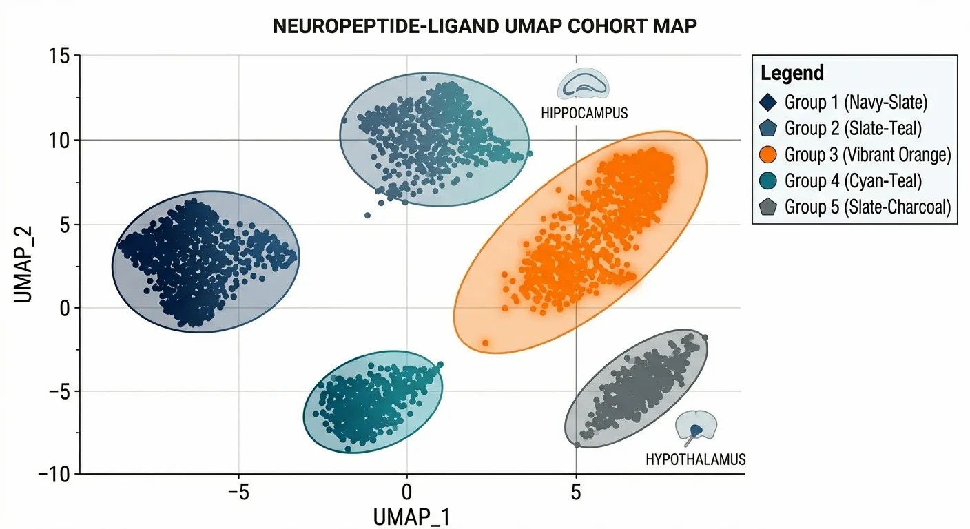
UMAP/t-SNE clustering of single cells showing distinct clusters of individual cells separated purely by their endogenous neuropeptide profiles.
Peptide Expression Heatmap
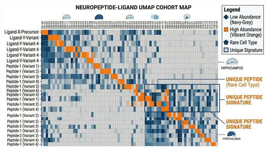
Peptide expression heatmap across cells detailing the presence, absence, and abundance of specific bioactive peptides across 100+ single cells.
Prohormone Processing Maps
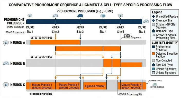
Prohormone processing maps (e.g., POMC) displaying how a single precursor is cleaved into different active isoforms depending on the cell type.
Quantitative Peptide Plots
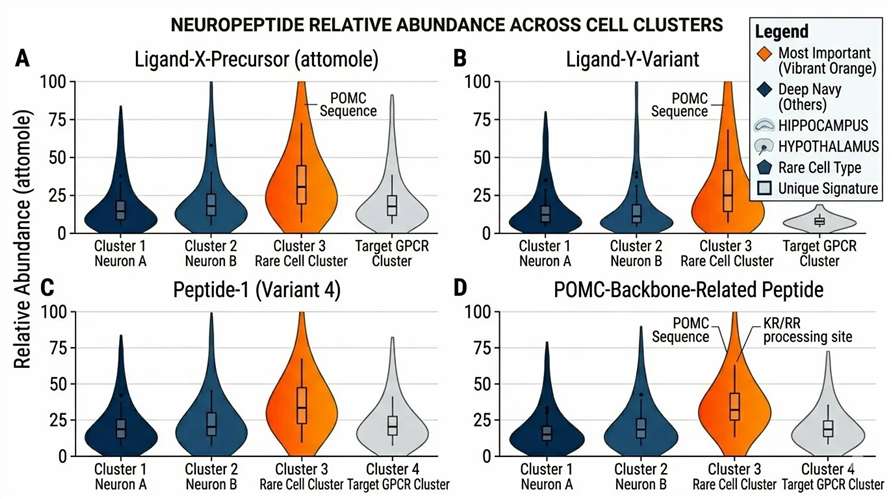
Quantitative peptide distribution plots comparing the abundance of a specific modified peptide (e.g., amidated vs. non-amidated) across different experimental groups.
Data Deliverables for Single-Cell Neuropeptidomics
We provide comprehensive data packages designed to eliminate bioinformatic uncertainty and accelerate your research:
- Single-cell peptide identification matrix (raw and normalized abundances).
- Cell clustering and visualization plots (UMAP/t-SNE).
- PTM annotation and comprehensive peptide variant mapping.
- Prohormone processing maps illustrating cell-type specific cleavage.
- Ranked candidate peptide biomarkers for your target populations.
Sample Requirements for Single-Cell Analysis
| Sample Type | Use Case | Minimum Input | Key Notes |
|---|---|---|---|
| Brain regions | Neuro-circuit mapping | ≥10 mg | Rapid thermal stabilization required immediately upon dissection. |
| Primary neurons | Functional studies | 105–106 cells | Live-cell handling preferred; requires gentle dissociation protocols. |
| Rare clinical samples | Translational research | Limited | Requires optimized, project-specific handling consultation. |
Disclaimer: All services and platforms described are for Research Use Only (RUO). Not for use in diagnostic procedures.
What is the success rate for single-cell detection? +
Our optimized nano-scale preparation and ultra-sensitive MS platforms (like Orbitrap Astral) yield a highly robust success rate. Provided the cells are properly stabilized to prevent ex vivo degradation, we routinely recover 50 to over 100 unique endogenous peptides per individual cell.
How many cells are required per project? +
While we can detect signals from a single cell, meaningful statistical clustering and biological interpretation typically require profiling between 100 to 500 individual cells per experimental condition or biological replicate.
Can you integrate with RNA-seq data? +
Yes. Our bioinformatics team can perform cross-omics correlation. We map the identified mature peptides to their precursor genes, allowing you to overlay peptidomic clustering data directly onto your existing scRNA-seq UMAP/t-SNE plots.
How do you ensure peptide stability? +
Neuropeptides are highly prone to rapid enzymatic degradation during sample handling. We provide strict protocols involving rapid thermal inactivation (e.g., heat stabilization) and specialized protease inhibitor cocktails applied immediately upon cell isolation or tissue microdissection.
How complex is the data interpretation? +
Single-cell MS data is inherently complex, but our deliverables are designed for clarity. We provide fully processed data matrices, annotated PTMs, and intuitive visual plots (heatmaps, processing maps) so your team can focus on biological insights rather than mass spectra interpretation.