Why Growth Factor Peptide Quantification Requires Specialized LC-MS/MS Methods
Growth factor peptides—including insulin-like growth factors (IGF-1, IGF-2), neurotrophins (NGF, BDNF), and mitogenic peptides (EGF, FGF-2, PDGF)—regulate cell proliferation, differentiation, survival, and migration. Their dysregulation underlies cancer, neurodegeneration, growth disorders, and impaired tissue repair.
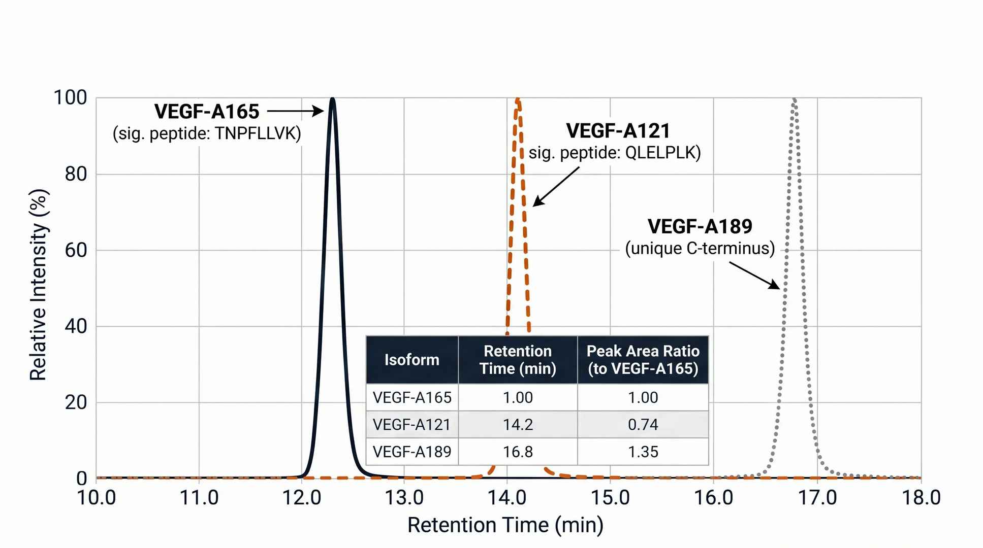
The analytical challenges are distinct from standard protein biomarkers. IGF-1 and IGF-2 circulate tightly bound to IGF-binding proteins (IGFBPs), requiring acid dissociation before LC-MS analysis. Neurotrophins like NGF and BDNF are present at pg/mL concentrations in plasma and contain multiple disulfide bonds that hinder tryptic digestion. Many growth factors exist as multiple isoforms (VEGF-A 165/121/189, FGF family members) with differential biological activities that immunoassays cannot resolve.
Our Growth Factor Peptide Quantification Panel uses targeted LC-MS/MS with optimized pre-analytical workflows—including acid dissociation for IGFBPs, disulfide reduction, and isoform-specific signature peptide selection—to deliver sequence-level quantification of each growth factor independently.
What This Panel Helps You Decide
Configurable Growth Factor Peptide Panel
Depending on your therapeutic area and disease model, our panel can be configured to cover the major growth factor families.
| Growth Factor Family | Selected Target Analytes | Relevance to Drug Discovery |
|---|---|---|
| Insulin-like Growth Factors | IGF-1, IGF-2, IGFBP-3 (adjusted free fraction) | Key mediators of GH axis. Essential for growth disorder research, oncology IGF-1R targeting, and metabolic disease. |
| Neurotrophins | NGF, BDNF, NT-3, NT-4, GDNF | Critical for neurodegenerative disease, peripheral neuropathy, and pain research. Present at pg/mL levels. |
| Mitogenic Peptides | EGF, HB-EGF, TGF-α, Amphiregulin, Epiregulin | EGFR ligand family. Quantification supports oncology and dermatology preclinical programs. |
| Angiogenic Factors | VEGF-A (isoforms 165/121/189), FGF-1, FGF-2, PDGF-AA, PDGF-BB | Core biomarkers for anti-angiogenic therapy, wound healing, and cardiovascular regeneration studies. |
This panel integrates with our RAAS and Vasoactive Peptide Panel, HPA Axis Stress Hormone Panel, Gut-Brain Axis Hormone Panel, and neuropeptidomics profiling platform for a holistic view of growth factor signaling in disease models.
LC-MS/MS Workflow for Growth Factor Peptide Quantification
Our targeted LC-MS/MS workflow addresses the unique pre-analytical and analytical challenges of growth factor peptides—from IGFBP interference to disulfide-rich neurotrophin structures.
Sample Prep & IGFBP Dissociation
Acid dissociation liberates IGF-1/2 from binding-protein complexes.
Reduction & Alkylation
DTT + IAA for disulfide-rich growth factors (NGF, BDNF, EGF).
Proteolytic Digestion & ISTD Spiking
Trypsin/Lys-C with isotope-labeled internal standards for each target.
SPE Cleanup & Enrichment
Solid-phase extraction for low-abundance neurotrophin enrichment.
LC-MS/MS Targeted Acquisition
Orbitrap Astral PRM/MRM with isoform-specific transition design.
Data Processing & Quantification
Skyline peak integration, ISTD normalization, isoform ratio reporting.
1
Sample Preparation & IGFBP Dissociation
Serum or plasma is treated with acid dissociation buffer to liberate IGF-1 and IGF-2 from their binding-protein complexes (IGFBP-1 through -6). This critical step ensures that total IGF concentrations reflect the true peptide pool rather than the fraction accessible by immunoassay. For neurotrophins, samples are processed under native conditions to preserve disulfide integrity until reduction.
2
Reduction & Alkylation
Growth factor peptides are rich in disulfide bonds—NGF contains 3, BDNF 3, EGF 3, and FGF-2 2 disulfide bridges. Complete reduction with DTT followed by iodoacetamide alkylation linearizes these structures, enabling full tryptic access and 100% sequence coverage. This step is essential for accurate quantification of disulfide-rich growth factors.
3
Proteolytic Digestion & ISTD Spiking
Reduced/alkylated samples are digested with trypsin or Lys-C to generate signature peptides unique to each growth factor. Isotope-labeled internal standards for each target are spiked prior to digestion, enabling absolute quantification with correction for recovery, matrix effects, and instrumental drift. Isoform-specific peptides are selected to distinguish closely related family members.
4
SPE Cleanup & Enrichment
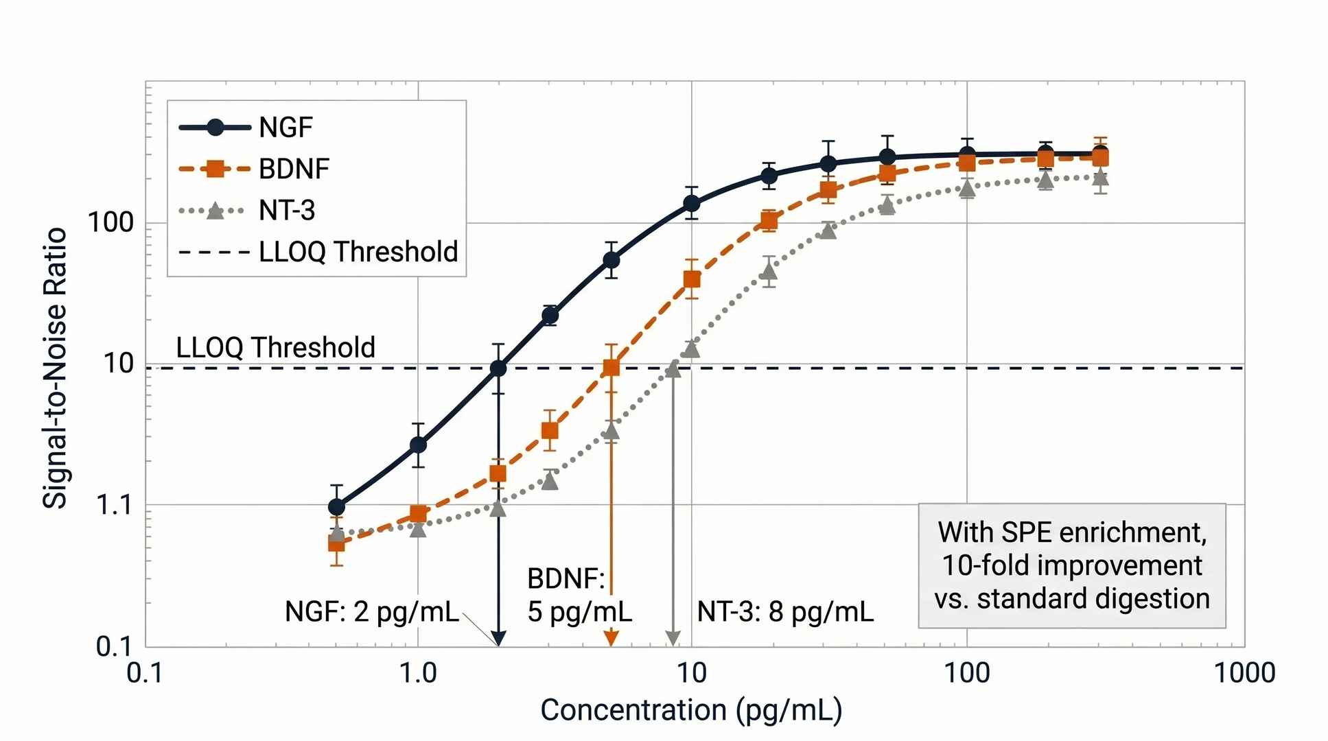
Post-digestion samples undergo solid-phase extraction (C18 or mixed-mode) to remove salts, detergents, and undigested proteins. For low-abundance neurotrophins (NGF, BDNF at low pg/mL), an additional enrichment step using cation-exchange SPE concentrates the target peptides, improving LLOQ by up to 10-fold.
5
LC-MS/MS Targeted Acquisition
Purified digests are analyzed on an Orbitrap Astral platform using parallel reaction monitoring (PRM) with scheduled acquisition windows. Each growth factor is monitored by 3-5 unique transitions, with isoform-specific peptides for VEGF-A165 vs VEGF-A121 and FGF-2 vs FGF-1. Retention time alignment against ISTDs ensures confident peak assignment.
6
Data Processing & Quantification
Raw data are processed in Skyline for peak integration, transition quality review, and ISTD-normalized quantification. Results are reported as absolute concentrations (pg/mL) with LLOQ, linear dynamic range, and intra-batch QC metrics. Isoform ratios (e.g., VEGF-A165/VEGF-A121) are calculated to provide functional insight beyond total concentration.
Platform Specifications and Detection Performance
We deploy high-resolution LC-MS/MS platforms optimized for growth factor peptide quantification.
- IGFBP-Independent Quantification
Acid dissociation step ensures total IGF-1/2 measurement independent of binding-protein interference. - Multi-Species Validation
Validated for rat, mouse, and human plasma/serum with species-specific MRM transitions for each target. - >90% MS/MS Peptide Coverage
HCD and ETD fragmentation enables detection of phosphorylated, amidated, and glycosylated growth factor peptide variants. - 1% FDR Stringent Filtering
Ensures data reliability and reproducibility across biological replicates. - Isoform-Specific Detection
Signature peptide selection distinguishes VEGF-A isoforms, FGF-1 from FGF-2, and PDGF-AA from PDGF-BB. - Low Input Compatibility
Deep coverage from as little as 100 μL plasma or 50 μL serum.
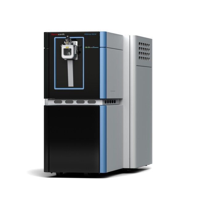 Orbitrap Astral™
(Fig from Thermo Scientific)
Instrument Capability Overview
| Feature | Orbitrap Astral™ | timsTOF Pro | Q Exactive HF-X |
|---|---|---|---|
| Scan Speed | Up to 200 Hz | ~100 Hz (PASEF) | ~20–25 Hz |
| Quantification | Label-free, PRM, SureQuant™ | Label-free, DIA, PRM | Label-free, TMT |
| Peptide Coverage | >90% MS/MS | >90% MS/MS | ~85% MS/MS |
| Low-Abundance | Single-peptide resolution | Ion mobility separation | Standard |
Comparative Analysis of Growth Factor Peptide Quantification Approaches
| Dimension | Creative Proteomics LC-MS/MS | Immunoassay | Standard MS-Based Service |
|---|---|---|---|
| IGFBP Interference | Acid dissociation liberates total IGF pool; binding-protein independent | Antibody may be blocked by IGFBP-bound epitopes | Not addressed in standard workflow |
| Isoform Resolution | Distinguishes VEGF-A165/121, FGF-1/2 by unique signature peptides | Antibody cross-reactivity between isoforms | Detects peptides; isoform assignment not routine |
| Multiplexing | 10+ growth factors per run | 1–2 per assay | Unlimited (discovery mode) |
| Sensitivity | pg/mL (PRM with SPE enrichment for neurotrophins) | pg/mL range with signal amplification | ng/mL range |
| Disulfide Handling | Complete reduction/alkylation for disulfide-rich peptides | Not relevant; antibodies detect folded protein | Partial; may miss disulfide-linked regions |
| Sample Input | ≥100 μL plasma or 50 μL serum | Varies by kit | ≥50 μg protein |
Growth Factor Research Applications in Drug Development
Our panel is designed to support specific R&D tasks across oncology, neurology, and regenerative medicine programs.
- Anti-Angiogenic Therapy Profiling. Quantify VEGF-A isoforms, FGF-2, and PDGF-BB in tumor xenograft models to confirm vascular target engagement and monitor compensatory angiogenic signaling.
- Neurotrophin Replacement Therapy. Track NGF, BDNF, and NT-3 levels in CNS and peripheral matrices following gene therapy or protein replacement in neurodegenerative disease models.
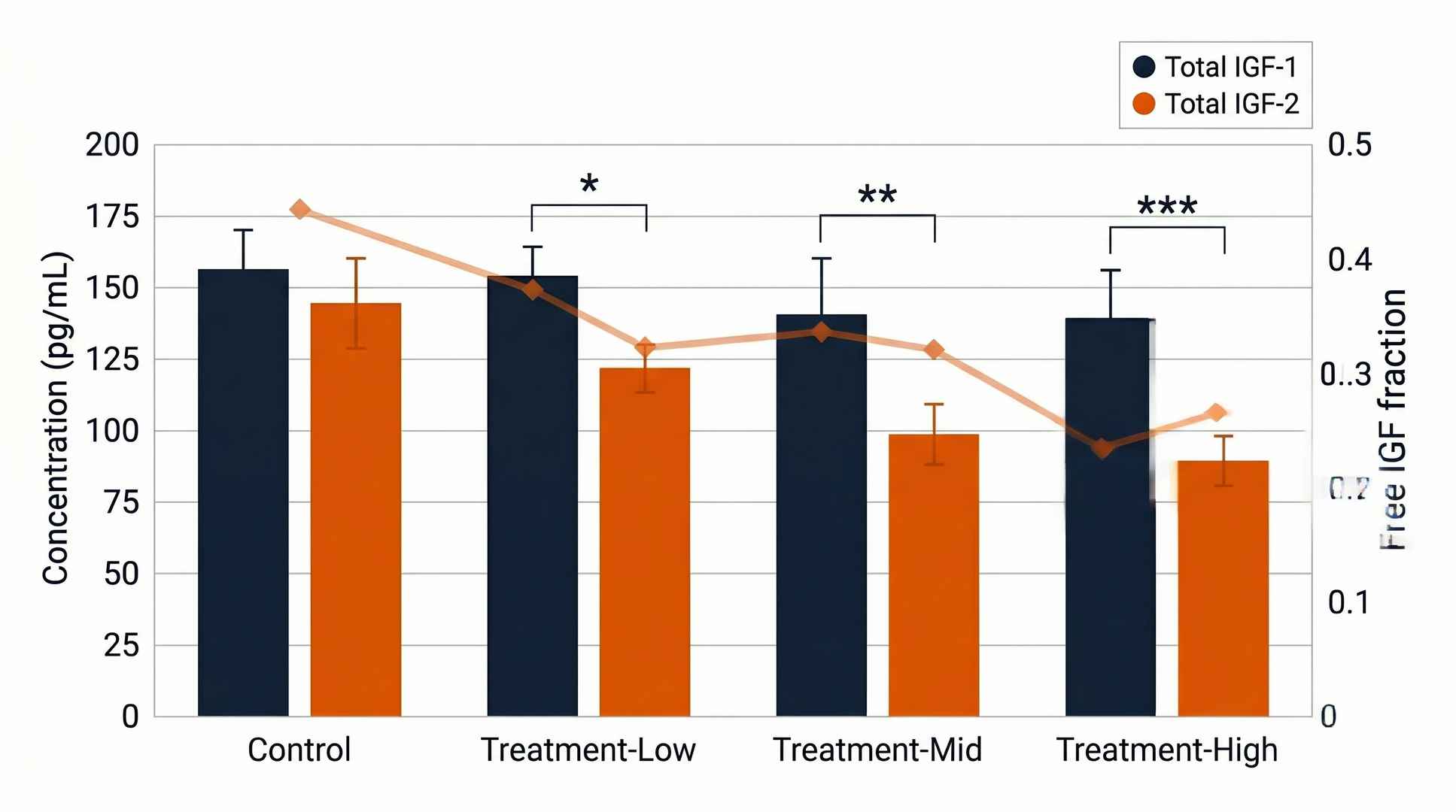
- Growth Hormone Axis Assessment. Profile IGF-1, IGF-2, and calculated free IGF fractions in growth disorder models and GH treatment optimization studies.
- Wound Healing & Tissue Regeneration. Monitor EGF, FGF-2, HB-EGF, and PDGF-BB in cutaneous wound, bone fracture, and myocardial infarction repair models.
- EGFR-Targeted Therapy PD. Track EGF, TGF-α, and amphiregulin levels in response to EGFR inhibitor treatment to assess feedback ligand upregulation.
- Biosimilar Comparability Studies. Compare growth factor isoform profiles and post-translational modification patterns between biosimilar candidates and reference products.
Growth Factor Peptide Panel Demo Data
IGF-1 and IGF-2 Multiplex Quantification
VEGF-A Isoform Ratio Analysis
Neurotrophin Panel Sensitivity
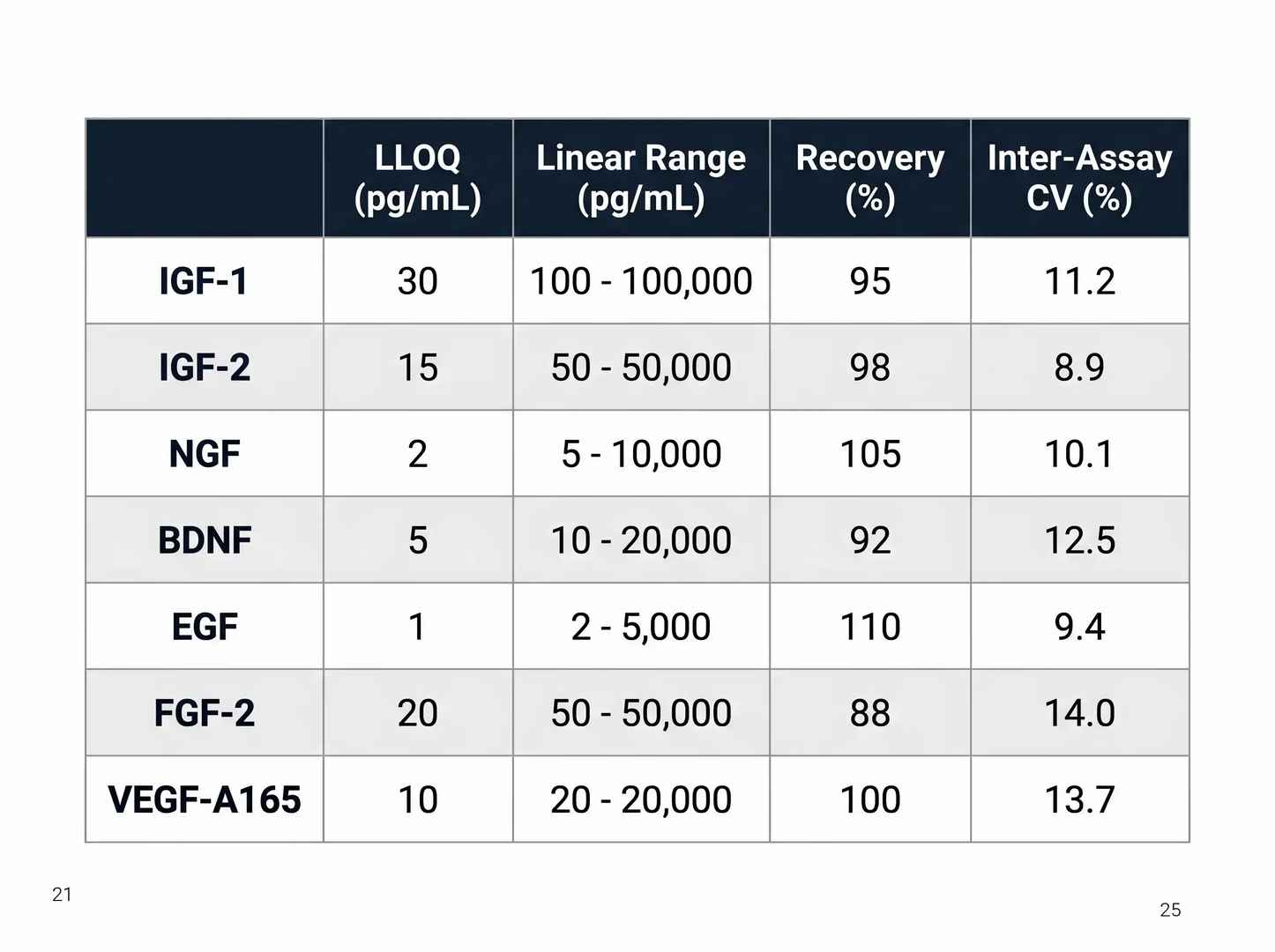
Growth Factor Panel QC Package
Sample Collection Requirements for Growth Factor Peptide Analysis
| Sample Type | Species / Context | Min Volume / Mass | Critical Pre-Analytical SOPs |
|---|---|---|---|
| Plasma (EDTA) | Rodent, NHP, Human | 100 μL | Standard EDTA collection. Avoid freeze-thaw cycles. For IGF quantification, acid dissociation is performed upon sample receipt. |
| Serum | Rodent, NHP, Human | 50 μL | Allow clotting at room temperature for 30 min. Serum is the preferred matrix for IGF-1/2 and growth hormone axis studies. |
| Tissue (Tumor / Brain) | Rodent Models | 20 – 50 mg | Snap-freeze in liquid nitrogen within 30 seconds. Homogenize in RIPA buffer with protease and phosphatase inhibitors. |
| Cell Supernatant | Ex vivo / In vitro | 500 μL | Centrifuge at 4°C to remove debris. Add protease inhibitor cocktail. For neurotrophins, add antioxidant to prevent oxidation. |
| CSF | Rodent, NHP, Human | 100 μL | Collect into polypropylene tubes; avoid glass which adsorbs neurotrophins. Centrifuge at 4°C, aliquot and flash-freeze within 30 minutes. |
(Consult with our scientific team prior to initiating your in vivo study for tailored guidance on collection protocols specific to your growth factor targets.)
Deliverables: What You Receive
- Growth Factor Quantification Report (PDF)
Complete summary of all detected and quantified growth factors across all samples, with statistical analysis and isoform ratio calculations. - Raw Data Files
Full LC-MS/MS raw data in standard format (Thermo .raw or Bruker .d) for archival and regulatory submission support. - Quantification Table (CSV)
Peptide IDs, retention times, peak areas, absolute concentrations (pg/mL), isoform ratios, and QC metrics for each growth factor. - Internal Standard Recovery Report
Recovery rates for each isotope-labeled internal standard, documenting assay performance across the analytical batch. - Comparative Statistics
Fold-change analysis, p-values, PCA plots, and dose-response curves for multi-group preclinical study designs. - Optional: IGFBP-Adjusted Free IGF Calculation
Computed free IGF-1 and IGF-2 fractions based on total IGF and IGFBP-3 levels for growth hormone axis assessment.
How do you handle IGF-binding protein interference? +
We apply acid dissociation buffer to liberate IGF-1 and IGF-2 from IGFBP-1 through IGFBP-6 complexes prior to digestion. This ensures that total IGF concentrations reflect the complete peptide pool, not just the immunoassay-accessible fraction.
Can you distinguish VEGF-A isoforms? +
Yes. VEGF-A165, VEGF-A121, and VEGF-A189 are distinguished by isoform-specific signature peptides generated after tryptic digestion. Each isoform produces unique MRM transitions that enable independent quantification.
What is the sensitivity for neurotrophins like NGF and BDNF? +
With SPE enrichment, we routinely achieve LLOQ in the low pg/mL range (1–10 pg/mL) for NGF, BDNF, and NT-3 in plasma and CSF. Without enrichment, the LLOQ is approximately 50–100 pg/mL.
How do you handle disulfide-rich growth factors? +
Complete reduction with DTT followed by iodoacetamide alkylation linearizes disulfide bonds before tryptic digestion. This ensures 100% sequence coverage and accurate quantification of NGF, BDNF, EGF, and FGF-2.
What sample types are compatible? +
Plasma (EDTA), serum, tissue (tumor, brain), cell culture supernatant, and CSF have all been validated. Serum is preferred for IGF-1/2 quantification.
Is the panel validated for both rodent and human samples? +
Yes. Rat, mouse, and human plasma/serum have been validated. Cross-species sequence differences are accounted for in MRM transition design.
Can I add custom growth factors to the panel? +
Yes. Additional growth factors or related signaling peptides can be added on request. Contact our scientific team with your targets of interest for feasibility assessment.
Disclaimer: All services and analytical platforms described are intended for translational research and preclinical support. Research Use Only (RUO). Not for use in diagnostic procedures.