Published Research
Novel Peptide Macoluxin from Madagascar Cat-Eyed Snake Venom Blocks Nicotinic Acetylcholine Receptor
Journal
Russ J Bioorg Chem
DOI
10.1134/S1068162023030159
Study Overview
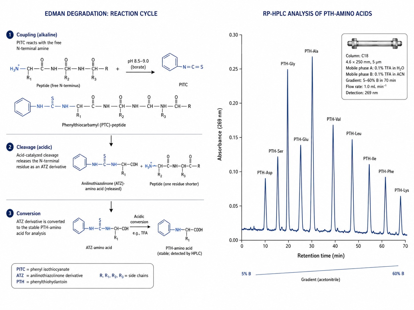
Kryukova et al. (Russian Journal of Bioorganic Chemistry, 2023) screened the venom of the Madagascar cat-eyed snake (Madagascarophis colubrinus) — a rear-fanged species — and discovered macoluxin, a novel peptide that competitively inhibits muscle-type nicotinic acetylcholine receptors (nAChR). This work marked the first identification of nAChR-inhibitory activity in any rear-fanged snake species, significantly expanding the known pharmacological diversity of snake venom components. The venom was fractionated by size-exclusion chromatography (Superdex 75) followed by reversed-phase HPLC on a Jupiter C18 column, isolating a single bioactive fraction. Because macoluxin had no prior sequence entry in any database, Edman degradation was the only viable method for primary structure determination.
Key Methods
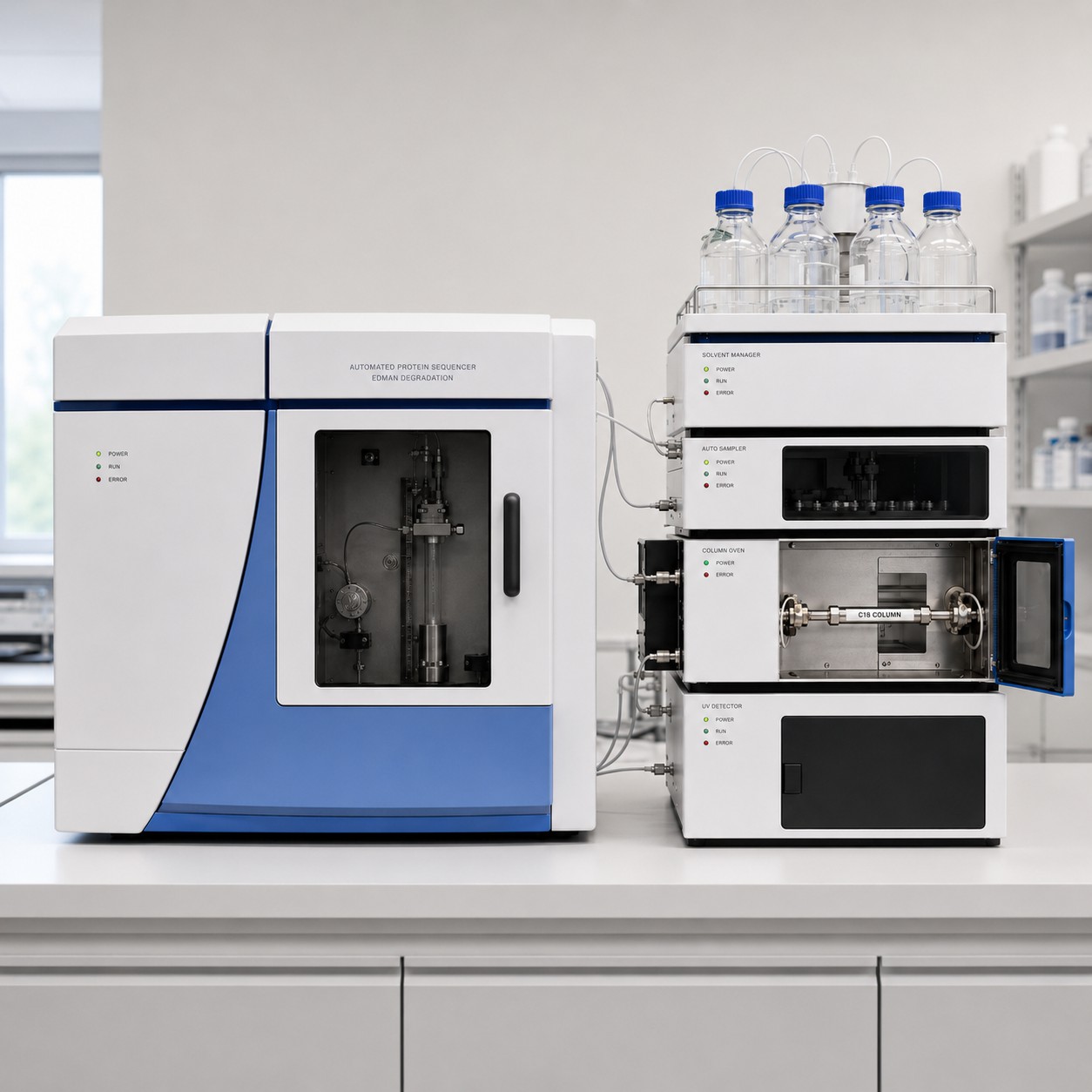
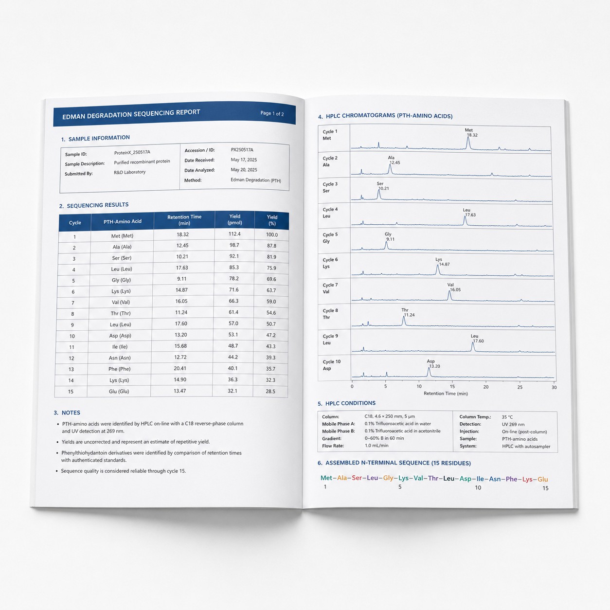
- N-terminal sequencing by Edman degradation — primary structure of macoluxin determined on a Shimadzu PPSQ-33A automated protein sequencer, producing a complete N-terminal read without database support
- Solid-phase peptide synthesis — macoluxin chemically synthesized to confirm the Edman-derived sequence and produce milligram quantities for biological validation
- Competitive fluorescence binding assay — Alexa Fluor 488-labeled α-bungarotoxin used to measure macoluxin's affinity for nAChR on Torpedo californica electroplaque membranes
- Two-electrode voltage clamp electrophysiology — Xenopus oocytes expressing muscle-type nAChR used to confirm macoluxin reversibly inhibited acetylcholine-evoked currents
- Intracellular calcium imaging — genetically encoded Case12 calcium sensor expressed in Neuro2a and HEK293 cells to monitor real-time nAChR activity changes upon macoluxin treatment
- [125I]-α-bungarotoxin radioligand competition assay — confirmed competitive, reversible binding at the nAChR orthosteric site
Relevance to Edman Degradation Sequencing
- This study exemplifies the irreplaceable role of Edman degradation when characterizing novel, unsequenced peptides — without a sequence database for MS/MS spectral matching, Edman degradation provided the definitive residue-by-residue N-terminal read that anchored the entire structural characterization.
- The Shimadzu PPSQ-33A sequencer used in this study belongs to the same instrument family as our PPSQ-51A/53A platform, demonstrating the workflow's direct transferability to a service-level protein sequencing environment.
- Edman sequencing revealed macoluxin's high sequence homology to a snake venom metalloproteinase fragment — a discovery that would have been far less certain by MS-based de novo sequencing alone, given the peptide's novelty and the absence of genomic context for this species.
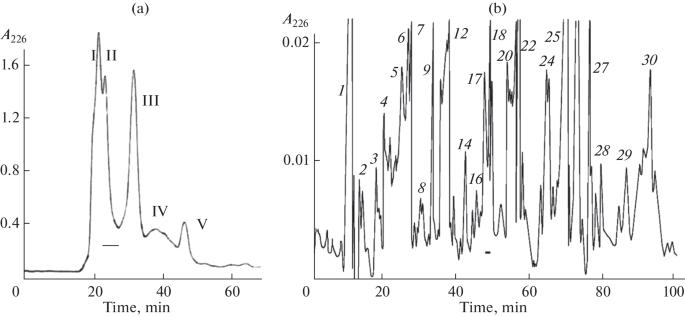
Fig. 1. Isolation of the active compound (macoluxin) from the venom of M. colubrinus. Fraction 18 was separated by reversed-phase HPLC on a Jupiter C18 column (4.6 × 250 mm) with a 20–30% acetonitrile gradient over 60 min at a flow rate of 1 mL/min. The horizontal line indicates the active fraction that was subsequently analyzed by N-terminal Edman degradation on a Shimadzu PPSQ-33A sequencer. (Adapted from Kryukova et al., 2023)
Key Finding
Edman degradation on the Shimadzu PPSQ-33A successfully determined the complete N-terminal sequence of macoluxin — a peptide with no prior sequence information in any public database. The Edman-derived sequence revealed that macoluxin is a proteolytic fragment of a larger snake venom metalloproteinase, demonstrating that venom protein processing can generate structurally distinct, biologically active peptides with pharmacological functions different from their parent molecules. Macoluxin competitively inhibited muscle-type nAChR with reversible kinetics, as confirmed by three orthogonal assays. Without Edman degradation, the primary structure of this novel peptide — and the mechanistic insight into its evolutionary origin — would have remained unresolved.
Publication Reference
Kryukova EV, Ivanov IA, Khochareva TA, Ziganshin RH, Starkov VG, Tsetlin VI, Utkin YN. A New Peptide from the Venom of the Madagascar Cat-Eyed Snake Madagascarophis colubrinus Blocks Nicotinic Acetylcholine Receptor. Russ J Bioorg Chem. 2023;49(3):529–537. DOI: 10.1134/S1068162023030159.