Recombinant monoclonal antibodies, as intricate biomacromolecules, manifest heterogeneity, rendering antibody drugs a consortium of various entities rather than a singular substance. Throughout processes such as production, storage, and transportation, these antibodies are prone to alterations like aggregation, post-translational modifications, and degradation, consequently instigating changes in protein surface charges. This phenomenon underscores the genesis of charge variants in recombinant monoclonal antibodies.
Several studies have illuminated the influence of antibody charge variations on the binding of antibodies to proteins/cell membranes, subsequently altering tissue penetration, distribution, and pharmacokinetic (PK) profiles of the antibodies. The impact of charge variants on protein functionality is intricately linked to the nature, location, and extent of their post-translational modifications. A thorough examination of the molecular basis and influencing factors behind the formation of charge variants contributes to the nuanced control and adjustment of charge heterogeneity in the development of antibody drugs. Consequently, the analysis and detection of variants in substances related to antibody drugs hold paramount significance. This review aims to provide an overview of the research progress on the modifications of monoclonal antibodies concerning the generation of charge variants and the analytical methods employed in this context.
Dedicated to establishing a comprehensive platform for the analysis and detection of antibody drugs, Creative Proteomics boasts state-of-the-art analytical instruments and a proficient technical team. Our platform aims to provide a wide array of analytical and detection services, including but not limited to iCIEF, IEC analysis, physicochemical analysis, content determination, heterogeneity studies, purity and product-related impurity analysis, process-related impurity assessment, biological function testing, excipient analysis, safety evaluation, as well as analysis of culture media and cell culture supernatants. Our core principles revolve around efficiency, precision, and professionalism, committed to delivering optimal analytical and detection solutions for your needs.
Select Service
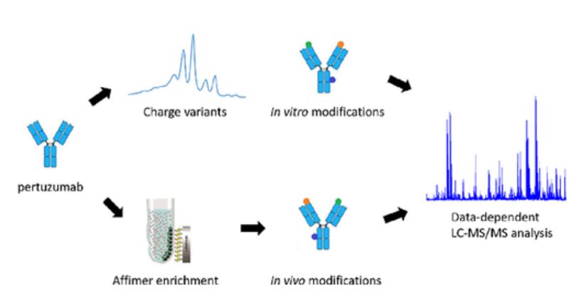 Pertuzumab Charge variants analysis (Anal. Chem. 2023, 95, 8, 3951-3958)
Formation of Charge Variants in Antibody
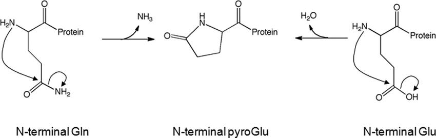
Formation of N-Terminal Pyroglutamate
The N-terminal regions of antibody molecules, encompassing both light and heavy chains, typically contain glutamic acid (Glu) or glutamine (Gln). Under the catalytic action of glutaminyl cyclase, Glu and Gln undergo dehydration or deamidation reactions, resulting in the formation and cyclization of N-terminal pyroglutamate (pyroGlu). Recombinant monoclonal antibodies often have N-terminal Gln that tends to cyclize into pyroGlu, while those with N-terminal Glu usually remain in their natural form.
During the cyclization process, the loss of the amino group accompanying Gln leads to a reduction in the positive charge, consequently causing a decrease in the net charge of the antibody. Antibodies with this modification are typically eluted in the main peak during chromatography. However, antibodies where Gln does not cyclize or cyclizes incompletely may form basic isoforms. In contrast, Glu undergoes dehydration condensation during cyclization, but its net charge remains unchanged.
 Pyroglutamate formation mechanism. (Y. Diana Liu et al,. 2011)
Removal of C-Terminal Lysine
The C-terminal region of the heavy chain of an antibody molecule typically concludes with the amino acid sequence proline-glycine-lysine (-Pro-Gly-Lys). During the cell culture process, the terminal lysine (Lys) is partially cleaved by carboxypeptidases, resulting in incomplete removal. This incomplete removal leads to the presence of residual lysine in some antibody proteins, referred to as lysine variants.
Considering lysine's alkaline nature, antibodies containing residual lysine are identified as basic isoforms due to the presence of this lysine residue. This phenomenon arises from the partial removal of lysine during the cellular cultivation process.
N-Glycosylation (Sialylation)
Glycosylation refers to the enzymatic process of attaching sugar chains to protein or lipid molecules, representing a crucial post-translational modification in biological organisms. In the context of recombinant monoclonal antibodies, N-glycosylation typically occurs at the Asn297 residue within the conserved sequence of the Fc segment CH2. This site consists of a core pentasaccharide structure composed of 2 N-acetylglucosamine (GlcNAc) and 3 mannose (Man) units. The addition of different monosaccharides (such as galactose, mannose, sialic acid, etc.) to its terminal end results in pronounced heterogeneity of the antibody.
While the glycan chain is generally neutral, the addition of negatively charged sialic acid residues at the terminal end, along with increased galactose residues, induces changes in the charge, leading to charge heterogeneity.
The deamidation of asparagine and isomerization to isoaspartic acid Upon deamidation, asparagine (Asn) transforms into succinimide (Asu), resulting in the formation of an acidic variant through an irreversible process. Succinimide, upon hydrolysis, can convert back to aspartic acid (Asp) or isomerize into isoaspartic acid, constituting a reversible reaction known as aspartate isomerization. While the isomerization of Asp does not alter the net charge of the antibody, it does modify the distribution of charges. Consequently, in cation exchange chromatography, this is reflected as an alkaline peak due to the altered charge distribution, highlighting the nuanced impact of aspartate isomerization on the chromatographic behavior of antibodies.
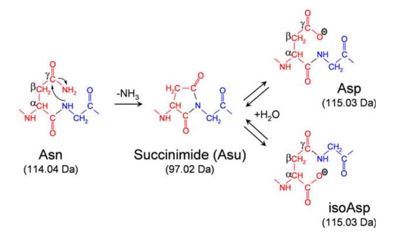 The mechanism of asparagine deamidation and aspartic acid isomerization via succinimide intermediate. (Josef Vlasak et al,. 2019)
Oxidation Methionine (Met) is a susceptible amino acid to oxidation within antibodies. Under elevated oxidative conditions, Met undergoes oxidation to form methionine sulfoxide. Despite the absence of net charge alteration during the oxidation process, the spatial charge distribution within the antibody is affected. Research has confirmed that the oxidation of Met can lead to the generation of alkaline variants. Additionally, the phenomenon of oxidation is commonly observed in other amino acids such as cysteine, histidine, and tryptophan. This underscores the broader spectrum of oxidative events impacting amino acid residues within antibodies.
Other Factors
Research indicates that the generation of charge variants in monoclonal antibodies is not solely attributed to the aforementioned five modification methods. It also involves glycation, a reaction between reducing sugars and lysine residue side chains or N-terminal primary amines. Additionally, atypical disulfide bond formations in IgG2 (IgG2B, IgG2A/B), the generation of tri-disulfide bonds, high mannose content, malic acid modifications, and cysteinylation induced by free thiol groups contribute to the diversity of phenomena that may result in the production of charge variants in antibody therapeutics. These findings underscore the intricate interplay of various biochemical processes influencing the charge heterogeneity of antibody drug products.
Select Service
Analysis Strategies for Antibody Protein Charge Heterogeneity
Methods for the analysis of protein charge variants primarily include isoelectric focusing (IEF) on flat-bed gels, capillary isoelectric focusing gel electrophoresis (cIEF), and ion exchange chromatography (IEC). These techniques collectively form the arsenal for scrutinizing the intricacies of antibody protein charge heterogeneity. Through isoelectric focusing, capillary electrophoresis, and ion exchange chromatography, researchers gain the ability to discern and characterize the diverse charge isoforms present in antibody proteins.
iCIEF
Two types of electrophoresis techniques, IEF and cIEF, operate on the principle of separating analytes based on differences in total charge. In conventional electrophoresis, acidic peaks correspond to substances with isoelectric points (pI) lower than the peak, while basic peaks correspond to substances with pI values higher than the peak. However, due to issues such as complexity in operation, low resolution, and inaccurate quantification, traditional IEF techniques have gradually been replaced by capillary electrophoresis, specifically cIEF.
cIEF, depending on differences in imaging and detection, can be categorized into traditional cIEF (which requires solute passage through the detector by means of pressure or altering the pH of the detector's reservoir at the end of the electrode) and whole-column imaging capillary electrophoresis (icIEF, which employs whole-column imaging for detection).
Compared to conventional capillary electrophoresis imaging (cIEF), intelligent capillary electrophoresis imaging (iCIEF) possesses numerous advantages, such as no need for post-focusing migration, fewer influencing factors, and greater versatility. Hence, it is considered a platform approach. In the field of biopharmaceuticals, this method has been widely employed for the determination of charge heterogeneity.
Select Service
Ion Exchange Chromatography (IEC)
Ion exchange chromatography (IEC) can be classified into anion exchange chromatography (AEX) and cation exchange chromatography (CEX). Given the weak alkalinity of most antibodies, CEX is particularly prevalent in practical applications. In IEC, the delineation between acidic and basic peaks is based on the retention time of chromatographic peaks. In cation exchange chromatography, peaks appearing earlier than the main peak are termed acidic peaks, while those appearing later than the main peak are referred to as basic peaks.
IEC methods boast high durability, excellent resolution, and require no specialized equipment, only necessitating high-performance liquid chromatography for implementation. Furthermore, this method can be broadly applied for the separation and preparation of charge variants, facilitating in-depth studies. However, in comparison to iCIEF, when applied to different antibodies, IEC requires the targeted development of new chromatographic methods. Simultaneously, when dealing with complex bioproducts such as fusion proteins, bispecific antibodies, antibody-drug conjugates (ADCs), the resolution of IEC may not always meet expected standards.
Differences Between iCIEF and IEC
Although capillary electrophoresis (iCIEF) and ion exchange chromatography (IEC) exhibit similar distributions and quantities of acidic and basic protein constituents, subtle disparities may arise due to the distinct principles underlying these two analytical methods. iCIEF, predominantly driven by differences in total charge (pI), contrasts with IEC, wherein variations in the inherent charge distribution of proteins also play a pivotal role. This is significant because charge distribution can impact the interaction between antibodies and the resin column in chromatography.
Discrepancies between cIEF and IEC have been repeatedly demonstrated and documented in the literature. For instance, separation of different retention time components of a murine monoclonal antibody using weak cation exchange (WCX) columns showed identical pI values during subsequent capillary electrophoresis analysis. In isoelectric focusing (IEF) analysis, recombinant monoclonal antibodies with aspartic acid (Asp) on both heavy chains or with one Asp on one heavy chain and one isoaspartic acid (isoAsp) on the other exhibited the same pI values. However, these variants could be resolved through cation exchange chromatography (CEX). Furthermore, Fab fragments of recombinant mouse/human chimeric antibodies containing pyroglutamic acid (pyroGlu) on either the light or heavy chains displayed no expected pI differences and could be resolved using strong cation exchange (SCX) columns.
These examples highlight that chromatography-based separation does not exclusively rely on total charge. Therefore, in investigating charge variants of antibody drugs, a combined analysis of proteins using capillary electrophoresis (iCIEF) and ion exchange chromatography (IEC), coupled with mass spectrometry for identifying isoform components, constitutes an effective approach for ensuring the quality control and stability of recombinant protein drug production.