Peptide Mass Fingerprinting Service
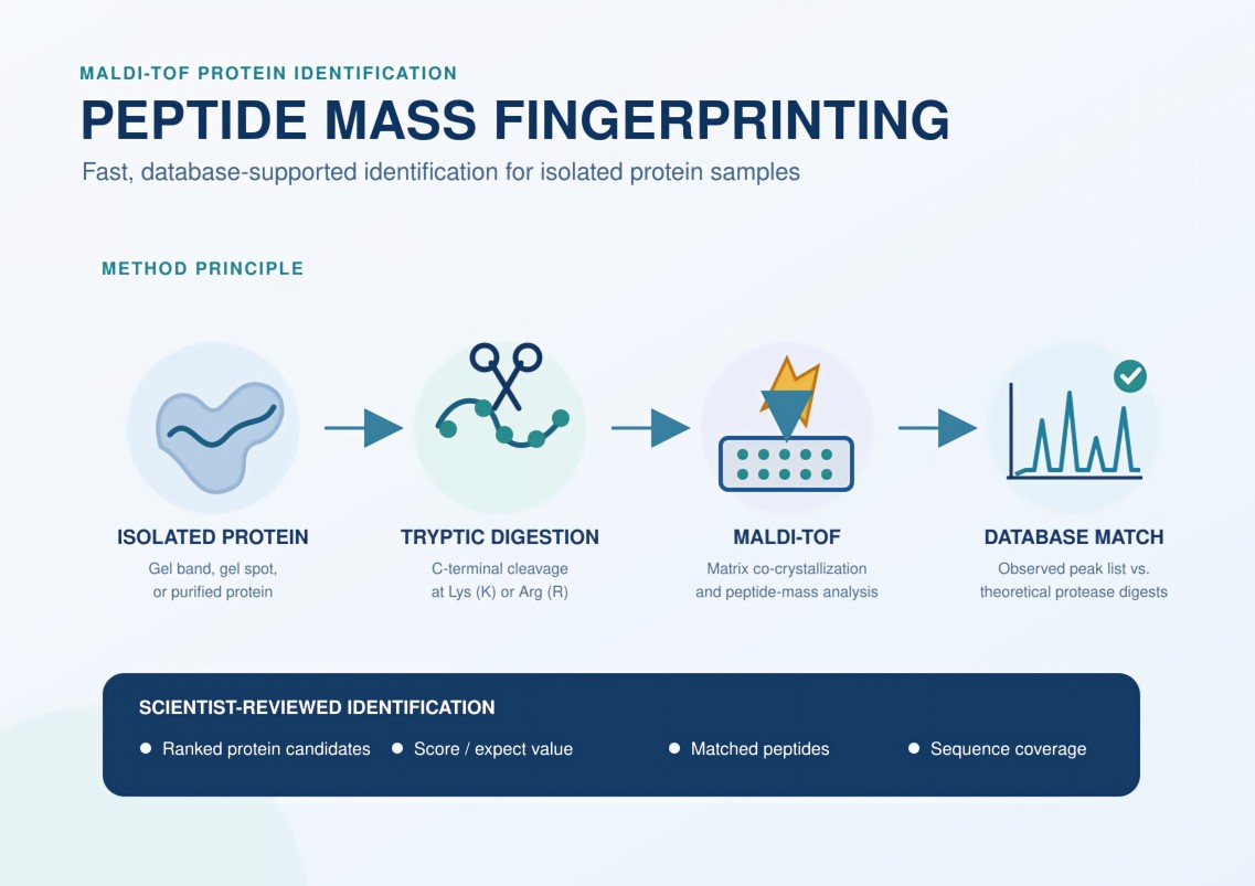
Our Peptide Mass Fingerprinting (PMF) service provides researchers and biotechnology professionals with fast, accurate, and cost-effective protein identification. Using matrix-assisted laser desorption/ionization time-of-flight (MALDI-TOF) mass spectrometry, we generate a characteristic peptide-mass pattern and compare it with theoretical digests in sequence databases.
Whether you are verifying a recombinant protein or identifying an unknown spot from a 2D gel, our team delivers traceable spectra, database-search results, and scientist-reviewed interpretation. Optional TOF/TOF MS/MS can provide additional sequence evidence when a mass fingerprint alone is ambiguous.
- Identify isolated proteins from Coomassie- or MS-compatible silver-stained gel bands and spots.
- Verify the identity of purified recombinant proteins before downstream studies.
- Escalate selected peptide ions to TOF/TOF MS/MS when additional confirmation is needed.
In line with ICH Q6B Guidance for biologic characterization
Jump to Section
- Why Peptide Mass Fingerprinting Matters
- Service Capabilities
- Research Applications
- Analytical Approaches
- Service Advantages
- Instrument Platform
- Workflow
- Report Deliverables
- FAQs
- Sample Requirements
Why Peptide Mass Fingerprinting Matters in Protein Research
Peptide mass fingerprinting begins by cleaving an unknown protein into peptides, usually with the sequence-specific protease trypsin. The absolute masses of the resulting peptides are then measured by MALDI-TOF mass spectrometry. Because the set of peptide masses reflects the protein sequence and the selected protease, it acts as a molecular fingerprint.
The experimental mass list is compared with theoretical peptide masses calculated from protein-sequence databases such as Swiss-Prot or NCBI resources. Probability-based search tools evaluate how well the observed peptides match candidate proteins. A statistically significant score, suitable mass accuracy, informative sequence coverage, and consistency with the sample context together support the identification.
PMF is especially useful when the sample contains one dominant protein, as in an excised 1D or 2D gel band or a purified recombinant-protein preparation. It provides a rapid route from a visible protein feature to a database-supported identity, making it valuable for proteomics screening, expression verification, biomarker follow-up, and pre-characterization quality control.
Unlike LC-MS/MS peptide mapping, which uses chromatographic separation and extensive fragment-ion data to characterize sequence coverage and modifications in greater depth, PMF primarily relies on accurate peptide masses. The two methods are complementary: PMF is streamlined for simple samples, while LC-MS/MS is generally more appropriate for complex mixtures, detailed PTM localization, or proteins absent from the selected database.
Peptide Mass Fingerprinting Service Capabilities
Our MALDI-TOF workflow supports protein identification, recombinant protein verification, gel-band or gel-spot analysis, alternative protease digestion, and optional MS/MS confirmation.
Protein Identification
Generate a peptide-mass fingerprint and search it against theoretical digests to identify an isolated or relatively simple protein sample.
Recombinant Protein Verification
Confirm whether a purified expression product is consistent with its expected protein identity before structural, functional, or formulation studies.
1D and 2D Gel-Spot Analysis
Excise visible protein bands or spots, perform in-gel digestion, and identify the dominant protein represented by the recovered peptide pattern.
Alternative Protease Digestion
Use trypsin as the standard enzyme or evaluate alternatives such as chymotrypsin or Glu-C when the sequence or project objective warrants another cleavage pattern.
Optional TOF/TOF MS/MS
Fragment selected precursor ions to obtain additional sequence evidence when the PMF score, coverage, or candidate ranking requires confirmation.
LC-MS/MS Escalation
Recommend a shotgun protein-identification or peptide-mapping workflow when the sample is complex, database coverage is limited, or detailed PTM analysis is required.
Peptide Mass Fingerprinting Across Research Applications
- Proteomics
Identify proteins excised from 1D or 2D SDS-PAGE gels when a band or spot contains one dominant protein or only a very simple mixture. - Quality Control
Verify the identity of purified recombinant proteins before downstream assays, structural studies, or broader characterization. - Biomarker Discovery
Support the initial identification of isolated candidate bands or spots emerging from comparative gel-based experiments. - Structural Biology
Confirm the identity of a purified protein before crystallography, NMR, binding, or other structure-function investigations.
Analytical Approaches for Protein Identification
The appropriate analytical depth depends on sample complexity, database representation, and the level of sequence evidence required.
| Approach | Best suited for | Typical information delivered |
| Standard MALDI-TOF PMF | Isolated gel bands or spots and purified proteins expected to be represented in a sequence database | Accurate peptide-mass list, probability-based database matches, protein score, sequence coverage, and matched-peptide assignments. |
| PMF with TOF/TOF MS/MS | Ambiguous PMF matches or projects needing additional evidence from selected peptides | PMF results plus fragment-ion spectra and sequence evidence for targeted precursor ions; de novo interpretation may be attempted where data quality permits. |
| Alternative-protease PMF | Proteins yielding an uninformative tryptic pattern or requiring complementary cleavage | A second peptide-mass fingerprint generated with an enzyme such as chymotrypsin or Glu-C. |
| LC-MS/MS protein identification | Complex mixtures, weak bands, database-poor targets, or projects requiring deeper sequence and PTM information | Chromatographically separated peptide identifications, extensive MS/MS evidence, and broader protein or modification coverage. |
Service Advantages
- High Sensitivity and Mass Accuracy
Optimized in-gel and in-solution protocols support low-abundance samples; practical sensitivity depends on protein properties, staining, recovery, and sample quality. - Rapid MALDI-TOF Acquisition
The 10 kHz smartbeam 3D laser on the Bruker rapifleX supports fast spectral acquisition and efficient analysis of peptide digests. - Probability-Based Bioinformatics
Search experimental peptide masses against suitable sequence databases using project-specific taxonomy, enzyme, tolerance, and modification settings. - Rigorous Quality Control
Use internal reference peaks such as trypsin autolysis products, together with external standards and spectrum review, to assess calibration and data quality. - MS/MS Backup Capability
Transition from peptide-mass measurement to TOF/TOF fragmentation of selected ions when a fingerprint alone does not provide sufficient discrimination. - Dedicated Scientific Support
Receive guidance on sample suitability, digestion strategy, database-search design, result interpretation, and escalation to LC-MS/MS when appropriate.
Instrument Platform for Peptide Mass Fingerprinting
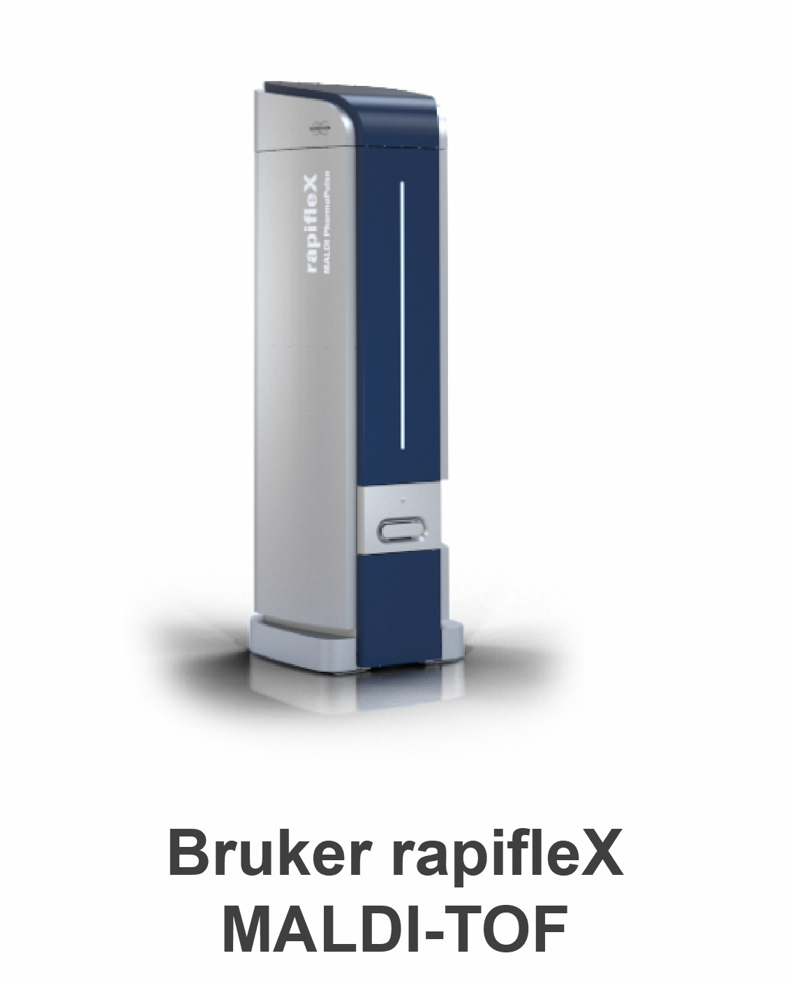
Our primary PMF platform is the Bruker rapifleX MALDI-TOF/TOF. Its high-speed 10 kHz smartbeam 3D laser, adaptable ion optics, and tandem-MS capability support rapid peptide-mass acquisition and optional fragmentation of selected precursor ions. These features help produce high-quality spectra for database searching while retaining a direct path to additional sequence evidence when required.
The broader mass-spectrometry platform also includes high-resolution LC-MS and triple-quadrupole LC-MS systems. These complementary instruments are selected when the research question extends beyond PMF—for example, complex-mixture identification, deeper peptide mapping, targeted quantification, or detailed PTM characterization.
Peptide Mass Fingerprinting Workflow
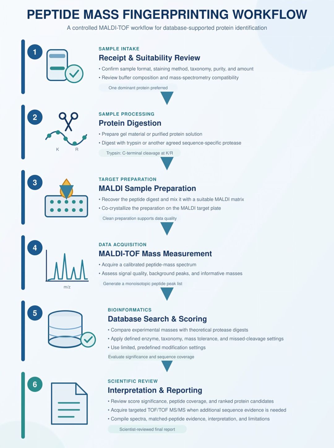
Our six-stage PMF workflow provides controlled, database-supported identification of isolated proteins.
- 1
Sample Receipt & Suitability Review
Sample suitability is assessed based on sample format, staining method, purity, amount, buffer composition, and identification objective.
- 2
Protein Digestion
Gel-derived or purified proteins are prepared and digested with trypsin or an agreed alternative protease.
- 3
MALDI Sample Preparation
Recovered peptides are combined with a suitable MALDI matrix and co-crystallized on the target plate.
- 4
MALDI-TOF Mass Measurement
Calibrated spectra are acquired and reviewed to generate an informative monoisotopic peptide-mass list.
- 5
Database Search & Scoring
Experimental peptide masses are searched against theoretical digests using predefined parameters, followed by significance and sequence-coverage assessment.
- 6
Interpretation & Reporting
Ranked candidates are scientifically reviewed, with optional targeted TOF/TOF MS/MS used when additional confirmation is required.
Report Deliverables
Upon completion, you will receive a comprehensive report package that includes:
- Detailed analytical report: methodology, database, taxonomy, enzyme, mass tolerance, modification settings, quality review, results, and interpretation.
- Database-search results: ranked identification hits, probability-based protein scores, sequence coverage, and lists of matched and unmatched peptide masses.
- Annotated MALDI-TOF spectra: labeled spectra showing informative peptide peaks and relevant calibration or background features.
- Optional MS/MS evidence: selected precursor and fragment-ion spectra when TOF/TOF confirmation is included in the project.
- Raw data: original mass-spectrometry files and agreed export formats for internal archiving or reanalysis.
Frequently Asked Questions About Peptide Mass Fingerprinting
Can PMF identify proteins in a complex mixture?
PMF is not suitable for genuinely complex mixtures. It performs best with one dominant protein and may support very simple mixtures containing only two or three proteins of similar abundance. For cell lysates or more complex samples, LC-MS/MS is recommended.
What enzymes do you use for digestion?
Trypsin is our standard enzyme because it produces peptides of ideal size for mass spectrometry. However, we can use alternative enzymes (e.g., chymotrypsin, Glu-C) depending on your protein's specific sequence.
What happens if my protein is not in the database?
PMF relies on existing databases. If your organism's genome is unsequenced or the protein is highly novel, traditional PMF may not yield a match. In such cases, we recommend de novo peptide sequencing via tandem mass spectrometry (MS/MS).
Does PMF detect post-translational modifications (PTMs)?
PMF may accommodate a limited number of predefined modification settings during database searching, but peptide-mass shifts alone do not provide confident PTM identification or site localization. LC-MS/MS is recommended for detailed PTM analysis.
Are there any limitations to PMF?
While PMF is highly effective for most standard applications, it can be less successful for exceptionally high molecular weight proteins, proteins with extreme pI values, or highly hydrophobic proteins (such as integral membrane proteins) due to challenges in solubility and enzymatic digestion. For these challenging samples, please consult our technical team for alternative LC-MS strategies.
How long is the turnaround time?
Our standard turnaround time is 2 weeks from sample receipt. It may vary depending on the specific requirements of each project.
Sample Requirements for Peptide Mass Fingerprinting
General sample-submission guidelines are summarized below. Contact our technical team before shipment when sample amount, staining chemistry, or buffer compatibility is uncertain.
| Parameter | General guideline | Additional notes |
| Accepted sample types | Coomassie-stained gel bands or spots; MS-compatible silver-stained gel pieces; purified protein solution | Gel bands or spots should be discrete and handled with clean tools to minimize keratin contamination. |
| Protein amount | A clearly visible gel feature; a faint Coomassie band may contain approximately 10-50 ng depending on protein size and staining | Recovery and identification success depend on protein abundance, staining, digestion, sequence, and background contamination. |
| Solution compatibility | Provide the complete buffer formulation and protein concentration | Avoid high levels of SDS, Triton, or other detergents, glycerol, and nonvolatile salts; dialysis, precipitation, or cleanup may be required. |
| Gel-piece shipping | Ship in a small volume of ultrapure water or 1% acetic acid in a clean, tightly sealed tube | Do not allow gel pieces to float in a large volume. Clearly label every tube and include sample metadata. |
| Solution shipping | Ship frozen on dry ice unless another condition has been agreed | Use low-binding, tightly sealed tubes and avoid repeated freeze-thaw cycles. |
Before submission. Provide the expected organism or taxonomy, approximate molecular weight, gel staining method, protein sequence if known, buffer composition, sample history, and whether optional TOF/TOF MS/MS or LC-MS/MS escalation should be considered.
References
- Henzel WJ, Billeci TM, Stults JT, Wong SC, Grimley C, Watanabe C. Identifying proteins from two-dimensional gels by molecular mass searching of peptide fragments in protein sequence databases. Proceedings of the National Academy of Sciences. 1993;90:5011-5015. doi:10.1073/pnas.90.11.5011.
- Perkins DN, Pappin DJC, Creasy DM, Cottrell JS. Probability-based protein identification by searching sequence databases using mass spectrometry data. Electrophoresis. 1999;20:3551-3567. doi:10.1002/(SICI)1522-2683(19991201)20:18<3551::AID-ELPS3551>3.0.CO;2-2.