In structural biology and biochemistry, protein secondary structure refers to the regularly repeated conformation in the protein. In 1951, Linus Pauling and colleagues accurately predicted the ideal helix and lamella structure based on the hydrogen bonds of the model protein skeleton. At the same time, they pointed out that the 310-helix could not appear in the protein due to the improper bond angle. But it was later discovered that 310-helix residues accounted for about 4% of the protein. Subsequently, in 1952, Kaj Ulrik Linderstrøm-Lang introduced the concept of secondary structure for the first time based on Pauling's work. At the same time, he also introduced the concepts of protein primary structure and tertiary structure.
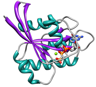
α-helix and β-sheet are the most important elements in the protein secondary structure. Residues with these two secondary structures account for half of the total number of protein residues. Other secondary structures include π-helix, 310-helix, left-handed helix, and PPII helix. The π-helix often appears at the end of the α-helix or the middle position of the α-helix, and the π-helix and the left-handed helix are found to be closely related to the function of the protein. Alpha-sheet, although rare in its natural state, is considered an important intermediate structure for protein folding. Turns and loops play the role of connecting regular elements in protein structure. The irregular coil is not a specific secondary structure, but a general term given to a structure when it cannot be designated as other regular secondary structures.
Protein secondary structure characterization methods:
Circular dichroism spectra (Far UV)
The circular dichroism of the protein and the difference in the absorption of left and right circularly polarized light by asymmetric molecules are used for structural analysis. The scanning pattern of the circular dichroism in the far ultraviolet region reflects the arrangement information of the peptide bonds of the protein, and the calculated ratio is the ratio of the protein secondary structure, that is, the ratio of α-helix, β-rotation folding, turning angle and irregular curling. This method is most suitable when samples are clear solutions with proteins and peptides in a narrow concentration range.
Ultraviolet and fluorescence spectroscopy is limited to the determination of a few proteins containing chromophores such as aromatic, heterocyclic or conjugated ring systems.
Infrared spectroscopy
Infrared spectroscopy has obvious advantages. It can identify complex systems macroscopically and measure proteins and peptides in different states, concentrations, and environments. Moreover, infrared spectroscopy is a fast non-destructive testing method. Specifically, one of the most powerful infrared spectroscopy for studying protein secondary structure is the fourier transform infrared spectroscopy (FTIR).
Nuclear magnetic resonance technology can determine the conformation of proteins in a solution, but it is generally limited to the study of smaller proteins with a molecular weight of not more than 20kD. The demand for the amount of sample is large, the purity requirements are high, and the measurement process is slow.
Although X-ray crystal diffraction can provide complete protein crystal structure information, it requires high-quality single-crystal samples, and the experimental process is complicated and takes a long time.


