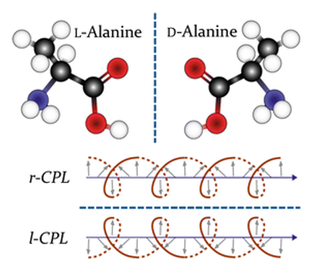
A beam of plane polarized light can be regarded as the superposition of left and right circularly polarized lights of the same frequency and amplitude. When plane-polarized light passes through an optically active medium that has an absorption peak in the ultraviolet region, the left-handed and right-handed circularly polarized light components contained in the region have different propagation speeds (due to different refractive indexes) and different intensities. This phenomenon is circular dichroism (CD).
Proteins are biological macromolecules with specific structures that are made up of amino acids connected by peptide bonds. Proteins have several main structural levels: primary, secondary, tertiary, and quaternary structures. Some also have domains or super secondary structures. In protein and polypeptide molecules, peptide bonds, aromatic amino acid residues and disulfide bridges in the peptide chain backbone are the main photoactive chromophores. When plane circularly polarized light passes through, these chromophores absorb left and right circularly polarized light differently, resulting in a difference in the amplitude of the polarized light vector. Turning the circularly polarized light into elliptically polarized light produces the circular dichroism of the protein.
The circular dichroic spectrum is a difference spectrum that shows the difference of the absorption spectrum of the sample under the irradiation of left and right polarized light. The circular dichroism is related to the wavelength. Take the absorbance difference of polarized light ΔA as the ordinate and the wavelength into the abscess coordinate to make the graph, then the circular dichrogram is obtained. The circular dichroism of chiral materials often has two manifestations: one is expressed by ΔA, ΔA=AL-AR; the other is expressed by molar ellipticity [θ], [θ]=3300ΔX=3300 [XL-XR]. Molar ellipticity [θ] is often used instead of absorbance difference ΔA.
The CD spectrum of a protein is mainly the sum of the circular dichroism of the active chromophore and the folded structure. According to the energy level of the electronic transition, the CD spectrum of a protein is divided into three wavelength ranges:
(1) In the far ultraviolet spectral region below 250 nm, the circular dichroism is mainly caused by the n→π electron transition of the peptide bond.
(2) In the near ultraviolet spectral region of 250 ~ 300 nm, the circular dichroism is mainly caused by the π→π electron transition of the side-chain aromatic group.
(3) In the ultraviolet-visible light spectrum region of 300~700 nm, the circular dichroism is mainly caused by external chromophores, such as prosthetic prosthetic groups.
The far UV circular dichroism spectroscopy reflects the direction and energy level transition of the peptide bond arrangement in the regular secondary structure of the protein or polypeptide. Through the analysis and comparison of the band position and absorption strength, the secondary structure of different proteins or peptides can be studied. The near UV circular dichroism spectroscopy reflects the circular dichroism of aromatic amino acid residues and disulfide bonds in an asymmetric environment. It mainly reveals the tertiary structure information of proteins, studies the changes and effects of the asymmetric microenvironment, and does not interfere with the CD signal of peptide bonds in the far ultraviolet region. Ultraviolet-visible spectroscopy is mainly used for coupling analysis of prosthetic groups.


