Flow cytometry (FCM) is a high tech that can simultaneously perform multi-parameter, rapid quantitative analysis and sorting of cells or biological particles in a fast linear flow state.
The characteristics of flow cytometry: 1. As long as the sample is prepared into a single cell or biological particle suspension, it can be analyzed. 2. The measurement speed is fast, thousands or even tens of thousands of cells can be measured per second. 3. It can simultaneously measure the multi-parameter characteristics of each cell. 4. While analyzing the cell characteristics, the cells with the specified characteristics can be separated, so as to further cultivate, clone, observe or perform certain experiments on these specific cells.
Flow cytometer system
Specimen → Laser system → Flow system → Signal processing system → Amplification system → Computer system → Result printing
Principles of flow cytometry
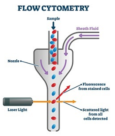
The specimen to be tested is prepared as a single cell suspension, which is then fluorescently stained and goes into a flow chamber filled with sheath fluid. The pressure of the sheath fluid is different from the pressure of the sample stream. When the pressure difference between the two reaches a certain level, the sheath fluid entrains the cells in the sample stream and arranges them in a single row to pass through the laser focus area one by one.
If we label the cells of interest with specific fluorescent dyes, these dyes will fluoresce at a specific wavelength when the cells pass through the laser detection area. Through a certain wavelength selective permeability color filter, we can distinguish the scattered light and fluorescent signals of different wavelengths and send them to different photomultiplier tubes. After a series of signal conversion, amplification, and digital processing, we can intuitively count the percentages of cells stained with various fluorescent dyes on the computer. Choosing different monoclonal antibodies and fluorescent dyes, we can use FC to measure many different characteristics on a cell at the same time. If you are interested in cells with certain characteristics, the flow sorting function can sort them out for further cultivation and research.
Workflow of flow cytometry sorting
1. Preparation of single-cell suspension
The cells or particles to be tested are made into a single cell suspension and stained with specific fluorescent dye-labeled antibodies. These cells enter the flow chamber under constant gas pressure. The buffer solution containing no cells or particles is ejected from the sheath fluid tube under high pressure. The inlet direction of the sheath liquid tube forms a certain angle with the sample flow to be measured. The sheath fluid flows around the sample at high speed, forming a cylindrical fluid stream. The cells or particles to be tested are arranged in a single row under the coating of the sheath fluid and pass through the detection area in sequence.
2. Collect fluorescent signals on single cells
A flow cytometer usually uses a laser as a light source, and the focused and shaped beam irradiates the sample stream vertically. The fluorescent dyes on the cells or particles are excited, generating scattered light and exciting fluorescence. The light scattering signal is detected at a small angle in the forward direction, and this signal reflects the size of the cell volume. The receiving direction of the fluorescent signal is perpendicular to the laser beam, and is separated by a series of dichroic mirrors and band-pass filters to form multiple fluorescent signals of different wavelengths.
3. Statistical results
The intensity of the fluorescent signal represents the intensity of the surface antigen of the measured cell membrane or the concentration of the substance in the cell, which is converted into a digital signal that can be recognized by a computer through photoelectric conversion. The computer processes the various signals and then displays the analysis results.


