Why Histone Modification Antibody Array Profiling
Histone post-translational modifications are uniquely well-suited for antibody-based array detection. Unlike many other PTM classes where modification-specific antibodies may have limited availability or cross-reactivity, the histone modification field has benefited from decades of antibody development and validation, producing a rich toolkit of well-characterized antibodies against specific histone marks. This makes antibody arrays a particularly powerful approach for histone PTM analysis, offering distinct advantages for targeted epigenetic profiling.
Complementary to Mass Spectrometry-Based Histone PTM Analysis
MS-based histone PTM analysis provides unparalleled depth for discovering and quantifying the full repertoire of histone modifications, including combinatorial PTM patterns on individual histone tails. However, for targeted screening of specific histone marks across many samples, antibody arrays offer complementary strengths: higher sample throughput (dozens of samples per batch), established antibody specificity for well-characterized marks, straightforward data analysis without specialized MS bioinformatics, and rapid turnaround from sample to results. For comprehensive histone PTM discovery and deep characterization, our Histone PTM Analysis Service provides MS-based discovery and quantification capabilities that can be combined with antibody array screening for an integrated histone PTM profiling strategy.
High-Throughput Epigenetic Screening for Drug Discovery
Epigenetic drug discovery programs targeting histone-modifying enzymes — including HDACs, HATs, histone methyltransferases, demethylases, and reader domain proteins — require robust, quantitative assays for monitoring histone mark changes in response to compound treatment. Histone modification antibody arrays provide a uniquely powerful platform for this application, enabling simultaneous measurement of dozens of histone marks across multiple drug concentrations, time points, and biological replicates in a single experiment. This makes them invaluable for assessing compound selectivity, identifying off-target epigenetic effects, and characterizing the comprehensive impact of epigenetic drugs on the histone code. For broader epigenetic PTM research beyond histones, our Epigenetic PTM Research Services provide integrated analysis solutions spanning histone and non-histone epigenetic modifications.
Histone Code Discovery and Biomarker Development
Altered histone modification patterns are hallmarks of cancer, neurodegenerative disorders, metabolic diseases, and aging. Antibody-based histone modification arrays enable systematic screening of histone mark changes in clinical samples, patient-derived materials, and disease models — providing a powerful tool for discovering epigenetic biomarkers of disease progression, treatment response, and prognosis. The high throughput and quantitative nature of array-based profiling makes it particularly suitable for clinical cohort studies where sample numbers are large and consistent across-batch normalization is essential.
Our Histone Modification Antibody Array Platform
Our histone modification antibody array platform deploys three complementary formats optimized for different research questions, sample types, and throughput requirements. Each format uses antibodies rigorously validated for histone modification specificity, with appropriate positive and negative controls embedded in every array.
Membrane-Based Histone Modification Arrays
Our standard membrane-based format uses nitrocellulose membranes printed with duplicate spots of capture antibodies against 10–50 specific histone marks. Acid-extracted or total histone preparations are incubated with the membrane, and bound histones are detected using a secondary detection system with chemiluminescent readout. This format is ideal for focused studies of 10–50 histone marks across multiple samples or conditions, with typical sample input of 1–5 µg of acid-extracted histones per membrane. The membrane format supports pre-configured panels for histone acetylation, methylation, and phosphorylation, as well as custom panel design.
Bead-Based Multiplex Histone PTM Assays
For higher-throughput quantitative profiling of histone marks, our bead-based multiplex assay format uses color-coded bead populations, each coupled to a specific histone modification antibody, with flow cytometric readout. This format enables simultaneous quantification of 20–50+ histone marks from as little as 0.5–2 µg of histone protein, with superior quantitative precision and dynamic range compared to membrane-based formats. The bead-based format is particularly well-suited for dose-response studies, time-course experiments, and clinical sample screening where sample availability is limited and quantitative accuracy is critical.
Integration with Complementary PTM Detection Platforms
Our histone modification antibody array platform is integrated within a comprehensive PTM analysis ecosystem. For broader PTM screening beyond histone marks, our Multiplex PTM Immunoassay Services extend antibody-based detection to acetylation, ubiquitination, and SUMOylation on non-histone proteins. For phospho-signaling focused studies, our Phospho-Signaling Antibody Array provides parallel analysis of kinase activation and signaling networks. Spatial Histone Modification Profiling extends our histone PTM analysis capabilities to tissue context, enabling spatially resolved mapping of epigenetic marks in tissue sections.
Available Histone Modification Antibody Array Panels
Our histone modification antibody array platform offers the following major panel categories, each customizable to your specific research needs. Multiple panels can be deployed in parallel for comprehensive histone code profiling.
Histone Methylation Arrays
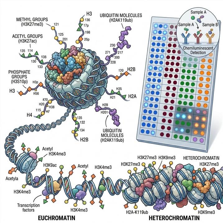
- H3 Lysine Methylation Panel — Comprehensive profiling of H3K4me1/2/3 (active promoters), H3K9me1/2/3 (heterochromatin), H3K27me1/2/3 (Polycomb repression), H3K36me1/2/3 (transcriptional elongation), and H3K79me1/2/3 (telomeric silencing), with distinction between mono-, di-, and tri-methylation states at each residue.
- H4 and Non-Canonical Histone Methylation Panel — Detection of H4K20me1/2/3 (DNA damage response, chromatin compaction), H4K3me, and methylation marks on histone variants (H2A, H2B, H3.3) for comprehensive coverage of the histone methylation landscape.
- Histone Arginine Methylation Panel — Profiling of H3R2me, H3R8me, H3R17me, H3R26me, H4R3me (asymmetric and symmetric dimethylation) catalyzed by PRMT family enzymes and involved in transcriptional regulation and RNA processing.
Histone Acetylation Arrays
- H3 Acetylation Panel — Quantitative detection of H3K9ac, H3K14ac, H3K18ac, H3K23ac, H3K27ac (active enhancers), H3K36ac, H3K56ac, and H3K79ac, covering the major acetylation sites on histone H3 that regulate transcription, chromatin structure, and DNA repair.
- H4 Acetylation Panel — Profiling of H4K5ac, H4K8ac, H4K12ac, H4K16ac (chromatin decompaction), and H4K91ac, providing comprehensive coverage of the H4 acetylation pattern that is combinatorially regulated during chromatin assembly and transcriptional activation.
- Pan-Acetyl Lysine and Multi-Histone Panel — Pan-acetyl-lysine detection combined with site-specific acetylation antibodies for global acetylation assessment alongside detailed site-level profiling.
Histone Phosphorylation and Additional Modification Arrays
- Histone Phosphorylation Panel — Detection of H3S10ph (mitosis, gene activation), H3S28ph (mitosis, stress response), H2AX S139ph (γH2AX, DNA damage marker), H2BS14ph (apoptosis), and H4S1ph (DNA damage, chromatin compaction).
- Custom Histone Code Panels — Flexible combination of methylation, acetylation, phosphorylation, and additional marks (H2BK120ub, H2AK119ub) tailored to your specific research question, with rapid panel development and validation.
All array panels can be customized to include specific modifications of interest. For MS-based deep profiling and absolute quantification of histone PTMs, our Histone PTM Identification service provides complementary discovery capabilities, while downstream PTM Bioinformatics Analysis offers pathway-level integration of histone PTM data with other epigenomic and transcriptomic datasets.
Workflow: From Sample to Histone PTM Array Data
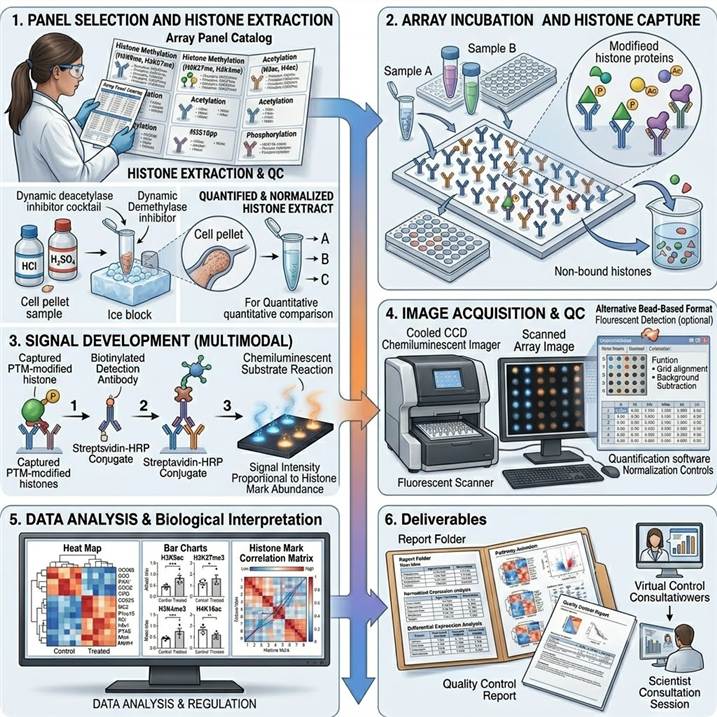
Step 1: Panel Selection and Histone Extraction
We collaborate with you to select the optimal histone modification antibody array panel(s) based on your research focus and histone marks of interest. Histones are extracted from cells or tissues using acid extraction (HCl or H₂SO₄) or high-salt extraction methods optimized for PTM preservation, with deacetylase and demethylase inhibitors included in extraction buffers.
Step 2: Array Incubation and Histone Capture
Quantified histone preparations are normalized and incubated with antibody array membranes, bead sets, or slides under optimized binding conditions. Histone proteins carrying specific PTM marks are captured by immobilized antibodies. After incubation, unbound material is removed through stringent washing optimized for histone-antibody interactions.
Step 3: Detection and Signal Development
Captured histones are detected using a detection antibody cocktail or directly labeled detection system. For membrane arrays, chemiluminescent signals are developed using HRP-based amplification. For bead-based assays, fluorescent detection antibodies enable quantitative flow cytometric readout. Signal intensity for each target is proportional to the abundance of the specific histone mark.
Step 4: Image Acquisition and Data Extraction
Array images are acquired using cooled CCD chemiluminescent imaging systems or fluorescent scanners with optimized exposure settings. Spot or bead intensity data are extracted using dedicated analysis software with background subtraction, positive control normalization, and intra-assay replicate quality assessment.
Step 5: Data Analysis and Biological Interpretation
Normalized signal intensities for each histone mark are compared across experimental groups using appropriate statistical tests. Histone mark correlation analysis reveals coordinated changes in the histone code. Pathway-level interpretation places histone mark changes in the context of known epigenetic regulatory mechanisms and biological processes relevant to your research question.
Step 6: Deliverables and Reporting
Histone modification array data package including raw and normalized signal intensities for each histone mark, fold-change and statistical significance tables, publication-quality heat maps and bar charts, histone mark correlation matrices, and a scientist consultation session for biological interpretation and integration with other epigenetic datasets.
Why Choose Our Histone Modification Antibody Array Service
Histone-Specific Antibody Validation
Every antibody used in our histone modification arrays undergoes rigorous validation specific to histone PTM detection: peptide competition ELISA to confirm mark-specific recognition, cross-reactivity testing against closely related modification states (e.g., H3K9me3 vs H3K27me3), dot blot and western blot confirmation of signal specificity, and ChIP-grade validation where applicable. This ensures that the array signals accurately reflect the abundance of each specific histone mark.
Integrated Histone PTM Analysis Ecosystem
Our antibody array platform is integrated within a comprehensive histone PTM analysis ecosystem that includes MS-based discovery and quantification (Histone PTM Analysis), Spatial Histone Modification Profiling for tissue-resolved epigenetic analysis, and bioinformatics integration (PTM Bioinformatics Analysis). This integration enables seamless multi-platform histone PTM analysis within a single service provider.
Flexible Panel Design and Multi-Platform Compatibility
We offer extensive pre-configured panels for common histone marks as well as custom array design to incorporate specific modification targets. Array data can be integrated with MS-based histone PTM datasets, ChIP-seq results, and transcriptomic data for comprehensive multi-omic epigenetic analysis, ensuring that your histone PTM profiling data is maximally valuable across your entire research program.
Technical Expertise and Reproducibility
Our team has extensive experience in histone extraction, PTM preservation, and antibody-based histone modification analysis across diverse sample types including cultured cells, fresh-frozen and FFPE tissues, and clinical specimens. Each project includes appropriate quality control steps — total histone normalization, positive control verification, and replicate analysis — to ensure robust, reproducible results across experiments and time points.
Case Study: Epigenetic Reverse Phase Protein Array for Global Histone Modification Fingerprinting in Cancer Cells
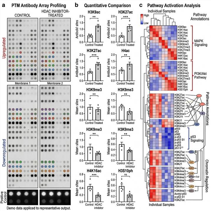
In a 2017 study published in Cell Death Discovery (Nature Publishing Group), Partolina et al. developed and validated the epigenetic reverse phase protein array (eRPPA) platform for simultaneous quantification of global histone modifications, demonstrating the power of antibody-based array technology for epigenetic profiling in drug discovery.
Background: Histone-modifying enzymes are important therapeutic targets in cancer, and drugs targeting these enzymes — particularly HDAC inhibitors — are approved for multiple cancer indications. However, assessing the comprehensive impact of epigenetic drugs on the histone code has been technically challenging. Mass spectrometry provides detailed information but is low-throughput and requires specialized expertise, while western blotting is limited to a few marks per experiment. A high-throughput, multiplexed method for quantifying multiple histone modifications simultaneously would significantly accelerate epigenetic drug development.
Approach: The team developed the eRPPA platform using a panel of well-characterized mouse monoclonal antibodies targeting specific acetylation and methylation marks on histones H3 and H4. Acid-extracted histones from HCT116 colorectal cancer cells were printed onto nitrocellulose-coated slides in replicate dilution series, and each slide was probed with a different histone modification antibody. The array format enabled simultaneous quantification of multiple histone marks from the same sample set, with signal intensity normalized to total histone H3 and H4 levels. The platform was validated by profiling histone mark changes in response to three structurally distinct HDAC inhibitors — HC toxin, trichostatin A (TSA), and CAY10603 — and further applied to investigate the effect of N-acetylated amino acids (ALCAR) as potential acetyl donors that could potentiate HDAC inhibitor effects.
Key Findings:
- The eRPPA platform successfully quantified 10+ distinct histone acetylation and methylation marks simultaneously, including H3K9ac, H3K14ac, H3K27ac, H4K5ac, H4K8ac, H4K12ac, H4K16ac, H3K4me2, H3K9me2/3, and H3K27me3, from as little as 2 µg of acid-extracted histones per sample
- HDAC inhibitor treatment produced dose-dependent increases in histone acetylation across multiple sites, with distinct selectivity profiles for each inhibitor: HC toxin preferentially increased H3K9ac and H3K14ac, while TSA more broadly affected H3 and H4 acetylation
- N-acetylated amino acids (ALCAR) were shown to serve as acetyl donors for histone acetylation and could potentiate the effects of HDAC inhibitors, suggesting a potential combinatorial therapeutic approach
- Correlation analysis of histone mark changes revealed coordinated regulation of specific acetylation sites, providing insights into the functional interplay between different histone modifications
- The eRPPA platform demonstrated robust technical reproducibility (inter-array CV < 15%) and quantitative accuracy across a broad dynamic range of histone mark abundance
Significance: This study established the eRPPA platform as a robust, quantitative, and high-throughput method for global histone modification profiling that bridges the gap between low-throughput western blotting and low-accessibility MS-based approaches. The platform's demonstrated utility for profiling HDAC inhibitor effects on the histone code established antibody arrays as a valuable tool for epigenetic drug discovery and development. The workflow and validation framework established in this study are directly applicable to contemporary antibody array platforms with expanded panel coverage and improved multiplexing capacity.

Figure 1 from Partolina et al. (2017). Epigenetic reverse phase protein array (eRPPA) platform for global histone modification fingerprinting in human cancer cells. (CC BY 4.0)
Representative Histone Modification Antibody Array Results
Our histone modification antibody array platform delivers comprehensive data packages designed for immediate biological interpretation and publication-ready visualization of histone code changes across experimental conditions.
Representative data outputs from our Histone Modification Antibody Array platform. Left: Array membrane images. Center: Quantitative comparison of histone mark intensities. Right: Heat map and correlation matrix of histone code changes.
Key deliverables included in every histone modification antibody array project:
- Raw and normalized signal intensities — For each histone mark on the array, with total histone normalization, positive control normalization, and replicate spot quality metrics
- Differential histone mark analysis — Fold-change, p-value, and false discovery rate for comparisons between experimental groups, with appropriate statistical tests for multi-group and time-course designs
- Data visualization — Publication-quality heat maps, bar charts, volcano plots, and histone mark correlation matrices for data exploration and presentation
- Histone mark correlation analysis — Pairwise correlation analysis revealing coordinated regulation patterns between different histone modifications across experimental conditions
- Quality control report — Array performance metrics including positive control signal intensity, background uniformity, replicate spot reproducibility, and detection sensitivity for each histone mark
Related Services
Our histone modification antibody array platform is part of a comprehensive PTM and epigenetic analysis service portfolio spanning antibody-based and MS-based detection platforms for integrated epigenetic research.
FAQs
What is the difference between histone modification antibody arrays and MS-based histone PTM analysis?
Histone modification antibody arrays use immobilized antibodies specific to individual histone marks (e.g., H3K4me3, H3K27ac) for targeted, multiplexed detection of predefined histone PTM panels. MS-based histone PTM analysis provides unbiased discovery of all histone modifications, including combinatorial PTM patterns on individual histone tails. Antibody arrays excel at high-throughput screening of known histone marks across many samples, while MS excels at comprehensive discovery and characterization of novel or low-abundance modifications.
How many histone modifications can be analyzed simultaneously in a single array?
Our histone modification antibody array platform covers a range of multiplexing capacities: membrane-based arrays typically profile 10–50 histone marks per sample, bead-based multiplex assays can quantify 20–50+ marks with superior precision, and custom arrays can be designed for specific combinations of methylation, acetylation, phosphorylation, and other marks. The optimal format depends on your research question and sample availability.
How do you distinguish between different methylation states (me1, me2, me3) at the same residue?
Our histone modification antibody arrays use antibodies that are rigorously validated to distinguish between mono-, di-, and tri-methylation states at specific residues (e.g., H3K4me1 vs H3K4me3). Each antibody undergoes peptide competition ELISA testing against all methylation states at the target residue to confirm state-specific recognition, and cross-reactivity testing against closely related sequence contexts to ensure specificity.
What is the minimum sample amount required for histone modification antibody array analysis?
Sample requirements depend on the array format. For membrane-based arrays, we typically recommend 1–5 µg of acid-extracted histones per membrane. For bead-based multiplex assays, 0.5–2 µg of histone protein is typically sufficient. For whole cell lysate analysis (without histone extraction), 10–50 µg of total protein per sample is recommended, depending on the cellular abundance of histone proteins and the specific marks of interest.
How do you ensure antibody specificity for histone modification detection?
Every antibody used in our histone modification arrays undergoes rigorous validation: peptide competition ELISA to confirm mark-specific recognition, cross-reactivity testing against closely related modification states and sequence contexts, dot blot confirmation of signal specificity on modified and unmodified histone peptides, batch-to-batch consistency testing, and correlation with orthogonal methods (western blot, MS) for key marks where possible.
Can histone modification antibody arrays detect changes in combinatorial histone marks?
Standard histone modification antibody arrays detect individual marks independently rather than combinatorial marks on the same histone tail. However, by profiling multiple marks in parallel and analyzing correlation patterns across samples, our arrays provide indirect insights into combinatorial mark relationships. For direct combinatorial analysis of multiple modifications on individual histone tails, we recommend our MS-based histone PTM analysis platform, which can detect and quantify co-occurring modifications on the same peptide.
How do I select which histone modification panel is right for my experiment?
Selection depends on your research focus. For broad epigenetic characterization, we recommend the combined methylation + acetylation panel for comprehensive coverage of the major regulatory histone marks. For drug discovery programs targeting specific histone-modifying enzymes, focused panels covering the relevant marks (e.g., acetylation panel for HDAC inhibitor studies, methylation panel for EZH2 or DOT1L inhibitor studies) are most appropriate. We provide complimentary scientific consultation to help you select or design the optimal panel.
How can antibody array results be validated by orthogonal methods?
We recommend orthogonal validation of key findings using complementary methods. For histone acetylation and methylation marks, validation can be performed using western blot with modification-specific antibodies, dot blot quantification, or targeted MS-based methods (PRM, MRM) for site-specific quantification. Our integrated platform can perform both antibody array screening and orthogonal validation within the same service workflow, providing cross-platform confidence in your results.
References
- Partolina M, Thoms HC, MacLeod KG, Rodriguez-Blanco G, Clarke MN, Venkatasubramani AV, Beesoo R, Larionov V, Neergheen-Bhujun VS, Serrels B, Kimura H, Carragher NO, Kagansky A. Global histone modification fingerprinting in human cells using epigenetic reverse phase protein array. Cell Death Discovery. 2017;3:16077.
- Masuda M, Nakagawa R, Kondo T. Harnessing the potential of reverse-phase protein array technology: Advancing precision oncology strategies. Cancer Science. 2024;115:1378-1387.
- Kawaguchi T, Hashimoto M, Nakagawa R, Minami R, Ikawa M, Nakayama JI, Ueda J. Comprehensive posttranslational modifications in the testis-specific histone variant H3t protein validated in tagged knock-in mice. Scientific Reports. 2024;14:21305.
For research use only. Not for use in diagnostic procedures.