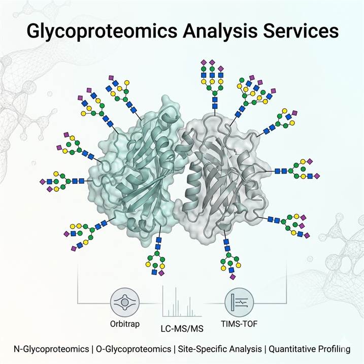
Meeting the Challenge of Glycosylation Complexity
Glycosylation presents a unique analytical challenge among post-translational modifications. Unlike phosphorylation or acetylation, where a single modification type has a defined mass shift, glycosylation encompasses hundreds of possible glycan structures that can be attached at different sites with variable occupancy — producing a combinatorial complexity that demands dedicated analytical strategies. Each glycoprotein in a biological sample exists as a distribution of glycoforms, and understanding this distribution is essential for connecting glycosylation to biological function.
Why the Glycoproteomics Approach Matters
Traditional glycomics — analyzing released glycans as a pool — provides information about total glycan diversity but loses all site-specific context. Glycoproteomics bridges this gap by analyzing intact glycopeptides, preserving the essential link between each glycan and its attachment site. For researchers investigating how glycosylation influences protein function, disease progression, or biotherapeutic activity, site-resolved glycoproteomic data is not merely complementary — it is indispensable. Changes in site-specific glycosylation have been implicated in tumor immune evasion, antibody efficacy, viral entry mechanisms, and metabolic signaling, making glycoproteomics a critical tool across biomedical research.
Our platform integrates multiple glycoproteomics strategies — from broad glycoproteome discovery to targeted site-specific analysis — allowing researchers to select the analytical depth that matches their biological question. Whether mapping the N-glycoproteome of a disease tissue, characterizing O-glycosylation sites on a recombinant protein, or quantifying glycoform ratios on a therapeutic antibody, our services deliver the molecular resolution needed for confident biological interpretation. For broader integration with other PTM types, our PTM Quantitative Analysis Services and PTM Crosstalk Analysis provide multi-modification co-regulation insights.
Our Glycoproteomics Service Portfolio
We offer a structured portfolio of glycoproteomics services designed to address specific research objectives — from comprehensive glycoproteome surveys to targeted characterization of individual glycoproteins. The table below maps common research goals to our recommended service modules.
| Research Objective |
Recommended Service |
Key Technology |
| Comprehensive N-glycosylation profiling — glycan identification and relative quantitation |
N-Glycosylation Profiling of Proteins |
PNGase F release, LC-MS/MS, HCD/EThcD fragmentation |
| Global N- and O-glycopeptide identification across the proteome |
Glycosylation Site Identification |
Multi-lectin/HILIC enrichment, stepped HCD, EThcD, TIMS-TOF |
| Site-resolved glycosylation analysis with glycoform distribution per site |
Site-Specific Glycosylation Analysis |
Glycopeptide-level LC-MS/MS, microheterogeneity profiling |
| Quantitative comparison of glycosylation across experimental conditions |
Quantitative Glycoproteomics Analysis |
Label-free quantification, TMT labeling, MRM/PRM validation |
| Specialized bioinformatics and glycopeptide data interpretation |
Glycoproteomics Data Analysis |
Byonic, pGlyco3, MSFragger-Glyco, O-Pair, manual validation |
Each service module is available independently, or services can be combined into an integrated glycoprotein characterization workflow. For biotherapeutic applications, we recommend including our Antibody Glycosylation Analysis service for comprehensive critical quality attribute assessment.
Integrated Technical Platform for Glycoproteomics
Reliable glycoproteomics data depends on optimized methods across the entire analytical pipeline — from glycopeptide enrichment through mass spectrometry acquisition to computational glycan assignment. Our platform integrates best-in-class approaches at each stage, configurable to match the complexity and objectives of your project.
Glycopeptide Enrichment Strategies
Glycopeptides constitute a minor fraction of total peptides in a typical digest and must be enriched for reliable LC-MS/MS detection. We deploy multiple complementary enrichment strategies, selected and combined based on sample type and analytical goal. HILIC-based enrichment provides broad, unbiased coverage of both N- and O-glycopeptides and is our default approach for global glycoproteome discovery. Lectin affinity capture offers targeted enrichment of specific glycan classes — ConA for high-mannose and complex N-glycans, WGA for hybrid and sialylated glycans, Jacalin for O-glycans, and AAL for fucosylated structures. For deep glycoproteome coverage, we combine multiple lectins in series or parallel. Our Glycopeptide Enrichment Service provides detailed information on enrichment strategy selection and optimization.
High-Resolution LC-MS/MS Acquisition
Glycopeptide analysis is performed on high-resolution Orbitrap and TIMS-TOF platforms, selected for their complementary strengths in fragmentation flexibility and ion mobility separation. Nano-flow and capillary-flow LC configurations are used to maximize sensitivity for limited sample amounts, while micro-flow systems provide the throughput and robustness needed for larger cohort studies. Data-dependent acquisition with dynamic exclusion is the standard for discovery glycoproteomics, with scheduled PRM methods developed for targeted glycopeptide quantification. Ion mobility separation (TIMS or FAIMS) is deployed when additional separation of isomeric glycopeptides is required — a common need in sialylated glycan analysis.
Multi-Mode Fragmentation for Complete Characterization
No single fragmentation method provides complete information for glycoproteomics. We employ a multi-mode strategy that tailors the fragmentation approach to the glycopeptide type and research objective. Stepped HCD produces diagnostic oxonium ions (e.g., m/z 204.09 for HexNAc, 366.14 for Hex-HexNAc, 292.10 for NeuAc) and generates peptide backbone fragments for identification, making it the primary method for N-glycopeptide discovery. EThcD is used to preserve labile glycan modifications during peptide backbone fragmentation, providing unambiguous O-glycosylation site localization. Supplemental activation methods are applied where additional fragmentation energy is needed for confident glycan structure assignment.
Computational Glycopeptide Identification and Glycan Assignment
Glycopeptide identification presents unique computational challenges due to the combinatorial space of peptide sequences, glycan compositions, and glycosylation sites. We deploy a multi-engine search strategy using Byonic (extensive glycan libraries with targeted FDR control), pGlyco3/pGlycoNovo (open-search capabilities for unexpected glycan discovery), MSFragger-Glyco (proteome-scale glycoproteomics), and O-Pair (specialized O-glycopeptide identification). Each identification candidate is validated against key quality criteria — oxonium ion presence and intensity, peptide backbone coverage, site localization confidence, and spectral quality — and ambiguous assignments are resolved through manual spectral interpretation by experienced glycoproteomics scientists.
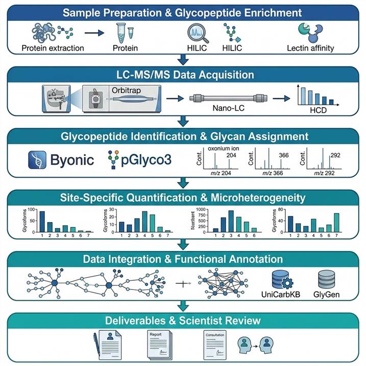
Glycoproteomics Workflow: From Sample to Publication-Ready Data
Step 1: Sample Preparation and Glycopeptide Enrichment
Proteins are extracted, reduced, alkylated, and digested using optimized protease combinations selected to maximize glycopeptide coverage. Glycopeptides are enriched by the method best suited to the research objective — HILIC for broad coverage, lectin affinity for targeted glycan class analysis, or chemical enrichment for specific glycan types. Parallel processing of non-enriched aliquots enables protein abundance normalization.
Step 2: LC-MS/MS Data Acquisition
Enriched glycopeptides are analyzed on Orbitrap or TIMS-TOF platforms with optimized LC gradients and data-dependent acquisition. Stepped HCD fragmentation provides glycan oxonium ion fingerprints and peptide sequence coverage. EThcD is applied for O-glycosylation site localization. Ion mobility separation is used for isomeric glycopeptide resolution when needed.
Step 3: Glycopeptide Identification and Glycan Assignment
Raw MS data are searched using multiple glycoproteomics search engines. Glycan databases are curated based on species, tissue type, and expected modification classes. Every glycopeptide identification is validated against oxonium ion quality, peptide backbone coverage (≥80%), and site localization confidence. Cross-validation across search engines maximizes coverage while maintaining stringent FDR control.
Step 4: Site-Specific Quantification and Microheterogeneity Analysis
For each identified glycosylation site, relative abundances of all detected glycoforms are calculated from extracted ion chromatogram areas. Glycoform distribution profiles are generated per site, displaying fractional abundance of each glycan structure. For multi-condition experiments, differential analysis identifies sites and glycoforms with statistically significant abundance changes across groups.
Step 5: Data Integration and Functional Annotation
Site-specific glycosylation data is integrated with protein-level information, functional domain context, and pathway analysis. For biotherapeutic samples, glycosylation profiles are assessed against established quality attributes. Results are cross-referenced with public glycoprotein databases (UniCarbKB, GlyConnect, GlyGen) for biological context and comparison with published datasets.
Step 6: Deliverables and Scientist Review
Complete glycoproteomics dataset including site-specific glycosylation tables with glycan assignments and relative abundances, annotated MS/MS spectra for identified glycopeptides, microheterogeneity bar charts per glycosylation site, differential analysis results for multi-condition studies, quality metrics (FDR, site localization confidence), and a scientist consultation session for biological interpretation.
For deeper exploration of glycosylation site mapping strategies, our MS-Based PTM Analysis service provides a comprehensive framework for multi-modification discovery workflows.
Glycoproteomics in Biomedical and Biopharmaceutical Research
Glycoproteomic analysis provides unique molecular insights across a broad spectrum of research areas. Our platform is configured to support the specific requirements of each application domain, from exploratory discovery in disease tissues to regulated characterization of biotherapeutic products.
Cancer Glycoproteomics and Biomarker Discovery
Altered glycosylation is a universal feature of malignant transformation. Tumor-specific glycan structures — including increased sialylation, fucosylation, and truncated O-glycans — are directly implicated in immune evasion, metastasis, and drug resistance. Glycoproteomics enables the systematic discovery of glycosylation changes on tumor cell surface proteins and secreted glycoproteins, providing both mechanistic insights and candidate biomarkers. Our global N- and O-glycoproteome profiling workflows are optimized for tumor tissue, biofluids, and conditioned media from cancer cell lines, delivering comprehensive site-specific glycosylation maps that capture the glycan alterations associated with cancer progression.
Biotherapeutic Glycosylation Characterization
Glycosylation is a critical quality attribute for therapeutic glycoproteins, directly impacting efficacy, pharmacokinetics, immunogenicity, and safety. Our glycoproteomics platform supports comprehensive characterization of antibody glycosylation, Fc glycan profiling, and site-specific glycosylation analysis of fusion proteins and biosimilars. Services are performed within a regulatory guidelines-compliant framework with full documentation suitable for ICH M10-aligned submissions. Our Biopharma PTM Characterization and ADC/Fusion Protein PTM Analysis services provide additional specialized capabilities for therapeutic glycoprotein characterization.
Glycosylation in Infectious Disease and Immunology
Viral glycoproteins mediate host cell entry and immune evasion, making their glycosylation patterns a critical area of investigation for vaccine development and antiviral therapy. Pathogen surface glycans are also key targets of the host immune response. Our glycoproteomics platform has been applied to characterize glycosylation of viral envelope proteins, bacterial adhesins, and host immune receptors, providing site-specific glycosylation data that informs both basic immunology and therapeutic development.
For spatial context in glycosylation analysis, our Spatial Glycan and Lectin Profiling service extends glycoproteomic analysis to the tissue microenvironment level with spatially resolved glycan mapping.
Case Study: Alternative Glycosylation Controls Endoplasmic Reticulum Dynamics and Tubular Extension
A 2021 study by Kerselidou et al. published in Science Advances demonstrated how alternative glycosylation of endoplasmic reticulum (ER) membrane proteins directly regulates ER morphology and tubular network formation, providing a compelling example of how N- and O-glycoproteomic profiling can uncover functional roles of glycosylation beyond protein folding and quality control.
Background: The ER is a highly dynamic organelle whose reticular morphology is regulated by membrane-shaping proteins, membrane contact sites, and lipid composition. While N-glycosylation was known to be important for protein folding in the ER lumen, whether the glycosylation state of ER-resident membrane proteins could directly influence ER structure and dynamics was poorly understood. The authors hypothesized that alternative glycosylation of ER membrane proteins could modulate their conformation, interactions, or localization to impact ER tubule formation and network architecture.
Approach: The team applied a combination of N-glycoproteomic profiling, site-directed mutagenesis of glycosylation sequons, live-cell ER imaging, and biochemical assays to identify glycosylation-dependent regulation of ER morphology. Glycoproteomic analysis was used to map N-glycosylation sites on ER membrane proteins, identifying which proteins carried glycans and how the glycosylation state of specific sites correlated with ER structural organization. The study integrated quantitative glycoproteomics with functional perturbation assays to establish causal relationships between glycosylation of individual proteins and ER network architecture.
Key Findings:
- Loss of N-glycosylation at specific sites on ER membrane-shaping proteins led to reduced ER tubular network complexity and compromised ER dynamics
- Site-specific glycosylation of the reticulon family proteins (key ER tubule-shaping proteins) was required for their proper membrane topology and tubule-forming activity
- Alternative glycosylation patterns — differences in glycan occupancy and processing at individual sites — correlated with functionally distinct ER subdomains, suggesting that glycosylation heterogeneity contributes to ER functional compartmentalization
- Disruption of glycosylation at specific sites did not affect protein folding or ER exit but specifically impaired ER membrane dynamics, revealing a direct structural role for glycans in ER architecture independent of their canonical chaperone-mediated quality control function
- The findings establish a previously unrecognized paradigm: glycosylation of ER membrane proteins directly regulates organelle architecture, expanding the functional repertoire of protein glycosylation beyond classical secretion and quality control roles
Significance: This study demonstrates that glycoproteomic analysis — particularly site-specific glycosylation mapping — can reveal unexpected functional roles of glycosylation in cellular architecture and organelle biology. The findings have implications for understanding disease mechanisms associated with ER dysfunction, including neurodegenerative disorders and congenital diseases of glycosylation, where ER morphology defects are increasingly recognized as contributing pathology. For researchers investigating the intersection of glycosylation and cellular organization, our glycoproteomics platform provides the analytical depth to map site-specific glycosylation changes and connect them to functional phenotypes.
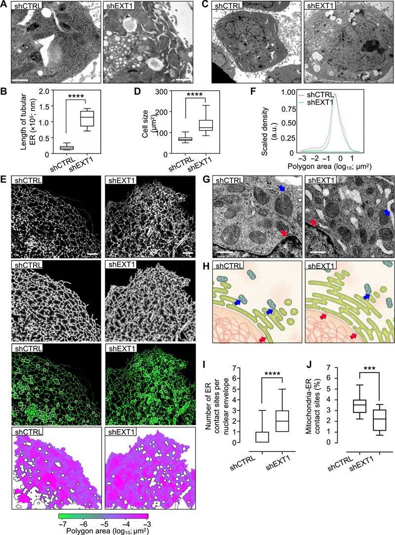
Figure from Kerselidou et al. (2021). Alternative glycosylation controls ER network morphology. Glycoproteomic profiling combined with functional perturbation assays revealed that site-specific N-glycosylation of ER membrane-shaping proteins is required for proper ER tubular network formation and dynamics. (CC BY 4.0)
Why Choose Our Glycoproteomics Analysis Services
Comprehensive Technology Portfolio
We offer the full spectrum of glycoproteomics capabilities — from N- and O-glycoproteome discovery to site-specific characterization and targeted quantification — within a single integrated service platform. This breadth eliminates the need to coordinate across multiple providers, ensuring consistent data quality, streamlined project management, and integrated reporting across all analytical modules.
Multi-Platform Mass Spectrometry
Our access to Orbitrap, TIMS-TOF, and triple quadrupole platforms allows us to match the instrumentation to the specific analytical requirements of each project. Nano-flow systems maximize sensitivity for limited samples, ion mobility provides isomeric glycopeptide resolution for challenging glycan structures, and triple quadrupole platforms deliver the robustness needed for routine MRM-based glycopeptide quantification.
Deep Glycoproteomics Expertise
Glycopeptide identification and glycan assignment require specialized knowledge that extends beyond conventional proteomics bioinformatics. Our team has extensive hands-on experience with glycoproteomics search engines, glycan database construction, manual spectral validation of glycan assignments, and biological interpretation of glycosylation data. Every project benefits from direct scientist involvement, not automated pipeline processing.
Regulatory Guidelines-Compliant Framework
For biopharmaceutical clients, our glycoproteomics services are performed within a documented quality framework aligned with ICH M10 guidelines, including full audit trails, sample chain-of-custody tracking, and instrument qualification records. This framework supports biotherapeutic characterization, biosimilarity studies, and lot-release testing workflows.
Related Services
Our glycoproteomics analysis services are supported by a broader PTM characterization platform offering complementary analytical capabilities. These services can be combined to address multi-dimensional characterization needs.
- PTM Bioinformatics Analysis — Comprehensive bioinformatics for PTM data integration, functional annotation, and pathway analysis across modification types
- Modified Peptide Absolute Quantification — AQUA-based absolute molar quantification of specific glycopeptides and glycosylated peptides using isotopically labeled synthetic standards
- O-Glycosylation of Proteins — Targeted O-glycoproteomic profiling with EThcD-based site localization for mucin-type and non-canonical O-glycans
- Protein Drug Glycosylation Analysis — Specialized glycosylation characterization of recombinant therapeutic proteins and glycoconjugate vaccines
- PRM PTM Verification — Targeted PRM-based validation and quantification of specific glycosylation sites and glycopeptide candidates
- Label-Free PTM Quantification — Versatile label-free quantification of glycopeptides and other modified peptides across multiple experimental conditions
- Open-Search PTM Discovery — Unbiased discovery of unexpected and novel PTMs including rare glycan structures through open-mass-search strategies
Frequently Asked Questions
What is the difference between glycoproteomics and glycomics?
Glycomics analyzes glycans released from proteins as a pooled mixture, providing information about the total glycan repertoire but losing all site-specific context — you know which glycans are present in the sample but not which proteins or which sites they came from. Glycoproteomics analyzes intact glycopeptides, preserving the connection between each glycan and its attachment site. This site-resolved information is essential for understanding how glycosylation at specific positions affects protein function, and for characterizing microheterogeneity — the distribution of different glycans at each individual modification site.
What types of glycosylation can your service analyze?
We analyze both N-linked glycosylation (via the N-X-S/T sequon) and all major classes of O-linked glycosylation including mucin-type O-GalNAc, O-GlcNAc, O-fucose, and O-mannose. For N-glycosylation, we distinguish high-mannose, complex, hybrid, and paucimannose types. For O-glycosylation, we use EThcD-based fragmentation for unambiguous site localization. We also support analysis of glycosaminoglycan attachment sites and glycation modifications. Methods are optimized for each glycosylation type, and multi-enzyme digestion strategies are employed to maximize glycopeptide coverage.
What sample types are compatible with your glycoproteomics workflow?
Our pipeline accepts a wide range of sample types including purified glycoproteins (≥10 μg), immunoprecipitated proteins (≥5 μg), total cell lysates (≥500 μg protein), tissue homogenates (≥20 mg wet weight), biofluids including serum, plasma, and CSF (≥50 μL), and FFPE tissue sections (≥5 sections). For limited or precious samples, we offer scaled-down protocols with optimized nano-flow LC-MS/MS acquisition to maximize glycoproteomic coverage from minimal starting material.
How do you handle quantitative glycoproteomics comparisons?
We offer multiple quantification strategies selected based on experimental design. Label-free quantification using extracted ion chromatogram (XIC) alignment is our default for discovery studies and is compatible with any sample type. For multiplexed comparisons, TMT labeling enables simultaneous analysis of up to 16 conditions with reduced missing data. For targeted validation of specific glycopeptides, MRM and PRM methods provide absolute quantification with AQUA peptide standards. Differential analysis across conditions includes both changes in site occupancy (macroheterogeneity) and shifts in glycoform distribution at individual sites (microheterogeneity).
What is microheterogeneity and why is it biologically important?
Microheterogeneity refers to the distribution of different glycan structures at a single glycosylation site. For example, a specific site on a therapeutic antibody may carry G0F, G1F, and G2F glycoforms in characteristic ratios that directly affect Fc receptor binding, complement activation, and immunogenicity. In disease contexts, shifts in microheterogeneity at specific sites on cell surface proteins can alter receptor signaling, cell adhesion, and immune recognition. Measuring these distributions at each site — rather than averaging across all sites — provides functionally relevant information that bulk glycan analysis cannot capture.
Can you analyze glycoproteins from non-mammalian species?
Yes — we have extensive experience with glycoproteomics across diverse species including plants, insects, yeast, bacteria, and non-human primates. Glycosylation pathways differ substantially across species, and our glycan databases and search strategies are customized for each organism type. For plant glycoproteins, we account for species-specific glycan features including core α1,3-fucose, β1,2-xylose, and arabinose modifications. For insect cell glycoproteins, we optimize for paucimannose and fucosylated glycan structures characteristic of lepidopteran expression systems.
How does your service handle biotherapeutic glycosylation characterization?
For biopharmaceutical clients, our glycoproteomics services are performed within a documented quality framework aligned with regulatory guidelines. Each project includes full sample traceability, instrument qualification documentation, and data integrity controls. We provide site-specific glycosylation characterization of therapeutic antibodies, fusion proteins, and biosimilars, including Fc glycan profiling, Fab glycosylation analysis, and comprehensive glycoform distribution assessment. Results are delivered with detailed documentation suitable for regulatory submissions. We also offer specialized bioinformatics support for glycoproteomics dataset analysis and biological annotation as part of our integrated service package.
References
- Kerselidou D, Dohai BS, Nelson DR, Daakour S, De Cock N, Hassoun Z, Kim DK, Olivet J, El-Assaad A, Al-Jaber H, Al-Mansoori A, Mahmoud L, Kandasamy RK, Larsen MR, Saleh MA, Saez-Rodriguez J, Merhi RA, Moustafa EA, Van Obberghen E, Jovanovic M, El Khallouki N, Falcon-Perez JM, Potes-Ares S, Bous J, Saab S, Rhee HW, Grice D, Antrobus R, Zitzler J, Wright R, Al Kubaisy G, Suleiman S, Rjab A, Eid AA, Ravid R, Lefebvre T, Kirmizitas FC, Pierredon S, Coute Y, Cuvelier M, Ivorra JL, Hammoud SM, Zallaa M, El-Sabban ME, Vo PH, Poteser M, Groschner K, Penninger JM, Rott R, Meriane M, Saleh S, King G, Schoenfeld R, Morelli X, Del Sol A, Drou N, Ghesquière B, De Bock PJ, Kim E, El Khoury R, Hachem S, Ghesquière B, De Bock PJ, Kim E, El Khoury R, Hachem S, Ghesquière B, De Bock PJ. Alternative glycosylation controls endoplasmic reticulum dynamics and tubular extension in mammalian cells. Sci Adv. 2021;7(37):eabe8349.
- Zeng WF, Yan G, Zhao HH, Liu C, Cao W. Uncovering missing glycans and unexpected fragments with pGlycoNovo for site-specific glycosylation analysis across species. Nat Commun. 2024;15:8055.
- Malaker SA. Glycoproteomics: charting new territory in mass spectrometry and glycobiology. J Mass Spectrom. 2024;59(6):e5034.
For research use only. Not for use in diagnostic procedures.