Comprehensive DNA and RNA Adductomics for Toxicology, Exposure Science, and Carcinogen Risk Assessment
Chemical modifications of DNA and RNA resulting from covalent binding of reactive chemical species — collectively termed adducts — represent the molecular intersection between environmental exposure and biological effect. These adducts arise from diverse sources including environmental and occupational carcinogens (polycyclic aromatic hydrocarbons, aflatoxins, aristolochic acid, tobacco-specific nitrosamines, heterocyclic amines, acrylamide), endogenous reactive metabolites (reactive oxygen and nitrogen species, lipid peroxidation aldehydes such as malondialdehyde and 4-hydroxynonenal, reactive carbonyl species), chemotherapeutic drugs (cisplatin, cyclophosphamide, temozolomide), and dietary genotoxins. DNA adducts, if not repaired before replication, can cause miscoding mutagenesis leading to permanent mutations in oncogenes and tumor suppressor genes, establishing adduct formation as a critical initiating event in chemical carcinogenesis. RNA adducts, while not directly mutagenic, can disrupt RNA function, translation, and cellular homeostasis, and may serve as more sensitive and dynamic biomarkers of recent or ongoing exposure than their DNA counterparts due to the higher turnover rate of RNA.
DNA/RNA adductomics — the comprehensive analysis of the nucleic acid adductome — addresses three fundamental challenges in toxicology and exposure science: (1) discovery of unknown adducts from novel or under-characterized exposures through untargeted high-resolution mass spectrometry, (2) sensitive and specific quantification of known adduct biomarkers for molecular epidemiology and biomonitoring studies, and (3) high-throughput screening of established adducts across large populations using immunoassay platforms. Our integrated service combines all three analytical modalities within a single workflow, enabling researchers to move from untargeted discovery to targeted validation to population-scale screening in a coordinated, analytically consistent framework. For comprehensive analysis of non-adduct RNA and DNA modifications, our DNA/RNA Modification LC-MS Analysis and RNA Modification Quantification by LC-MS/MS services provide complementary profiling of epigenetic and epitranscriptomic modifications without the adduct-focused enrichment and detection strategies described here.
Find Your Solution: Research Goal → DNA/RNA Adductomics Strategy
| Your Research Goal |
Recommended Approach |
Key Techniques |
| Untargeted discovery of known and novel DNA/RNA adducts in tissue or biofluid samples from exposed populations or experimental models |
LC-HRMS untargeted adductomics with DIA acquisition and computational adduct discovery |
DNA/RNA extraction, enzymatic hydrolysis, SPE enrichment, C18 HILIC or C30 LC separation, Orbitrap/QTOF DIA acquisition, nLossFinder or FeatureHunter data processing, database matching (MODOMICS, in-house adduct library), neutral loss filtering for ribose/deoxyribose-specific adduct identification |
| Absolute quantification of specific carcinogen-DNA adducts in human biospecimens for molecular epidemiology or biomonitoring |
Isotope dilution LC-MRM/MS (triple quadrupole) with adduct-specific enrichment |
DNA extraction, nuclease P1/BAL31 digestion, immunoaffinity or SPE enrichment of target adduct, C18 LC-MRM/MS with stable isotope internal standard (¹³C, ¹⁵N, or ²H-labeled adduct), quantification against calibration curve, detection limits down to 1–100 amol on-column |
| High-throughput screening of oxidative stress adducts (8-oxo-dG, 8-oxo-rG) across large clinical cohorts or longitudinal studies |
Competitive ELISA with colorimetric or fluorometric detection |
DNA/RNA extraction and enzymatic digestion, competitive ELISA in 96-well plate format using validated 8-oxo-dG monoclonal antibody (clone 483.15), TMB colorimetric or fluorometric detection, quantification by standard curve, normalization to DNA/RNA content by fluorometric method |
| Assessing chemotherapeutic drug-DNA adduct levels in preclinical or clinical samples for PK/PD correlation |
Targeted LC-MS/MS quantification of drug-DNA adducts with isotope internal standards |
Tissue or cell DNA extraction, enzymatic digestion preserving drug-adducted dinucleotides (cisplatin-d(GpG)), SPE desalting and enrichment, LC-MRM/MS with drug-adduct-specific MRM transitions, quantification relative to ¹⁵N-labeled internal standard, correlation with drug concentration in plasma |
| Simultaneous analysis of DNA and RNA adducts from the same biological sample for integrated exposome-biomarker studies |
Combined DNA/RNA extraction with parallel LC-HRMS adductomics |
Simultaneous DNA/RNA extraction from same sample, parallel enzymatic hydrolysis (DNase/RNase-specific), split-stream LC-HRMS DIA analysis, ribose vs. deoxyribose neutral loss discrimination for RNA vs. DNA adduct assignment, integrated data analysis with adduct class and nucleic acid origin annotation |
| Validation of candidate adduct biomarkers discovered in untargeted screening using orthogonal analytical methods |
Multi-platform cross-validation: LC-HRMS discovery → LC-MRM/MS quantification → ELISA population screening |
Tiered approach: initial adduct discovery by LC-HRMS DIA, synthesis or purchase of authentic adduct standards, targeted LC-MRM/MS method development and validation, cross-platform correlation analysis, ELISA development for validated adducts requiring population-scale throughput |
Complementary Platforms for Comprehensive DNA/RNA Adductomics Analysis
Our DNA/RNA adductomics service portfolio encompasses three complementary analytical platforms — untargeted LC-HRMS adductomics, targeted LC-MS/MS quantification, and immunoassay-based detection — each optimized for specific stages of the adduct biomarker discovery-validation-implementation pipeline. Platform selection depends on the research objective (discovery vs. quantification vs. screening), sample throughput requirements, adduct characteristics, and desired limits of detection.
Untargeted LC-HRMS Adductomics (Orbitrap/QTOF-DIA) for Broad-Spectrum Adduct Discovery
For comprehensive, untargeted characterization of the DNA/RNA adductome, we employ high-resolution accurate mass (HRAM) mass spectrometry — using Orbitrap (Q Exactive HF-X, Orbitrap Fusion Lumos) and QTOF (TripleTOF 6600, timsTOF flex) platforms — operating in data-independent acquisition (DIA) mode for broad-spectrum adduct detection without pre-selection of target analytes. DIA acquisition systematically fragments all precursor ions within sequential isolation windows (typically 25 Da windows across m/z 200–1000), generating complete fragment ion maps that enable retrospective data mining for any adduct without the need for re-acquisition. The characteristic neutral loss of 2'-deoxyribose (116.047 Da) from DNA adducts and ribose (132.042 Da) from RNA adducts upon CID fragmentation serves as a diagnostic filtering strategy — implemented in open-source software such as nLossFinder and FeatureHunter — to distinguish nucleic acid adducts from background chemical noise and to assign adducts to DNA versus RNA origin. Untargeted LC-HRMS adductomics can detect 50–150+ putative adducts per sample, with identification confidence levels ranging from accurate mass-based elemental composition assignment (Level 5, <3 ppm mass accuracy) through MS/MS spectral matching with reference standards (Level 1). This discovery platform is the method of choice for exposome-wide association studies, novel adduct identification, and characterization of adduct profiles in understudied exposure scenarios, tissue types, or model organisms.
Targeted Isotope Dilution LC-MRM/MS for Absolute Quantification of Known Adduct Biomarkers
For sensitive and specific quantification of validated adduct biomarkers — including aflatoxin B1-N7-guanine, BPDE-N2-dG, aristolochic acid-dA, 8-oxo-dG, 1,N6-etheno-dA, M1dG, cisplatin-d(GpG), and N7-methylguanine — we employ triple quadrupole (QQQ) mass spectrometry operating in multiple reaction monitoring (MRM) mode with isotope dilution quantification. In this configuration, the first quadrupole (Q1) selects the adduct-specific precursor ion, the collision cell (q2) generates characteristic fragment ions (typically the base loss or adduct-specific fragmentation product), and the third quadrupole (Q3) selects a diagnostic fragment ion, providing two stages of mass filtering that achieves limits of detection as low as 1–100 amol on-column. Quantification is performed by reference to stable isotope-labeled internal standards (¹³C, ¹⁵N, or ²H-labeled adducts) added at the earliest possible step of sample processing, correcting for extraction efficiency, matrix effects, and ionization variability. Adduct enrichment before LC-MS/MS — using offline solid-phase extraction, immunoaffinity columns, or nuclease P1 digestion followed by butanol extraction — is employed to concentrate adducted nucleosides from the vast excess of unmodified nucleosides (typical enrichment factor 100–1000-fold), enabling detection of adducts present at frequencies as low as 1 adduct per 10⁸–10⁹ unmodified nucleosides from DNA inputs of 10–100 μg. For targeted quantification of oxidative damage adducts specifically, our Oxidative DNA/RNA Damage Assay provides dedicated LC-MS/MS quantification of 8-oxo-dG, 8-oxo-rG, and related oxidative damage markers.
Competitive ELISA for High-Throughput Screening of Established Adduct Biomarkers
For established adduct biomarkers that are well-characterized and have demonstrated clinical or epidemiological utility — particularly 8-oxo-dG as the most widely studied oxidative stress biomarker, and to a lesser extent N7-methylguanine and 5-hydroxymethylcytosine as DNA damage markers — our competitive ELISA platform provides high-throughput (80–200 samples per week), cost-effective ($5–20 per sample) quantification in a standard 96-well plate format. The competitive ELISA format uses an adduct-specific monoclonal antibody (e.g., clone 483.15 for 8-oxo-dG, validated for specificity against unmodified guanosine and structurally related oxidative products) to bind adducted nucleosides in competition with an immobilized adduct conjugate on the plate surface. Colorimetric (TMB, 450 nm) or fluorometric (HRP-fluorophore) detection provides limits of detection suitable for typical 8-oxo-dG levels in human urine (0.5–5 ng/mL), tissue DNA (1–50 adducts/10⁶ dG), and plasma (10–100 pg/mL). While ELISA cannot match the chemical specificity of LC-MS/MS (potential cross-reactivity is the primary limitation), its throughput, minimal instrument requirements, and compatibility with standard clinical laboratory workflows make it the method of choice for large-scale epidemiological studies, clinical trials, and longitudinal biomonitoring programs where thousands of samples require analysis. For comprehensive immunoassay-based modification detection, our DNA/RNA Modification Immunoassays service provides a broader panel of antibody-based detection platforms beyond 8-oxo-dG.
Why Choose Our DNA/RNA Adductomics Service
Integrated Discovery-to-Screening Pipeline Under a Single Service Relationship
We offer the only fully integrated DNA/RNA adductomics pipeline spanning untargeted LC-HRMS discovery, targeted LC-MRM/MS validation, and ELISA-based population screening within a single service provider. This integration eliminates the fragmentation inherent in coordinating separate discovery and quantification platforms across different laboratories, ensures analytical continuity across all stages of the biomarker pipeline, and enables researchers to seamlessly transition from adduct discovery in model systems to targeted quantification in human biospecimens to large-scale screening in epidemiological cohorts without the method transfer and cross-validation challenges that typically plague multi-laboratory adductomics studies.
100+ Adduct Coverage Spanning Environmental, Oxidative, Lipid Peroxidation, and Chemotherapeutic Classes
Our combined adductomics platform detects and quantifies adducts across the full spectrum of chemical classes relevant to human health: environmental carcinogen adducts (polycyclic aromatic hydrocarbons, aflatoxins, aristolochic acid, aromatic amines, tobacco-specific nitrosamines, heterocyclic amines, acrylamide), oxidative stress adducts (8-oxo-purines, 8-oxo-pyrimidines, 5-hydroxy-pyrimidines, spiroiminodihydantoin, guanidinohydantoin), lipid peroxidation-derived etheno adducts (1,N6-etheno-dA, 3,N4-etheno-dC, N2,3-etheno-dG) and propano adducts (M1dG, 4-HNE-dG, acrolein-dG, crotonaldehyde-dG), endogenous alkylation products (N7-alkyl-dG, O6-alkyl-dG, N3-alkyl-dA), and chemotherapeutic drug-DNA adducts (platinum-based, nitrogen mustards, alkyl sulfonates, triazenes).
Sub-fmol Detection Limits with Isotope Dilution Quantitative Accuracy
Our targeted LC-MRM/MS platform achieves limits of detection of 1–100 amol on-column for most adducts through a combination of optimized enrichment strategies (immunoaffinity, SPE, butanol extraction), efficient chromatographic separation (narrow-bore C18 columns, 0.3–1 mm ID, with low-flow or microflow gradients), and sensitive MRM detection. Isotope dilution quantification using ¹³C-, ¹⁵N-, or ²H-labeled internal standards — added at the earliest possible step of sample processing — provides absolute quantification accuracy (typically 85–115% recovery, <15% inter-day CV) that is directly comparable across samples, studies, and laboratories, a critical requirement for regulatory-relevant toxicology and molecular epidemiology data.
Simultaneous DNA and RNA Adductomics from Single-Sample Input
We offer a unique simultaneous DNA/RNA adductomics workflow that extracts both DNA and RNA from the same biological sample, processes them through parallel hydrolysis and enrichment protocols, and analyzes them in a single LC-HRMS DIA injection — using the diagnostic neutral loss difference between 2'-deoxyribose (116.047 Da for DNA adducts) and ribose (132.042 Da for RNA adducts) to computationally assign detected adducts to their nucleic acid origin. This simultaneous DNA/RNA adductomics approach maximizes information yield from precious limited samples, enables direct comparison of adduct levels and profiles between DNA and RNA from the same tissue or biofluid, and provides complementary perspectives on exposure, damage, and biological effect within a unified analytical framework.
Workflow: From Sample to Comprehensive DNA/RNA Adductome Characterization
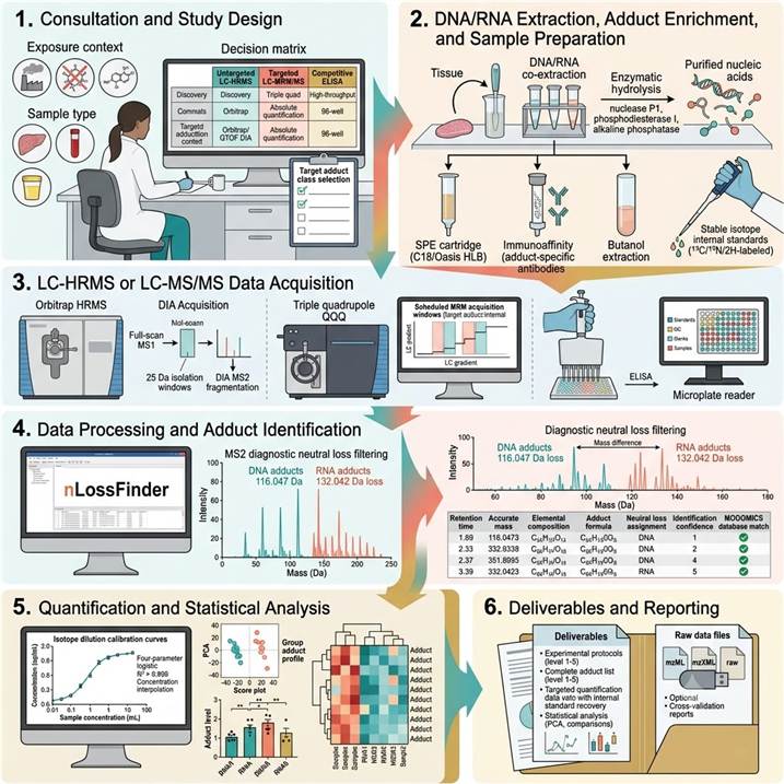
Step 1: Consultation and Study Design
We consult with you to define the adductomics strategy based on research objective (untargeted discovery, targeted quantification, or population screening), exposure or treatment context (environmental, occupational, therapeutic, dietary), target adduct classes (carcinogen adducts, oxidative stress adducts, lipid peroxidation products, drug-DNA adducts), sample types (tissue, blood, urine, buffy coat, FFPE, cells), and throughput requirements. We provide a detailed project proposal with platform recommendations, expected adduct coverage, limits of detection, and sample requirements.
Step 2: DNA/RNA Extraction, Adduct Enrichment, and Sample Preparation
DNA and RNA are co-extracted or separately extracted from provided samples using optimized protocols that preserve adduct integrity. For carcinogen adducts: DNA/RNA is enzymatically hydrolyzed to nucleosides (nuclease P1, phosphodiesterase I, alkaline phosphatase) or to nucleobases (thermal or acid hydrolysis for depurinating adducts). Adduct enrichment is performed by SPE (C18, Oasis HLB, carbograph), immunoaffinity chromatography (adduct-specific antibody columns), or butanol extraction (nuclease P1 digestion enriches adducted species). Stable isotope internal standards are added at the earliest possible step. Samples are desalted and concentrated for LC-MS analysis.
Step 3: LC-HRMS or LC-MS/MS Data Acquisition
For untargeted adductomics: enriched samples are analyzed by LC-HRMS on Orbitrap or QTOF instruments with full-scan MS¹ and DIA MS² acquisition over m/z 200–1000. For targeted quantification: samples are analyzed by LC-MRM/MS on triple quadrupole instruments with scheduled MRM acquisition windows for each adduct-internal standard pair. For ELISA: samples are analyzed in 96-well competitive ELISA format with colorimetric or fluorometric readout. Each analytical batch includes calibration standards, QC samples, blanks, and positive/negative controls.
Step 4: Data Processing and Adduct Identification
For untargeted LC-HRMS data: raw files are processed using nLossFinder, FeatureHunter, or Compound Discoverer software. Features are detected, aligned across samples, and filtered using the diagnostic neutral loss filtering strategy — searching for the characteristic loss of 2'-deoxyribose (116.047 Da) for DNA adducts or ribose (132.042 Da) for RNA adducts in MS² spectra. Putative adducts are assigned elemental compositions from accurate mass (<3 ppm error) and identified by MS/MS spectral matching against MODOMICS, PubChem, and in-house adduct libraries. For targeted data: peaks are integrated, retention times aligned, and analyte/internal standard ratios calculated.
Step 5: Quantification and Statistical Analysis
For targeted LC-MS/MS: absolute adduct concentrations are calculated using isotope dilution calibration curves and normalized to DNA/RNA input (expressed as adducts per 10⁶ or 10⁸ unmodified nucleosides, or fmol/μg DNA/RNA). For untargeted data: relative adduct abundance is compared across groups using peak area normalization. For ELISA: absorbance values are converted to adduct concentration using four-parameter logistic standard curves. Statistical analysis includes appropriate group comparisons (t-test, ANOVA, non-parametric alternatives), multiple testing correction, PCA for global adduct profile analysis, and correlation analysis with exposure metrics or clinical endpoints.
Step 6: Deliverables and Reporting
Comprehensive adductomics report including: complete experimental protocols with DNA/RNA extraction, hydrolysis, enrichment, and LC-MS or ELISA methods; all detected/quantified adducts with identification confidence levels, retention times, accurate masses, MS/MS spectra, and database matches (untargeted) or absolute concentrations with internal standard recovery and QC metrics (targeted); statistical analysis results with group comparisons, PCA, heat maps, and correlation analyses; and raw data files in standard formats (mzML, mzXML, raw) for independent re-analysis and regulatory submission.
Applications in DNA/RNA Adductomics Research
DNA/RNA adductomics has become an indispensable analytical platform for toxicology, exposure science, carcinogenesis research, and drug development, providing molecular-level evidence of nucleic acid damage from environmental, endogenous, and therapeutic sources. The following application areas represent the most active and impactful use cases for our adductomics platforms.
Environmental and Occupational Exposure Biomonitoring: Carcinogen-DNA Adducts as Molecular Exposure Dosimeters
DNA adducts of environmental and occupational carcinogens — including aflatoxin B1-N7-guanine (aflatoxin exposure, liver cancer risk), benzo[a]pyrene diol epoxide-N2-deoxyguanosine (BPDE-N2-dG, polycyclic aromatic hydrocarbon exposure from air pollution and tobacco smoke), aristolochic acid-dA (aristolochic acid exposure, urothelial cancer), 4-aminobiphenyl-dG (tobacco smoke and textile dye exposure, bladder cancer), and acrylamide-dG (dietary and occupational acrylamide exposure) — serve as molecular dosimeters that integrate exposure across all routes and over the lifetime of the DNA molecule (weeks to months in non-dividing tissues). Targeted LC-MRM/MS quantification of specific carcinogen-DNA adducts in human tissue, blood, or urine provides the most direct and specific measure of biologically effective dose — the amount of carcinogen that has reached the target tissue and formed covalent DNA lesions — making adduct quantification an essential tool for molecular epidemiology, regulatory toxicology, and occupational health surveillance. For comprehensive analysis of oxidative DNA damage in exposure contexts, our Oxidative DNA/RNA Damage Assay provides targeted LC-MS/MS quantification of 8-oxo-dG and related oxidative damage products.
Oxidative Stress and Lipid Peroxidation Adducts in Disease Pathogenesis and Aging Research
Reactive oxygen species (ROS) and reactive nitrogen species (RNS) generated during inflammation, mitochondrial dysfunction, ischemia-reperfusion, and aging produce a characteristic spectrum of oxidative DNA and RNA adducts including 8-oxo-7,8-dihydro-2'-deoxyguanosine (8-oxo-dG), 8-oxo-7,8-dihydroguanosine (8-oxo-rG), 5-hydroxymethyl-2'-deoxycytidine (5-HMdC), spiroiminodihydantoin (Sp), and guanidinohydantoin (Gh). Additionally, ROS-driven lipid peroxidation of polyunsaturated fatty acids generates reactive aldehydes — malondialdehyde (MDA), 4-hydroxynonenal (4-HNE), acrolein, and crotonaldehyde — that form exocyclic etheno and propano adducts with DNA bases (1,N6-etheno-dA, 3,N4-etheno-dC, M1dG, 4-HNE-dG, acrolein-dG). These oxidative and lipid peroxidation-derived adducts are implicated in the pathogenesis of cancer, cardiovascular disease, neurodegeneration (Alzheimer's, Parkinson's), diabetes, and aging. Simultaneous quantification of multiple oxidative adduct classes by LC-HRMS or targeted LC-MS/MS provides a comprehensive molecular readout of oxidative stress status across the DNA-RNA continuum, enabling mechanistic studies of oxidative damage in disease models and clinical cohorts.
Chemotherapeutic Drug-DNA Adduct Quantification for Pharmacodynamic and Toxicity Assessment
Many chemotherapeutic drugs exert their anti-cancer activity through the formation of covalent DNA adducts that block replication and transcription, triggering cell cycle arrest and apoptosis. Quantification of drug-DNA adduct levels in tumor tissue, peripheral blood mononuclear cells, or surrogate tissues provides a direct pharmacodynamic readout of drug target engagement at the DNA level, enabling correlation of adduct formation with therapeutic response, toxicity, and resistance. Key drug-DNA adducts quantified by our platform include: cisplatin-1,2-d(GpG) intrastrand crosslinks (the major cisplatin-DNA adduct, responsible for the drug's anti-cancer activity and measurable in patient blood cells for pharmacokinetic-pharmacodynamic correlation), oxaliplatin-d(GpG) adducts, cyclophosphamide-N7-guanine adducts (formed by phosphoramide mustard, the active metabolite of cyclophosphamide), temozolomide-N7-methylguanine and O6-methylguanine (the cytotoxic lesion that triggers mismatch repair-dependent cell death in MGMT-deficient tumors), and busulfan-DNA crosslinks. These measurements are critical for dose optimization, schedule selection, combination therapy design, and early assessment of treatment efficacy in preclinical and clinical drug development.
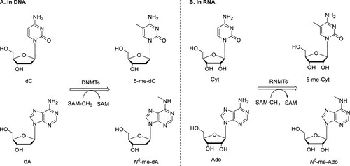
Endogenous Adductome Mapping: Characterizing the Background DNA Damage Landscape in Human Tissues
The endogenous adductome — comprising DNA and RNA adducts formed by normal cellular metabolism without exogenous exposure — represents the background damage landscape upon which environmental and therapeutic exposures are superimposed. Endogenous adducts include N7-methylguanine (the most abundant endogenous DNA adduct, formed by S-adenosylmethionine-mediated non-enzymatic methylation), N3-methyladenine, O6-methylguanine, 1,N6-etheno-dA (formed by lipid peroxidation products), 8-oxo-dG (from mitochondrial ROS), and various oxidative and deamination products. Untargeted LC-HRMS adductomics provides the comprehensive view of this endogenous adductome, revealing age-related, tissue-specific, and sex-specific differences in background adduct levels that establish baseline references for exposure studies. Guilbaud et al. (Nucleic Acids Research, 2023) used untargeted LC-MS/MS adductomics to characterize 114 putative DNA adducts in rat tissues and 111 in human heart and brain, revealing profound tissue-specific, age-dependent, and sex-specific differences in the endogenous adductome that provide essential reference data for biomarker studies.
RNA Adductomics for Dynamic Exposure Biomarker Discovery and Functional Impact Assessment
RNA adducts offer distinct advantages over DNA adducts as exposure biomarkers: RNA is not subject to excision repair pathways that remove DNA adducts, RNA turnover is more rapid (hours to days vs. days to weeks for DNA in many tissues), providing a shorter and more dynamic exposure window, and RNA can form adducts with the same reactive species that modify DNA. Yet RNA adductomics has historically been understudied compared to DNA adductomics. Recent methodological advances — particularly the simultaneous DNA/RNA adductomics approach developed by Martella et al. (Chemical Research in Toxicology, 2023) using LC-HRMS DIA with diagnostic neutral loss filtering — now enable parallel analysis of both nucleic acid adductomes from a single sample, revealing that RNA adducts are often more abundant and more sensitive to recent exposure than their DNA counterparts. Our platform incorporates these advances, offering simultaneous DNA/RNA adductomics that maximizes information yield from precious limited samples and provides complementary perspectives on nucleic acid damage from both endogenous and exogenous sources.
Case Study: Simultaneous RNA and DNA Adductomics Using Single Data-Independent Acquisition Mass Spectrometry Analysis for Environmental Sentinel Organism Exposure Assessment
In a 2023 study published in Chemical Research in Toxicology (CC BY 4.0), Martella et al. developed and applied the first single-injection LC-HRMS method for simultaneous untargeted detection of both DNA and RNA adducts, demonstrating the feasibility and power of integrated nucleic acid adductomics for environmental exposure assessment using Baltic amphipods as sentinel organisms.
Background: DNA adductomics has been established as a powerful approach for detecting nucleic acid damage from environmental exposures, but RNA adductomics has lagged behind due to methodological challenges in distinguishing RNA adducts from DNA adducts in complex biological samples. Since RNA and DNA adducts carry different biological implications — DNA adducts have mutagenic potential through replication errors, while RNA adducts may affect translation, splicing, and cellular homeostasis — simultaneous analysis of both nucleic acid adductomes from the same sample would provide complementary and distinct information about exposure effects. However, existing methods required separate analyses for DNA and RNA, doubling sample requirements and preventing direct comparison. The authors addressed this gap by developing a single-injection LC-HRMS method that exploits the characteristic mass difference between 2'-deoxyribose (116.047 Da, DNA) and ribose (132.042 Da, RNA) neutral losses upon CID fragmentation to computationally separate DNA and RNA adduct signals within a single DIA acquisition.
Approach: Nucleic acids were co-extracted from Baltic amphipods (Monoporeia affinis) — benthic invertebrates used as environmental sentinels for monitoring sediment contamination in the Baltic Sea — and enzymatically hydrolyzed to individual nucleosides. The hydrolysate was analyzed by LC-HRMS on an Orbitrap Q Exactive instrument operating in full-scan MS¹ mode (m/z 200–1000, resolution 70,000) with sequential DIA MS² acquisition (25 Da isolation windows, resolution 17,500, stepped HCD collision energy). Open-source nLossFinder software was used to screen MS² spectra for the characteristic neutral loss of 2'-deoxyribose (116.047 Da) for DNA adducts and ribose (132.042 Da) for RNA adducts, enabling automated assignment of each detected adduct to its nucleic acid origin. Putative adduct identifications were made by accurate mass matching (<3 ppm), retention time correlation, and MS/MS spectral comparison against the MODOMICS RNA modification database and in-house adduct reference libraries.
Key Findings:
- Simultaneous DNA and RNA adduct detection from a single injection: The single-injection LC-HRMS DIA method successfully detected both DNA and RNA adducts from the same nucleic acid hydrolysate, with the diagnostic neutral loss filtering strategy achieving clear separation of deoxyribose-conjugated (DNA) and ribose-conjugated (RNA) adducts without the need for separate DNA and RNA extraction and analysis — a methodological advance that halves sample requirements and enables direct comparison of DNA and RNA adduct profiles from the same biological specimen
- Comprehensive adduct coverage in environmental sentinel organisms: The method detected approximately 150 putative DNA adducts and 60 putative RNA adducts in Baltic amphipods collected from contaminated sediment sites, demonstrating that environmental sentinel organisms accumulate a complex mixture of both DNA and RNA adducts reflecting their exposure to complex environmental contaminant mixtures — including polycyclic aromatic hydrocarbons, heavy metals, and persistent organic pollutants — and highlighting the value of adductomics for integrated environmental exposure assessment
- Identification of known and novel adducts: Putative adduct identifications included simple mono- and dimethylation adducts on both DNA and RNA nucleosides (reflecting endogenous methylation by S-adenosylmethionine), larger functional group modifications, and the deaminated product inosine on RNA (reflecting RNA editing or hydrolytic deamination). Notably, 54 of the 60 detected RNA adducts remained structurally unidentified — compared to a larger proportion of identifiable DNA adducts — suggesting that the RNA adductome represents a largely unexplored frontier in adductomics with many novel nucleic acid modifications awaiting structural characterization
- Methodological validation using MODOMICS database: RNA adduct identification was performed by reference to the MODOMICS database — the comprehensive database of RNA modification structures — providing a standardized nomenclature and structural reference framework for RNA adduct annotation that had not been previously applied in the adductomics context, establishing a new bridge between the epitranscriptomics and adductomics research communities
- nLossFinder open-source data processing pipeline: The nLossFinder software was validated as an effective open-source platform for automated screening of diagnostic neutral losses in DIA data, enabling reproducible and transferable adduct detection across different laboratories and instrument platforms without reliance on proprietary software, critical for the broader adoption of simultaneous DNA/RNA adductomics in the environmental health research community
Significance: This landmark study established the first single-injection LC-HRMS method for simultaneous untargeted detection of DNA and RNA adducts, demonstrating that the characteristic neutral loss difference between deoxyribose (116.047 Da) and ribose (132.042 Da) in CID fragmentation provides a robust and generalizable strategy for computational assignment of adducts to their nucleic acid origin. The application to Baltic amphipod environmental sentinels demonstrated the method's utility for real-world exposure assessment in complex environmental matrices, while the identification of 60 putative RNA adducts — the majority structurally uncharacterized — highlighted the vast unexplored landscape of the RNA adductome. The open-source nLossFinder pipeline ensures that this approach is accessible and reproducible for the broader adductomics research community. Our service platform directly incorporates this methodological framework — simultaneous LC-HRMS DIA analysis with neutral loss-based DNA/RNA adduct discrimination — as a core component of our untargeted adductomics workflow.
Adapted from Martella et al. (2023). Simultaneous RNA and DNA Adductomics Using Single Data-Independent Acquisition Mass Spectrometry Analysis. Chemical Research in Toxicology 36(9):1471–1482. (CC BY 4.0)
Representative Results: DNA/RNA Adductomics Data Outputs and Platform Performance
Our DNA/RNA adductomics platform delivers integrated data packages combining untargeted adduct discovery, targeted quantification, and statistical analysis for comprehensive adductome characterization. The representative data below illustrates the typical output quality and performance specifications achieved across our three complementary platforms.
Platform Performance Specifications
| Performance Parameter |
Untargeted LC-HRMS (Orbitrap/QTOF DIA) |
Targeted LC-MRM/MS (Triple Quadrupole) |
Competitive ELISA (Colorimetric/Fluorometric) |
| Adduct coverage |
50–150+ putative adducts per sample |
1–30 targeted adducts per method |
1 adduct per assay (single-plex) |
| Detection mode |
Discovery (untargeted, full-scan + DIA MS²) |
Targeted (MRM, scheduled acquisition) |
Antibody-based (competitive binding) |
| Limit of detection |
10–100 fmol on-column (full-scan) |
1–100 amol on-column (MRM, adduct-dependent) |
0.1–1 ng/mL (8-oxo-dG ELISA) |
| Quantification type |
Semi-quantitative (relative peak area) |
Absolute (isotope dilution, fmol/μg DNA) |
Relative (% modification or ng/mL) |
| Identification confidence |
Level 2–5 (accurate mass, MS/MS, database) |
Level 1 (reference standard confirmation) |
Antibody specificity-dependent |
| Sample requirement |
5–50 μg DNA/RNA per analysis |
10–100 μg DNA/RNA (with enrichment) |
0.5–5 μg DNA/RNA per well |
| Throughput |
10–20 samples/week |
20–40 samples/week |
80–200 samples/week |
| Nucleic acid specificity |
Simultaneous DNA + RNA (neutral loss assignment) |
DNA or RNA (pre-selected by hydrolysis method) |
DNA or RNA (pre-selected by sample type) |
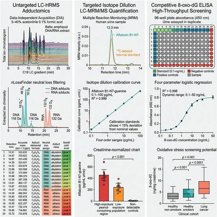
Representative data outputs from our DNA/RNA Adductomics platform. Left: Untargeted LC-HRMS DIA adductome profile with nLossFinder DNA/RNA adduct discrimination. Center: Targeted isotope dilution LC-MRM/MS quantification of aflatoxin B1-N7-guanine in human urine. Right: High-throughput 8-oxo-dG ELISA screening across clinical cohorts.
Key data deliverables included in every DNA/RNA adductomics project:
- Untargeted adductomics data package (LC-HRMS) — Complete list of all detected adduct features with retention time, accurate mass (<3 ppm), elemental composition, adduct class assignment (DNA vs. RNA by neutral loss filtering), MS/MS spectra with diagnostic fragment annotation, database match results (MODOMICS, PubChem, in-house library), identification confidence level, and relative abundance across all analyzed samples, with raw data files in mzML format
- Targeted quantification data package (LC-MRM/MS) — Absolute adduct concentrations (adducts per 10⁶ or 10⁸ unmodified nucleosides, or fmol/μg DNA/RNA) for all target adducts with individual internal standard recovery values (85–115% acceptance criteria), calibration curve parameters (R², linear range, LOD, LOQ), QC sample results, and inter-batch reproducibility metrics
- ELISA screening data package — Absorbance values for all samples, standards, and controls with 96-well plate layout documentation, calibration curve (four-parameter logistic regression parameters), calculated adduct concentrations with dilution factor correction, normalization to creatinine (urine samples) or DNA/RNA input (tissue samples), and inter-plate QC metrics
- Statistical analysis and visualization package — Group comparison results with appropriate statistical testing (t-test, ANOVA, Mann-Whitney, Kruskal-Wallis) and multiple testing correction (Benjamini-Hochberg FDR), with PCA score plots, hierarchical clustering heat maps, volcano plots, box plots, and correlation analysis with exposure metrics or clinical endpoints
- Methods documentation — Complete protocols for DNA/RNA extraction, enzymatic hydrolysis, adduct enrichment, LC-MS acquisition parameters, ELISA conditions, and data analysis methods, formatted for publication methods sections, grant applications, and regulatory reference
Related Services
Our DNA/RNA adductomics platform is part of a comprehensive nucleic acid modification analysis service portfolio spanning LC-MS quantification, oxidative damage analysis, immunoassay-based detection, and multi-platform integration for toxicology, exposure science, epigenetics, and translational research programs.
- Oxidative DNA/RNA Damage Assay — Targeted LC-MS/MS quantification of 8-oxo-dG, 8-oxo-rG, and related oxidative damage products for oxidative stress research and toxicology studies
- DNA/RNA Modification Immunoassays — Antibody-based ELISA, dot blot, fluorometric, and qPCR-compatible detection platforms for high-throughput screening of established modification and adduct biomarkers
- DNA/RNA Modification LC-MS Analysis — Integrated LC-MS analysis platform covering both DNA and RNA modifications in a single analytical workflow for comprehensive nucleoside modification profiling
- RNA Modification Quantification by LC-MS/MS — Mass spectrometry-based absolute quantification of 40+ modified ribonucleosides using isotope dilution LC-MS/MS for epitranscriptomics research
- m⁶A Modification LC-MS Analysis — Dedicated LC-MS analysis of N⁶-methyladenosine (m⁶A) and related adenine modifications with optimized enrichment and quantification protocols
- mRNA Modification LC-MS Analysis — Targeted LC-MS/MS profiling of modified ribonucleosides in purified mRNA with polyA selection and rRNA depletion workflows
FAQs
What is the difference between DNA/RNA adductomics and DNA/RNA modification analysis?
While the analytical techniques overlap, adductomics and modification analysis address distinct biological questions. DNA/RNA modification analysis typically focuses on epigenetically or epitranscriptomically relevant modifications — such as 5mC, 5hmC, m⁶A, m⁵C, and pseudouridine — that are enzymatically introduced by dedicated writer enzymes (methyltransferases, demethylases) and have regulatory functions in gene expression. Adductomics focuses on adducts formed by the non-enzymatic reaction of nucleic acids with reactive chemical species — including environmental carcinogens, endogenous electrophiles, ROS, and chemotherapeutic drugs — which represent damage rather than regulation. Methodologically, adductomics requires more extensive enrichment (since adducts are typically 10⁴–10⁹-fold less abundant than unmodified nucleosides) and employs different sample preparation strategies (adduct-specific SPE, immunoaffinity, butanol extraction) compared to modification analysis. Our service covers both domains with platform-specific expertise and optimized workflows.
What types of DNA and RNA adducts can be detected by untargeted LC-HRMS adductomics?
Untargeted LC-HRMS adductomics can detect any modification to a canonical nucleoside that is preserved during sample preparation and generates a detectable molecular ion and diagnostic neutral loss upon CID fragmentation. This includes alkyl adducts (methyl, ethyl, propyl, butyl, 2-hydroxyethyl), aryl and aralkyl adducts (BPDE-dG, 4-ABP-dG, PhIP-dG), etheno adducts (1,N6-etheno-dA, 3,N4-etheno-dC, N2,3-etheno-dG), propano adducts (M1dG, 4-HNE-dG, acrolein-dG, crotonaldehyde-dG), oxidative adducts (8-oxo-dG, 5-HMdC, Sp, Gh), and drug-DNA adducts (cisplatin-d(GpG), cyclophosphamide-dG). The detection coverage depends on the adduct's ionization efficiency, stability during sample preparation, and abundance relative to the analytical detection limits. A typical untargeted DIA experiment detects 50–150+ putative adduct features per tissue or biofluid sample, with each sample's adductome profile reflecting its unique exposure history and endogenous damage status.
What are the minimum DNA/RNA amounts required for adductomics analysis?
Sample requirements vary by platform and target adduct. For untargeted LC-HRMS adductomics: 5–50 μg of DNA or RNA is recommended for comprehensive adduct coverage, with lower inputs (1–5 μg) possible for abundant endogenous adducts (N7-meG, 8-oxo-dG) using optimized nanoflow LC-HRMS methods. For targeted LC-MRM/MS quantification of specific adducts with enrichment: 10–100 μg of DNA or RNA is typically required, with lower inputs (1–10 μg) achievable for high-abundance adducts using immunoaffinity enrichment. For competitive 8-oxo-dG ELISA: 0.5–5 μg of DNA or 5–50 μL of urine per well provides sufficient signal for quantification within the physiological range. For simultaneous DNA/RNA adductomics from limited samples (core needle biopsies, laser capture microdissected tissue, rare cell populations), we offer optimized microextraction and nanoflow LC-HRMS methods that can reduce sample requirements to 100 ng–1 μg total nucleic acid input.
How do you prevent artefactual adduct formation during sample processing?
Artefactual adduct formation during sample processing — particularly oxidation of guanine to 8-oxo-dG — is a well-recognized challenge in adductomics that can lead to overestimation of oxidative damage levels. Our protocols incorporate several measures to minimize artefactual adduct formation: (1) inclusion of antioxidants (butylated hydroxytoluene, deferoxamine, TEMPO) in all extraction and hydrolysis buffers; (2) use of chaotropic DNA/RNA extraction methods (guanidine-based) that minimize air exposure and reduce oxidation during purification; (3) sample processing under argon or nitrogen atmosphere for oxidative adduct quantification; (4) rapid sample handling at 4°C with minimal light exposure; (5) inclusion of ¹⁵N-labeled internal standards for each target adduct at the earliest possible step to correct for any artefactual formation; (6) monitoring of artefactual adduct formation using control samples (commercial DNA/RNA with known low adduct levels) processed in parallel with study samples; (7) validation of key findings using orthogonal analytical methods (LC-MS/MS validation of ELISA results).
Can DNA/RNA adductomics be performed on FFPE tissue samples?
Yes — FFPE tissue samples are compatible with our adductomics platform, with specific modifications to extraction protocols. FFPE DNA/RNA extraction requires extended proteinase K digestion and de-crosslinking (reversal of formaldehyde-induced nucleic acid-protein crosslinks) to release intact nucleic acids. However, FFPE processing introduces several considerations: (1) some adducts — particularly depurinating adducts like N7-alkyl-dG — may be partially lost during the prolonged heating at elevated temperatures required for de-crosslinking; (2) the formalin fixation process itself can introduce formaldehyde-DNA adducts (hydroxymethyl-dG, methylene crosslinks) that appear as additional adduct features requiring discrimination from biologically relevant adducts; (3) the quantity and quality of DNA/RNA from FFPE tissues are typically lower than from fresh frozen tissues. We validate adduct recovery from FFPE samples for each adduct class of interest and recommend using matched fresh frozen tissue controls where possible. Despite these challenges, FFPE-compatible adductomics enables retrospective analysis of the vast archives of clinically annotated FFPE tissues in hospital pathology departments for molecular epidemiological studies of environmental carcinogen exposure and cancer risk.
How are adducts identified and structurally characterized in untargeted adductomics?
Adduct identification in untargeted LC-HRMS adductomics follows a confidence level framework adapted from metabolomics standards (modified Schymanski scale). Level 1 (confirmed structure) requires matching retention time, accurate mass (<3 ppm), and MS/MS spectrum against an authentic standard analyzed under identical conditions. Level 2 (probable structure) requires MS/MS spectral matching against public or in-house libraries (MODOMICS, PubChem, METLIN, mzCloud, nLossFinder databases). Level 3 (tentative candidate) uses accurate mass and elemental composition to propose one or more candidate structures, with diagnostic neutral loss (116.047 Da for DNA, 132.042 Da for RNA) confirming nucleic acid origin. Level 4 (unknown adduct) is assigned to features with clear nucleic acid origin (neutral loss match) but insufficient spectral information for structural assignment. Level 5 (unknown feature) has uncertain adduct identity. Our reporting clearly states the identification confidence level for each detected adduct, and we offer optional targeted method development and synthesis of reference standards to advance Level 3–4 identifications to Level 1 confirmation for the most biologically significant adducts discovered in your study.
How do you distinguish between adducts arising from exogenous exposure vs. endogenous processes?
Distinguishing exogenous from endogenous adduct origins is a fundamental challenge in adductomics that requires multiple lines of evidence. Exogenous adducts — such as aflatoxin B1-N7-guanine, BPDE-N2-dG, and aristolochic acid-dA — have unique chemical structures not formed by any known endogenous process and are unambiguously assignable to specific exposures when confirmed by reference standards. Endogenous adducts — such as 8-oxo-dG, N7-methylguanine, and 1,N6-etheno-dA — are formed by normal cellular metabolism (ROS, lipid peroxidation, SAM-mediated non-enzymatic methylation) and are present in all individuals at baseline levels. The attribution of elevated endogenous adduct levels to specific exposures requires: (1) comparison with appropriate unexposed control groups; (2) correlation analysis with exposure metrics (questionnaire data, environmental monitoring, urinary metabolite levels); (3) adduct profile analysis (the pattern of multiple adducts provides more specific exposure signatures than individual adducts); (4) isotope labeling studies in model systems to trace adduct origin; (5) longitudinal sampling to establish temporal relationships between exposure and adduct levels. Our bioinformatics platform supports these comparative analyses with appropriate statistical methods for exposure-adduct association studies.
References
- Martella G, Motwani NH, Khan Z, Sousa PFM, Gorokhova E, Motwani HV. Simultaneous RNA and DNA Adductomics Using Single Data-Independent Acquisition Mass Spectrometry Analysis. Chemical Research in Toxicology. 2023;36(9):1471–1482.
- Guilbaud A, et al. Discovery adductomics provides a comprehensive portrait of tissue-, age- and sex-specific DNA modifications in rodents and humans. Nucleic Acids Research. 2023;51(20):10829–10845.
- Hu CW, Chang YJ, Chang YR, Cooke MS, Chen YR, Chao MR. A Novel Adductomics Workflow Incorporating FeatureHunter Software: Rapid Detection of Nucleic Acid Modifications for Studying the Exposome. Environmental Science & Technology. 2024;58(1):75–89.
For research use only. Not for use in diagnostic procedures.
![Scientific infographic of DNA/RNA adductomics concept: a DNA double helix and RNA strand showing various colored covalent adduct modifications from different sources — aflatoxin B1 (red), benzo[a]pyrene diol epoxide (orange), 8-oxo-dG (gold), cisplatin crosslinks (teal), etheno-dA (purple), and aristolochic acid-dA (coral) — on a white background. Right side shows three detection platforms: an Orbitrap HRMS for untargeted DIA acquisition, a triple quadrupole MS for targeted MRM quantification, and an ELISA plate reader for high-throughput adduct screening. The exposome-to-disease continuum is represented as a gradient arrow from environment (cigarette smoke, grilled food, pollution) to DNA adducts to mutation to disease. Teal-coral-gold color scheme, photorealistic scientific illustration style, clean minimal composition.](upload/image/pic-dnarna-adductomics-1.jpg)