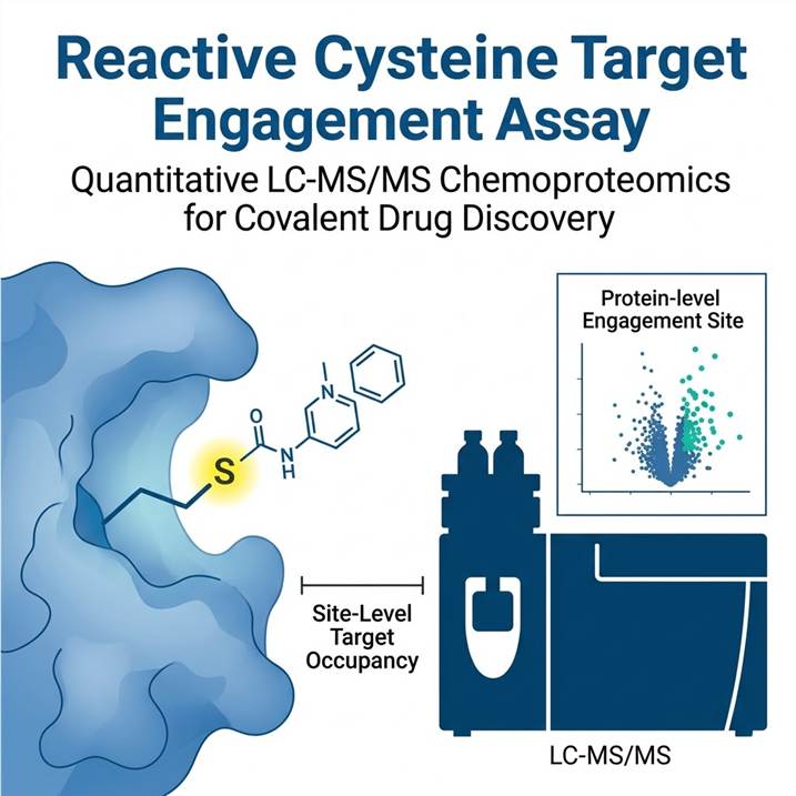
Why Measure Cysteine Target Engagement — The Covalent Drug Discovery Challenge
Covalent inhibitors offer unique advantages over non-covalent drugs: sustained target occupancy, the ability to target shallow binding sites, and potential for improved selectivity through targeted covalent warheads. However, these advantages come with heightened scrutiny from reviewers and regulators on two fronts: proving that the intended cysteine is actually modified in a complex proteome, and demonstrating that off-target engagement is minimal.
Traditional approaches to measuring target engagement — gel-based ABPP, intact protein MS, and thermal shift assays — each have critical limitations. Gel-based methods lack site-level resolution and quantitative accuracy. Intact protein MS requires purified protein and cannot assess proteome-wide selectivity. Thermal shift assays measure stability changes rather than direct occupancy, and they miss the majority of cysteine engagement events that do not alter protein thermal stability.
Our LC-MS/MS chemoproteomics platform addresses all three gaps simultaneously. Using competitive activity-based protein profiling (ABPP) with label-free quantification, we measure cysteine engagement at single-residue resolution across the entire expressed proteome, delivering the site-level occupancy data that covalent drug discovery programs need.
For research programs studying cysteine modifications beyond drug engagement, our Redox PTM Proteomics platform provides complementary cysteine oxidation and S-nitrosylation profiling. The Protein Oxidation Analysis service extends the assessment to other oxidation-sensitive residues.
Competitive ABPP Workflow — From Probe Labeling to Site-Level Quantification
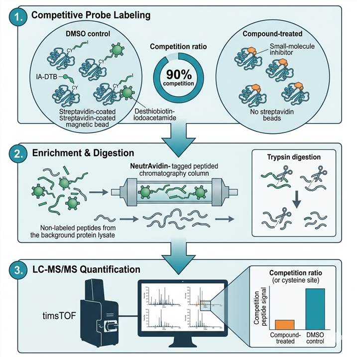
Our assay is built on a competitive ABPP design that uses a broadly reactive cysteine probe (iodoacetamide-desthiobiotin, IA-DTB) to quantify how much each cysteine is occupied by your compound versus the probe alone.
Step 1: Competitive Probe Labeling
- Compound or DMSO control incubated with cell lysate or live cells at specified concentrations
- IA-DTB probe added at saturating concentration to label all unoccupied cysteines
- Probe-labeled cysteines represent the fraction NOT engaged by the test compound
- Multiple concentrations and time points supported for dose-response and kinetics
Step 2: Enrichment and Digestion
- Probe-labeled proteins precipitated and digested with trypsin
- Desthiobiotin-tagged peptides enriched on NeutrAvidin resin
- Stringent washes remove non-labeled peptides
- Eluted cysteine-containing peptides prepared for LC-MS/MS
Step 3: LC-MS/MS Acquisition and Quantification
- Label-free quantification (LFQ) using DIA on timsTOF Pro 2 or Orbitrap platforms
- Peptide identification and quantification via Spectronaut or DIA-NN
- Competition ratio (CR) calculated as: compound signal ÷ DMSO control signal
- CR ≤ 0.5 indicates ≥50% target occupancy at that cysteine site
Typical project turnaround is 4–6 weeks from sample receipt to final report. For streamlined proteome-wide mapping of multiple PTM types in parallel, our Global PTMs Profiling service and Pan PTM Proteomics provide complementary analytical breadth.
Cysteine Reactivity & Ligandability Profiling Across the Proteome
Not all cysteines are equally reactive. Understanding the intrinsic reactivity landscape of the cysteinome is essential for prioritizing targets and interpreting engagement data. Our assay simultaneously reports on cysteine reactivity by measuring the baseline IA-DTB probe labeling intensity across >30,000 cysteine sites in each experiment.
Quantifying the Druggable Cysteinome
In a standard experiment using mammalian cell lysates, our platform routinely quantifies 23,000–32,000 unique cysteine sites derived from 8,000+ proteins. The detected cysteinome covers approximately 40% of all computationally predicted cysteine residues, enriched for surface-accessible and reactive thiols — precisely the subset most relevant for covalent drug targeting. This coverage depth enables the identification of ligandable hotspots, frequently engaged sites, and differentially reactive cysteines across cell states or tissues.
Ligandability Mapping from Fragment Screens
When applied to covalent fragment or electrophile libraries, the assay generates proteome-wide ligandability maps that reveal which cysteines are most readily engaged by different warhead chemotypes. Published benchmark studies using libraries of 80 chloroacetamide fragments identified 742 unique liganded cysteine events across 438 sites in 413 proteins, with most liganded proteins (>80%) belonging to the Tbio or Tdark categories — proteins with little or no existing chemical tool coverage. This makes the assay particularly valuable for expanding the druggable proteome into previously inaccessible target space.
Quantifying Target Occupancy — What Percentage of Your Cysteine Target Is Engaged?
Knowing that a compound binds to a cysteine is not enough. Medicinal chemists need to know how much of the target is occupied at a given concentration — the occupancy value that directly determines pharmacodynamic effect. Our assay delivers this through the competition ratio (CR), which is directly proportional to fractional target occupancy.
From Competition Ratios to Occupancy Values
The competition ratio (compound ÷ DMSO) decreases as compound concentration increases and more target cysteines become occupied. A CR of 0.2 means 80% occupancy; a CR of 0.5 means 50% occupancy. By running 6–8 compound concentrations, we generate full concentration-response curves and calculate pTE₅₀ values (the −log₁₀ of the concentration achieving 50% target engagement). These values provide direct, quantitative SAR feedback that correlates with functional activity.
Selectivity Profiling — Distinguishing On-Target from Off-Target Cysteine Engagement
For covalent drug programs, selectivity is the single most important determinant of clinical success. Off-target cysteine engagement can lead to mechanism-based toxicity, adverse pharmacology, and failed development programs. Our assay provides a comprehensive selectivity map for each compound in a single experiment, revealing every cysteine site engaged across the proteome.
Proteome-Wide Selectivity Metrics
We report three complementary selectivity metrics: (1) total number of engaged cysteine sites per compound at the screening concentration, (2) competition ratio distribution across all engaged sites, and (3) a selectivity score that weights engagement events by their potency relative to the intended target. In a typical 50 µM single-concentration screen, well-behaved fragments engage an average of 5 cysteine sites, while promiscuous compounds may engage 50 or more. These data are presented as intuitive volcano plots and interaction heatmaps that immediately highlight on-target engagement versus off-target hits.
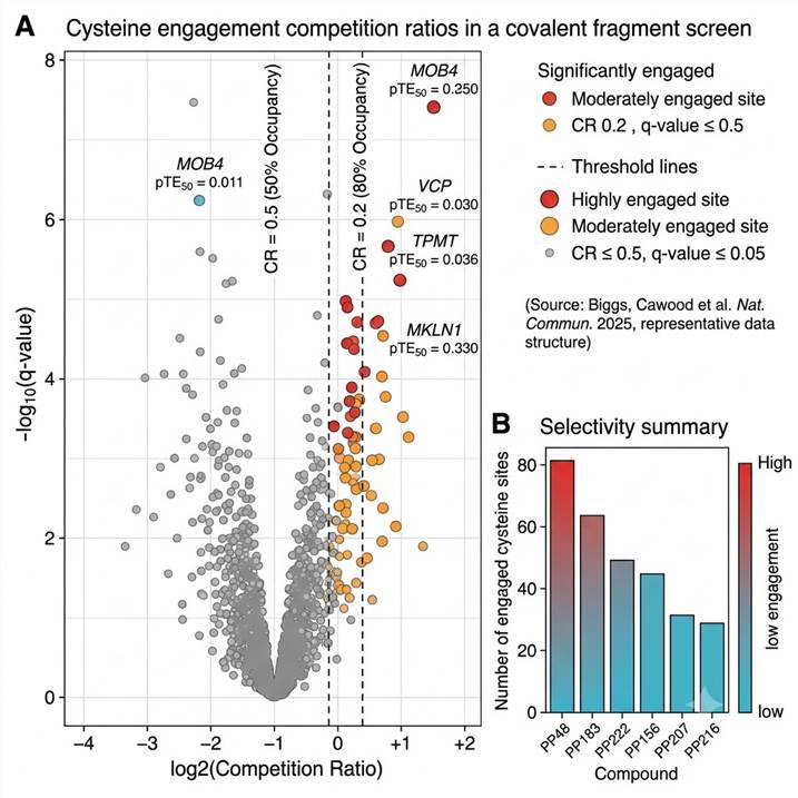
Differentiating Specific from Promiscuous Engagement
The ability to distinguish specific, saturable engagement from promiscuous reactivity is essential. Through concentration-response profiling, specific interactions show clear dose-dependence, while promiscuous reactivity typically reaches saturation at low concentrations. In the benchmark 80-fragment study, four high-confidence specific interactions were identified and validated across two cell lines: MOB4 Cys134–PP48, MKLN1 Cys82–PP156, VCP Cys522–PP183, and TPMT Cys70–PP222. Concentration-response experiments confirmed that these four interactions were not detected for any of the other 79 fragments, and each compound showed at least 10-fold selectivity for its primary target over the next most potently engaged site. For mapping phosphorylation-dependent signaling networks alongside target engagement, our Kinase-Substrate Network Analysis service provides complementary orthogonal data.
Case Study: Label-Free Chemoproteomics Profiling of 80 Cysteine-Reactive Fragments
To demonstrate the performance and biological utility of competitive ABPP for cysteine target engagement, we highlight a landmark study by Biggs, Cawood et al. (Nature Communications, 2025) that applied the same label-free chemoproteomics workflow used in our service to profile 80 chloroacetamide fragments against the native HEK293T and Jurkat proteomes. This study provides a comprehensive template for the type of data our service delivers.
Study Design and Analytical Performance
Eighty chloroacetamide fragments spanning a range of molecular weights and electrophilic reactivities were screened at 50 µM in both HEK293T and Jurkat cell lysates using competitive IA-DTB probe labeling followed by DIA-based LC-MS/MS. The platform achieved a median inter-replicate Pearson correlation of 0.96 and a median CV of 24.8%, with two-thirds of all quantified cysteine peptides detected in ≥75% of analyzed samples. Across both cell lines, the study quantified 32,000 unique cysteine sites from 8,000+ proteins, covering approximately 40% of all computationally predicted cysteine residues in the human proteome.
Key Findings: Ligandability and Selectivity
Screening the 80-fragment library identified 742 unique liganded cysteine events spanning 438 sites across 413 proteins. Each fragment engaged an average of 5 cysteine sites. Notably, >80% of the liganded proteins belonged to the Tbio or Tdark target development levels — proteins with little or no existing chemical tool coverage — demonstrating the ability of competitive ABPP to expand the druggable proteome. Four high-confidence specific interactions were validated across both cell lines: MOB4 Cys134–PP48, MKLN1 Cys82–PP156, VCP Cys522–PP183, and TPMT Cys70–PP222. Each of these interactions was exclusive to its cognate fragment among all 80 compounds tested.
Concentration-Response Validation
Four fragments with distinct selectivity profiles — PP207 (promiscuous), PP156 (moderate), PP152 (selective), and PP216 (highly selective) — were selected for concentration-response analysis across 6–8 concentrations. PP207 engaged >200 cysteine sites at 200 µM, while PP216 engaged fewer than 10. The resulting concentration-response curves yielded pTE₅₀ values that differentiated the fragments by potency: MOB4 Cys134–PP48 (pTE₅₀ 5.4 ± 0.1, ~4 µM), VCP Cys522–PP183 (pTE₅₀ 4.9 ± 0.1, ~13 µM), TPMT Cys70–PP222 (pTE₅₀ 5.7 ± 0.4, ~2 µM), and MKLN1 Cys82–PP156 (pTE₅₀ 5.2 ± 0.2, ~6 µM). Concentration-response data confirmed at least 10-fold selectivity for each primary target over the next most potently engaged off-target site. Structure-activity relationship (SAR) analysis around the PP48 scaffold further demonstrated that chemical modifications could tune selectivity between homologous targets, providing direct guidance for medicinal chemistry optimization.
Source
Biggs, G. S.; Cawood, E. E.; Rittinger, K.; Bush, J. T. et al. Nat. Commun. 2025, 16, 73. DOI: 10.1038/s41467-024-55057-5. Figure 4 (concentration-response and selectivity profiling) provides the primary data visualization for this study.
Sample Requirements & Project Planning for Cysteine Engagement Studies
Each project is scoped to the specific compound series, target hypothesis, and decision point. The table below provides general guidelines. Contact our scientific team for a project-specific feasibility assessment.
| Sample Type |
Recommended Amount |
Storage & Shipping |
Critical Notes |
| Cell lysate (treated/untreated) |
2–5 mg protein per condition |
−80 °C, ship on dry ice |
Include protease inhibitors; avoid freeze-thaw cycles. DMSO concentration ≤ 1% for compound treatment |
| Live cells (for in situ labeling) |
2–5 × 10⁷ cells per condition |
Snap-freeze pellet after compound incubation; −80 °C, ship on dry ice |
Compound incubation time and concentration must be specified. Include DMSO-only control |
| Tissue homogenate |
10–50 mg tissue equivalent |
Snap-freeze in liquid nitrogen; −80 °C, ship on dry ice |
Homogenize in lysis buffer with protease inhibitors; aliquot to avoid refreezing |
| Recombinant protein spike-in |
1–10 µg per condition + lysate background |
−80 °C, ship on dry ice |
For targeted occupancy assays where the target protein is expressed at low endogenous levels |
Deliverables — From Raw MS Data to Prioritized Target Lists
Every project delivers a complete data package designed to support medicinal chemistry decisions and regulatory-grade documentation. Our reporting is structured to answer the specific question that motivated the study — not just to deliver a list of identified cysteines.
Standard Deliverables
- Raw LC-MS/MS data files — instrument-native format (.d or .raw) for independent reanalysis
- Processed peptide identification tables — all identified cysteine-containing peptides with sequence, protein accession, and localization scores
- Quantification results — competition ratios (CR) and pTE₅₀ values for every engaged cysteine site in every condition
- Target engagement volcano plots — compound vs. DMSO comparison highlighting significantly engaged sites (CR ≤ 0.5, q-value ≤ 0.05)
- Selectivity heatmaps — compound-by-cysteine interaction matrices for multi-compound comparisons
- Prioritized target list — ranked by engagement potency and selectivity, annotated with protein function, tissue expression, and known pharmacology
- Interpretive project summary — contextual narrative linking engagement data to your specific drug discovery question, with recommended follow-up experiments
Representative Results — Cysteine Engagement Data in Action
The volcano plot below shows representative data from a competitive ABPP experiment, illustrating how target engagement results are visualized and interpreted. Significantly engaged cysteine sites are plotted by competition ratio (fold-change) versus statistical significance, with annotated pTE₅₀ values for top hits. For a detailed case study with full experimental context and concentration-response data, see the Case Study section above.
The engagement volcano plot displays the competition ratio (compound ÷ DMSO) on the x-axis (log₂ scale) and statistical significance (−log₁₀ q-value) on the y-axis. Sites in the upper-left quadrant (CR ≤ 0.5, q ≤ 0.05) represent high-confidence engagement events where ≥50% of the cysteine is occupied by the compound. Each project receives interactive versions of these plots alongside annotated target lists for downstream analysis.
Reactive Cysteine Target Engagement Assay: Frequently Asked Questions
How does this assay differ from standard activity-based protein profiling (ABPP)?
Standard ABPP uses activity-based probes to map enzyme activity states. Our Reactive Cysteine Target Engagement Assay extends this concept by adding a competitive compound incubation step before probe labeling, which allows us to quantify how much of each cysteine site is occupied by your compound versus the probe. This competition format delivers target occupancy values (competition ratios) that standard ABPP cannot provide.
What is the minimum sample amount required?
For cell lysate experiments, we recommend 2–5 mg total protein per condition. For live-cell labeling, 2–5 × 10⁷ cells per condition. These amounts support quantification of 15,000–30,000 cysteine sites. Smaller amounts are possible for targeted experiments where only a subset of cysteines needs to be monitored.
How is target occupancy calculated?
Target occupancy is derived from the competition ratio (CR) = compound signal ÷ DMSO control signal. A CR of 0.2 means 80% of that cysteine site is occupied by the compound; CR of 0.5 means 50% occupancy. For concentration-response experiments, occupancy-versus-concentration curves are fitted by logistic regression to calculate pTE₅₀ (the −log₁₀ of the concentration achieving 50% engagement).
Which covalent warhead chemotypes are compatible?
The assay primarily uses an iodoacetamide-desthiobiotin probe that broadly labels reactive cysteine thiols. This probe is compatible with most electrophilic warheads including acrylamides, chloroacetamides, vinyl sulfonates, Michael acceptors, and other cysteine-reactive groups. Compounds must be soluble in DMSO at the required screening concentration (final DMSO ≤ 1% v/v). For warheads that modify non-cysteine residues, alternative probe chemistries can be developed on a project-specific basis.
What is the typical turnaround time?
Standard projects are completed within 4–6 weeks from sample receipt. Single-concentration screening experiments are faster (3–4 weeks); multi-concentration dose-response studies may require 5–6 weeks. Rush timelines are available for time-sensitive programs.
How does this compare to thermal shift-based engagement assays?
Both methods assess target engagement, but they measure different biophysical properties and have complementary strengths. Thermal stabilization assays (PISA-based) detect compound-induced changes in protein thermal stability; our ABPP-based assay directly measures cysteine occupancy. Thermal stabilization approaches can be applied to any protein but require that compound binding alters thermal stability — a property that is not guaranteed for covalent engagement. Our assay provides direct, site-level evidence of cysteine engagement regardless of whether thermal stability is affected. When both methods agree, confidence in target engagement is substantially strengthened.
References
- Biggs, G. S.; Cawood, E. E.; Vuorinen, A.; McCarthy, W. J.; Wilders, H.; Riziotis, I. G.; van der Zouwen, A. J.; Pettinger, J.; Nightingale, L.; Chen, P.; Powell, A. J.; House, D.; Boulton, S. J.; Skehel, J. M.; Rittinger, K.; Bush, J. T. "Robust Proteome Profiling of Cysteine-Reactive Fragments Using Label-Free Chemoproteomics" Nat. Commun. 2025, 16, 73.
- Boatner, L. M.; Palafox, M. F.; Schweppe, D. K.; Backus, K. M. "CysDB: A Human Cysteine Database Based on Experimental Quantitative Chemoproteomics" Cell Chem. Biol. 2023, 30, 683–698.e3.
- Yang, K.; Whitehouse, R. L.; Dawson, S. L.; Zhang, L.; Martin, J. G.; Johnson, D. S.; Paulo, J. A.; Gygi, S. P.; Yu, Q. "Accelerating Multiplexed Profiling of Protein-Ligand Interactions: High-Throughput Plate-Based Reactive Cysteine Profiling with Minimal Input" Cell Chem. Biol. 2024, 31, 565–576.e4.