Activity-Based DUB and Ubiquitin Enzyme Assays for Targeted Protein Degradation and Drug Discovery
The ubiquitin system — comprising the hierarchical E1→E2→E3 ubiquitination cascade that installs ubiquitin on substrate proteins, and the ~100 deubiquitinating enzymes (DUBs) that remove ubiquitin to reverse or edit the ubiquitin signal — regulates nearly every aspect of eukaryotic cell biology, including protein stability, proteasomal degradation, DNA damage repair, NF-κB signaling, endocytic trafficking, autophagy, and innate immune responses. Dysregulation of ubiquitin enzymes is implicated in a broad spectrum of diseases: DUB overexpression or mutation drives oncogenesis (USP7 in multiple myeloma, USP14 in ovarian cancer, USP28 in colorectal cancer), E3 ligase dysfunction underlies neurodegenerative disorders (Parkin/PINK1 in Parkinson's disease, UBE3A in Angelman syndrome), and aberrant ubiquitin signaling contributes to inflammatory and autoimmune conditions (A20/TNFAIP3 in rheumatoid arthritis, NOD2 ubiquitination in Crohn's disease). The emergence of targeted protein degradation — particularly PROTACs (PROteolysis TArgeting Chimeras) that recruit E3 ligases to ubiquitinate and degrade disease-relevant proteins — has elevated the ubiquitin enzyme system from a therapeutic target class to a central enabling technology for pharmacological intervention across the proteome.
Activity-based DUB and ubiquitin enzyme assays differ fundamentally from abundance-based methods (western blot, immunofluorescence, RNA-seq) in that they measure the functional catalytic activity of the enzyme — not its presence or quantity. This distinction is critical because DUBs and E3 ligases are frequently regulated not at the expression level but through post-translational modifications (phosphorylation, ubiquitination itself), protein-protein interactions (adaptor proteins, substrate binding), and conformational changes that modulate activity independently of protein abundance. Activity-based probes (ABPs) — suicide inhibitors that form a covalent bond with the active-site cysteine of DUBs (or the catalytic cysteine of certain E3 ligases) and are equipped with detection tags (biotin, fluorophores, TMT) — capture the functional state of the enzyme at the time of probe treatment, providing a direct readout of catalytic activity that is orthogonal to and more mechanistically informative than abundance-based measurements. Our service integrates these activity-based approaches with complementary antibody-based and MS-based ubiquitination analysis platforms to deliver a complete picture of ubiquitin enzyme function in any biological context. For comprehensive ubiquitination site mapping, our Ubiquitylomics Analysis service provides global K-ε-GG ubiquitin remnant profiling by LC-MS/MS.
Find Your Solution: Research Goal → DUB and Ubiquitin Enzyme Activity Assay Strategy
| Your Research Goal |
Recommended Approach |
Key Techniques |
| Multiplexed DUB activity profiling across the full cysteine protease DUB family to identify hyperactive or dysregulated DUBs in disease models |
Activity-based DUB profiling (ABPP) with biotin-Ub-VME/biotin-Ub-PA probes and TMT-LC-MS/MS multiplexed quantification |
Cell/tissue lysate preparation, biotin-Ub-VME or biotin-Ub-PA probe labeling, streptavidin pull-down, on-bead trypsin digestion, TMT labeling (11-plex), nanoLC-MS/MS (Orbitrap), database searching for DUB identification, activity ratio quantification across experimental groups |
| Biochemical characterization and inhibitor screening for a specific DUB target |
Fluorogenic DUB activity assay using Ub-AMC or ubiquitin-PL substrate in 384-well plate format |
Recombinant DUB enzyme (full-length or catalytic domain), Ub-AMC substrate (7-amido-4-methylcoumarin), kinetic fluorescence readout (λex 380 nm / λem 460 nm) in microplate reader, dose-response curves for compound IC50 determination, Z′-factor for assay quality assessment, counter-screen against panel DUBs for selectivity profiling |
| E3 ligase activity assessment for PROTAC hijacking feasibility |
Reconstituted E1/E2/E3 ubiquitination cascade assay with biochemical or fluorescence readout |
E1 (UBA1/6×His-UBA1), E2 (UbcH5c, UbcH7, or target-specific E2), E3 ligase (CRBN-VHL-MDM2-IAP-RNF4-Parkin), ubiquitin ± biotin/FLAG/His tag, ATP/Mg²⁺ regeneration system, anti-ubiquitin antibody or streptavidin-HRP detection, autoubiquitination vs. substrate-specific ubiquitination readout |
| Cell-based DUB target engagement and proteome-wide selectivity for a DUB inhibitor candidate |
Cell-permeable activity-based probe (cR10-Ub-PA or Ub-PA) with pull-down and LC-MS/MS or AlphaLISA target occupancy readout |
Live cell treatment with cell-permeable Ub-PA probe, cell lysis, streptavidin pull-down, LC-MS/MS identification of probe-labeled DUBs, target occupancy calculation (labeled DUB signal in inhibitor-treated vs. untreated), proteome-wide selectivity profiling, deconvolution of off-target cysteine reactivity by competitive ABPP |
| Global ubiquitination analysis to assess degrader-induced ubiquitin signaling changes |
Ubiquitin remnant (K-ε-GG) LC-MS/MS proteomics with TMT quantification |
Cell/tissue lysate, trypsin digestion (generates K-ε-GG remnant on formerly ubiquitinated lysines), anti-K-ε-GG antibody (PTMScan) immunoaffinity enrichment, TMT labeling, nanoLC-MS/MS, MaxQuant/Proteome Discoverer with ubiquitination site localization, differential ubiquitination analysis, pathway enrichment |
| PROTAC ternary complex-induced target ubiquitination efficacy assessment |
Target-specific ubiquitination assay in reconstituted biochemical system or cellular context |
Recombinant target protein + E3 ligase + PROTAC compound, E1/E2/ubiquitin/ATP, anti-target antibody capture, anti-ubiquitin detection (AlphaLISA or MS), PROTAC concentration-response for ubiquitination induction, ternary complex stabilization (SPR or ITC), cellular ubiquitination by K-ε-GG MS after PROTAC treatment |
Integrated Assay Platforms for DUB and Ubiquitin Enzyme Activity Profiling
Our DUB and ubiquitin enzyme activity assay service portfolio encompasses four complementary platforms — biochemical activity assays, E1/E2/E3 cascade reconstitution, cell-based activity probes, and ubiquitin remnant proteomics — each optimized for specific stages of the drug discovery pipeline from target identification and validation through hit identification, lead optimization, and preclinical candidate characterization. Platform selection depends on the enzyme target (DUB vs. E3 ligase vs. full cascade), throughput requirements, desired mechanistic information, and the biological context of the study.
Biochemical DUB Activity Assays — Fluorogenic and MS-Based Readout Platforms
For direct, quantitative measurement of DUB catalytic activity in purified enzyme systems, we offer a comprehensive suite of biochemical activity assays using ubiquitin-based substrates with fluorogenic, chemiluminescent, and mass spectrometry readout formats. The Ub-AMC (ubiquitin-7-amido-4-methylcoumarin) assay represents the most widely used biochemical DUB activity format: upon DUB-catalyzed hydrolysis of the C-terminal amide bond between ubiquitin Gly76 and AMC, the free AMC fluorophore emits fluorescence at 460 nm (excitation 380 nm), providing a continuous real-time readout of DUB activity that is compatible with high-throughput screening (384-well and 1536-well plate formats, Z′ > 0.7 for optimized assays). For DUBs that show poor activity against Ub-AMC, alternative fluorogenic substrates including Ub-PL (ubiquitin-phospholipase A₂ substrate, which generates a larger fluorescence increase upon cleavage due to relief of self-quenching) and FRET-based diubiquitin probes (donor-acceptor fluorophore pair flanking the isopeptide bond, providing linkage-specific DUB activity readout) are available. For non-fluorogenic readout, activity-based probes (ABPs) — including biotin-Ub-VME (vinyl methyl ester) and biotin-Ub-PA (propargyl amide) — form an irreversible covalent bond with the DUB active-site cysteine, enabling pull-down and LC-MS/MS-based identification and quantification of active DUBs. All biochemical assays include appropriate positive controls (recombinant USP2 catalytic domain, 1–10 nM), negative controls (N-ethylmaleimide-treated DUB, 10 mM), and DUB inhibitor controls (PR-619, 10 μM) for assay validation.
E1/E2/E3 Ubiquitination Cascade Assays — Reconstituted Systems for Ligase Activity Assessment
For assessing E3 ligase activity — a critical requirement for PROTAC degrader development where the hijacked E3 ligase must retain catalytic competence to ubiquitinate the target-PROTAC-E3 ternary complex — we offer fully reconstituted E1/E2/E3 ubiquitination cascade assays. The complete cascade is assembled with purified recombinant components: E1 activating enzyme (UBA1 or UBA6, 50–100 nM), cognate E2 conjugating enzyme (selected from a panel of ~40 E2 enzymes, 0.5–2 μM), E3 ligase of interest (10–500 nM, purified catalytic domain or full-length FLAG-tagged), ubiquitin (10–50 μM, ±biotin or FLAG tag for detection), and ATP (2 mM) in an optimized buffer system (50 mM Tris pH 7.5, 5 mM MgCl₂, 1 mM DTT). Autoubiquitination activity (the E3 ligase ubiquitinating itself, measured by anti-ubiquitin immunoblot or streptavidin-HRP detection of biotin-ubiquitin incorporation into the E3) serves as a first-line activity readout. Substrate-specific ubiquitination assays — where a specific substrate protein (e.g., p53 for MDM2, HIF-1α for VHL, NRF2 for KEAP1, IκBα for SCF-β-TrCP) is added to the cascade — provide target-relevant ubiquitination activity measurement. For PROTAC ternary complex-induced ubiquitination, the PROTAC compound, target protein, and E3 ligase are co-incubated in the cascade to measure compound-dependent ubiquitination with PROTAC concentration-response curves (1 nM–10 μM, 6-point or 10-point titration). For detailed analysis of ubiquitination site specificity, our Ubiquitylomics Analysis service provides LC-MS/MS-based mapping of ubiquitinated lysine residues.
Cell-Based DUB Target Engagement and Proteome-Wide Selectivity Profiling
For assessing DUB target engagement in physiologically relevant cellular environments — a critical translational step that bridges biochemical potency and cellular efficacy — we offer cell-based DUB activity profiling using cell-permeable activity-based probes (ABPs). The cell-permeable ubiquitin-PA probe (typically conjugated with a poly-arginine (cR10) cell-penetrating peptide sequence) enters live cells and covalently labels active DUBs in their native cellular context, capturing the endogenous activity state of each DUB as modulated by cellular signaling, protein-protein interactions, and pharmacological intervention. Following probe treatment (1–10 μM, 1–4 hours, 37°C), cells are lysed, probe-labeled DUBs are captured by streptavidin pull-down, and identified/quantified by label-free or TMT-LC-MS/MS. Target occupancy — the percentage of a DUB's active sites that are occupied by a pre-administered inhibitor and thus unavailable for probe labeling — is calculated as the ratio of probe-labeled DUB signal in inhibitor-treated vs. vehicle-treated cells, providing a quantitative, activity-based measure of cellular target engagement that directly informs the pharmacokinetic-pharmacodynamic (PK-PD) relationship. Proteome-wide selectivity profiling by competitive ABPP compares the probe-labeling signal across all detectable DUBs between inhibitor-treated and untreated samples, enabling unbiased assessment of selectivity against the entire DUB family — a critical requirement for avoiding off-target toxicity in DUB-targeted drug development programs.
Ubiquitin Remnant (K-ε-GG) LC-MS/MS Proteomics for Global Ubiquitination Analysis
While activity-based assays directly measure DUB and E3 enzyme function, ubiquitin remnant proteomics provides a complementary global view of the downstream consequences of ubiquitin enzyme activity — the ubiquitination state of the proteome. Upon trypsin digestion of cellular proteins, ubiquitinated lysine residues retain a di-glycine remnant (K-ε-GG, +114.043 Da) on the formerly modified lysine, which serves as a stable mass spectrometry signature for site-level ubiquitination quantification. Our K-ε-GG ubiquitin remnant proteomics workflow uses anti-K-ε-GG antibody (PTMScan) enrichment from tryptic digests (2–5 mg total protein per IP), TMT-11-plex labeling for multiplexed quantification, and nanoLC-MS/MS on Orbitrap Fusion Lumos or Q Exactive HF-X platforms with MaxQuant or Proteome Discoverer databases searching, ubiquitination site localization (Andromeda score > 40, localization probability > 0.75), and differential ubiquitination analysis between experimental conditions. This platform identifies and quantifies 8,000–20,000 unique ubiquitination sites per experiment, providing a comprehensive view of ubiquitin signaling changes induced by DUB inhibition, E3 ligase modulation, or PROTAC-mediated degradation — enabling researchers to connect changes in ubiquitin enzyme activity directly to the proteome-wide ubiquitination response.
Why Choose Our DUB and Ubiquitin Enzyme Activity Assay Service
Integrated Activity-Proteomics Platform Under a Single Service Relationship
We offer the industry's only fully integrated DUB and ubiquitin enzyme activity platform that combines biochemical activity assays, cell-based target engagement (cR10-Ub-PA, Ub-PA ABPP), and global ubiquitin remnant (K-ε-GG) proteomics within a single service relationship. This integration eliminates the fragmentation common in coordinating activity-based measurements with a separate proteomics provider, ensures that activity data and ubiquitination site-level data are generated from matched biological samples using consistent experimental conditions, and enables researchers to directly correlate enzyme activity changes with proteome-wide ubiquitination consequences — providing mechanistic depth that neither platform alone can achieve.
Comprehensive DUB and E3 Ligase Target Coverage
Our DUB activity-based profiling platform covers 40+ cysteine protease DUBs across all major subfamilies — USP (USP2/4/5/7/8/9x/10/11/14/15/16/20/21/22/24/25/28/30/34/36, 20+), UCH (UCHL1, UCHL3, UCHL5), OTU (OTUB1/2, OTUD1/2/3/4/5/6/7B, A20/TNFAIP3, Cezanne/OTUD7B), MJD (USP30, ATXN3/ATXN3L, JOSD1/2), and JAMM/MPN+ (BRCC36, CSN5, AMSH) — using validated activity-based probes with confirmed specificity. For E3 ligases, we offer single-protein activity assays for 30+ E3 ligases including CRBN, VHL, MDM2, XIAP/cIAP1/2, RNF4, RNF214, MARCH5, Parkin, NEDD4, and SCF complexes, with custom assay development available for novel or under-characterized E3 ligases.
PROTAC-Focused Assay Design with Ternary Complex Readout
Our assay platform is uniquely designed to support PROTAC and targeted protein degradation programs, with specialized assays including: (1) E3 ligase recruitment and catalytic competence assessment for evaluating candidate E3 ligases for PROTAC hijacking; (2) PROTAC ternary complex-induced ubiquitination assays that directly measure compound-dependent target ubiquitination in reconstituted biochemical systems; (3) cellular target engagement assays that quantify the fraction of E3 ligase or DUG enzyme occupied by a PROTAC or inhibitor in live cells; and (4) global K-ε-GG ubiquitin remnant profiling to map the proteome-wide ubiquitination response to degrader treatment and assess target degradation selectivity and off-target ubiquitination signatures.
Orthogonal Assay Validation Across Biochemical, Cellular, and Proteomic Readouts
Every DUB and ubiquitin enzyme activity assay we perform is validated through orthogonal readouts wherever possible: biochemical Ub-AMC results are cross-validated with Ub-PA ABPP pull-down LC-MS/MS, cell-based target engagement data are confirmed with anti-ubiquitin immunoblot or AlphaLISA in parallel wells, and K-ε-GG ubiquitin remnant signatures are validated against known ubiquitination sites from PhosphoSitePlus and ubiquitin pathway databases. This multi-platform validation approach ensures that activity measurements are robust, reproducible, and interpretable in the context of the biological question being addressed.
Workflow: From Research Question to Activity-Based Ubiquitin Enzyme Insights
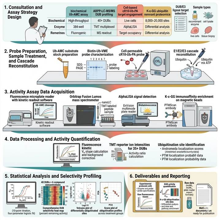
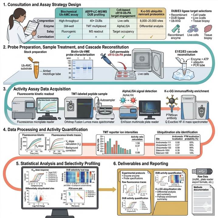
Step 1: Consultation and Assay Strategy Design
We consult with you to define the DUB and ubiquitin enzyme activity assay strategy based on research objective (target identification, inhibitor screening, PROTAC efficacy assessment, mechanistic study), enzyme target (specific DUB, E3 ligase, or full cascade), sample type (recombinant enzyme, cell lysate, live cells, tissue), throughput requirements (single enzyme to full DUB family), and desired readout (biochemical activity, cellular target engagement, global ubiquitination). We provide a detailed project proposal with platform recommendations, assay design specifications, positive/negative control strategies, and data analysis plan.
Step 2: Probe Preparation, Sample Treatment, and Cascade Reconstitution
For biochemical assays: recombinant DUB or E3 ligase is prepared at appropriate concentration. Activity-based probes (Ub-AMC, Ub-PL, biotin-Ub-VME, biotin-Ub-PA) are prepared and verified for activity. For cell-based assays: cells are treated with cell-permeable probe (cR10-Ub-PA, 1–10 μM, 1–4 h) with or without pre-incubation with test compounds. For E1/E2/E3 cascade: each enzyme component is optimized for concentration and activity, and the cascade is assembled with ubiquitin, ATP, and detection reagents. For K-ε-GG proteomics: cells/tissues are lysed, proteins are extracted, quantified, and prepared for trypsin digestion.
Step 3: Activity Assay Acquisition
For fluorogenic assays (Ub-AMC, Ub-PL): kinetic fluorescence readout at 1–5 minute intervals for 30–120 minutes at 25°C or 37°C on a microplate reader (BioTek Synergy H1, Tecan Spark, or Molecular Devices FlexStation). For ABPP-LC-MS/MS: probe-labeled DUBs are captured by streptavidin pull-down, on-bead digested, TMT-labeled, and analyzed by nanoLC-MS/MS (Orbitrap Fusion Lumos, 120-minute gradient). For K-ε-GG proteomics: K-ε-GG peptides are immunoaffinity enriched and analyzed by nanoLC-MS/MS. For E3 cascade assays: reaction products are analyzed by anti-ubiquitin immunoblot, AlphaLISA, or MS.
Step 4: Data Processing and Activity Quantification
For fluorogenic assays: initial velocity (V₀) is calculated from the linear phase of fluorescence increase, V₀ values are corrected for background (no enzyme control), and activity is expressed as relative activity (% of control) or specific activity (pmol AMC released/min/mg enzyme). IC₅₀ values are calculated by nonlinear regression (four-parameter logistic fit). For ABPP-LC-MS/MS: DUBs are identified from MS/MS spectra, TMT reporter ion intensities are quantified, and activity ratios (treated/control) are calculated for each DUB. For K-ε-GG: ubiquitinated peptides are identified with site localization, and differential ubiquitination is calculated.
Step 5: Statistical Analysis and Selectivity Profiling
For DUB inhibitor screening: IC₅₀ values are reported with 95% confidence intervals, Z′-factors assess assay quality (>0.5 acceptable, >0.7 excellent), and selectivity ratios (IC₅₀ off-target / IC₅₀ primary target) quantify DUB family selectivity. For cell-based engagement: target occupancy (%) is calculated for each DUB at each inhibitor concentration, and proteome-wide selectivity is visualized as a heat map of residual activity across all detected DUBs. For ubiquitination data: statistical testing (Moderated t-test, ANOVA with Benjamini-Hochberg FDR correction) identifies significantly changed ubiquitination sites and proteins, with pathway enrichment (KEGG, Reactome) annotation.
Step 6: Deliverables and Reporting
Comprehensive activity assay report including: complete experimental protocols with enzyme preparations, probe specifications, and assay conditions; raw fluorescence traces or MS chromatograms with quality control metrics; quantified activity data with IC₅₀ values, dose-response curves, target occupancy percentages, and selectivity heat maps; K-ε-GG ubiquitination site identification and quantification with differential analysis and pathway enrichment; and raw data files, analysis parameters, and methods documentation formatted for publication and regulatory reference.
Applications in DUB and Ubiquitin Enzyme Activity Assay Research
Activity-based DUB and ubiquitin enzyme assays have become indispensable tools in drug discovery and basic research, providing functional readouts of enzyme activity that inform target identification, hit validation, lead optimization, and mechanistic understanding across oncology, neurodegeneration, inflammation, and targeted protein degradation. The following application areas represent the most active use cases for our assay platforms.
DUB Inhibitor Discovery and Characterization for Oncology and Immuno-Oncology
DUBs have emerged as promising therapeutic targets across multiple cancer types, with roles in stabilizing oncoproteins (USP7 stabilizes MDM2 to suppress p53, USP28 stabilizes MYC and ΔNp63, USP14 stabilizes the proteasome to enhance degradation of pro-apoptotic factors), regulating DNA damage repair (USP1 stabilizes FANCD2 in the Fanconi anemia pathway, USP7 regulates the MRN complex), and modulating anti-tumor immune responses (USP22 deubiquitinates histone H2B to regulate immune checkpoint gene expression, A20 restricts NF-κB signaling in the tumor microenvironment). Our DUB activity-based profiling platform supports the full inhibitor discovery pipeline: target identification by comparing DUB activity between tumor and normal tissues, hit identification by high-throughput Ub-AMC screening (up to 50,000 compounds/week in 384-well format), lead optimization by IC₅₀ determination and selectivity profiling against active DUB family members, and mechanism-of-action studies using cell-based target engagement and proteome-wide selectivity profiling, enabling the development of selective, potent, and cell-active DUB inhibitors for oncology and immuno-oncology applications.
E3 Ligase Activity Profiling for PROTAC and Targeted Protein Degradation
The clinical success of PROTACs — particularly ARV-110 (bavdegalutamide, targeting AR with CRBN) and ARV-471 (vepdegestrant, targeting ER with CRBN) — has established hijacked E3 ligase activity as a critical determinant of degrader efficacy. Successful PROTAC development demands that the recruited E3 ligase retains catalytic ubiquitination activity within the target-PROTAC-E3 ternary complex. Our E1/E2/E3 cascade assay platform directly measures this catalytic competence: (1) baseline E3 ligase autoubiquitination activity to confirm that the recombinant E3 is functional; (2) E3 ligase panel screening to identify the most active pair for a given target-PROTAC combination; (3) PROTAC-induced ubiquitination assays that quantify compound-dependent target ubiquitination in reconstituted biochemical systems with PROTAC titration (1 nM–10 μM, 10-point curve); and (4) cellular ubiquitination efficacy assessment by K-ε-GG proteomics after PROTAC treatment. For advanced degrader characterization, our Protein Degrader Ubiquitination Efficacy and AP-MS for Protein Degrader services provide complementary cellular and proteomic readouts.
Deubiquitination in Neurodegenerative Disease: Parkinson's and Alzheimer's Research
Ubiquitin enzyme dysfunction is centrally implicated in neurodegenerative disorders: loss-of-function mutations in the E3 ligase Parkin (PARK2) and the Ser/Thr kinase PINK1 (PARK6) cause early-onset autosomal recessive Parkinson's disease through defective mitophagy, while DUBs such as USP30 (which opposes Parkin-mediated ubiquitination of mitochondrial outer membrane proteins) and USP14 (which modulates proteasomal degradation of tau and other aggregation-prone proteins) represent therapeutic targets for restoring proteostasis in Parkinson's and Alzheimer's diseases. Our activity-based assays enable: (1) Parkin E3 ligase activity assessment (using the Parkin activation assay with phospho-ubiquitin Ser65⁹⁺ PINK1-generated substrate) to evaluate Parkin activation by small molecules or peptide activators; (2) USP30 DUB activity profiling and inhibitor screening (Ub-AMC assay and Ub-PA ABPP) to identify compounds that enhance mitophagy in cellular Parkinson's models; (3) measurement of ubiquitination changes in patient-derived iPSC-derived neurons using K-ε-GG proteomics to assess the impact of genetic mutations or therapeutic interventions on the neuronal ubiquitin landscape; and (4) tau ubiquitination and proteasomal degradation assessment in Alzheimer's models, providing activity-based readouts that complement established neurodegeneration research platforms.
Ubiquitin Signaling in Inflammatory and Immune Regulation
The ubiquitin system is a master regulator of inflammatory and immune signaling pathways, with ubiquitin enzymes controlling NF-κB activation (by the LUBAC E3 ligase complex generating linear/Met1-linked ubiquitin chains, the A20 DUB/OTU family deubiquitinating RIPK1 and NEMO, and the E3 ligase TRAF6 catalyzing K63-linked ubiquitination), interferon signaling (by the E3 ligase RNF125 promoting RIG-I degradation, the DUB USP18 removing ISG15 from IRF3), and inflammasome activation (by the E3 ligase Parkin restricting NLRP3 activation). Our activity-based assay platforms support immune-oncology and inflammatory disease drug discovery by: (1) measuring LUBAC linear ubiquitination activity using Ub-AMC or diubiquitin FRET probes in the presence of HOIL-1/HOIP/Sharpin components; (2) profiling A20 DUB activity against K63-linked diubiquitin substrates to identify modulators of A20's anti-inflammatory activity; (3) assessing TRAF6 E3 ligase activity and its modulation by small molecules or peptides; and (4) quantifying global ubiquitination changes in immune cells after cytokine stimulation or drug treatment using K-ε-GG proteomics, providing a system-level view of ubiquitin signaling in inflammation.
Mechanistic Studies of the Ubiquitin Proteasome System in Cancer Biology
Beyond individual DUB and E3 targets, our multi-platform approach enables comprehensive mechanistic studies of how the ubiquitin-proteasome system coordinates cancer-relevant processes including cell cycle progression (APC/C E3 ligase controlling mitotic exit, SCF complexes regulating G1/S transition), DNA damage repair (RNF168/RNF8 E3 ligases assembling ubiquitin signals at double-strand breaks, OTUB1 DUB restricting RNF168 activity), apoptosis (XIAP/cIAP1/2 E3 ligases inhibiting caspase activity, SMAC mimetics inducing IAP autoubiquitination), and metastasis (A20 DUB modulating EMT transcription factor stability). By combining activity-based DUB profiling with K-ε-GG ubiquitin remnant quantification in cancer cell lines, patient-derived xenografts, and clinical tumor specimens, our integrated platform enables cancer biologists to map the functional landscape of ubiquitin enzyme activity in tumor biology, identify dysregulated DUBs and E3 ligases as candidate therapeutic targets, and characterize the ubiquitin signaling consequences of genetic and pharmacological perturbations with unprecedented mechanistic depth.
Case Study: Neutron-Encoded Diubiquitins to Profile Linkage Selectivity of Deubiquitinating Enzymes by Multiplexed Intact Mass Spectrometry
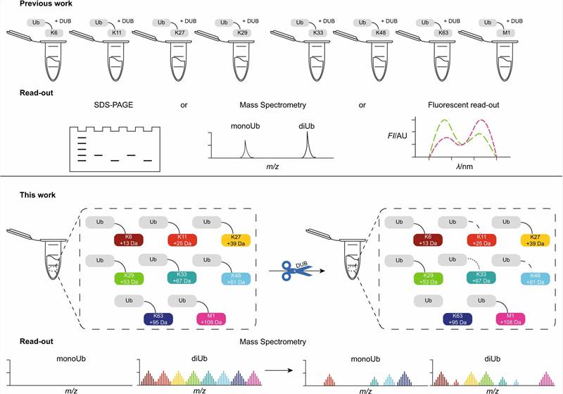
In a 2023 study published in Nature Communications (CC BY 4.0), van Tol et al. developed a groundbreaking mass spectrometry-based platform for multiplexed profiling of DUB linkage selectivity, using neutron-encoded (¹³C/¹⁵N) diubiquitin probes that enable simultaneous detection of DUB activity across all eight ubiquitin chain linkage types from a single LC-MS experiment — a methodological advance that transforms our ability to characterize DUB specificity, substrate preference, and inhibitor selectivity in a single, unified assay.
Background: Deubiquitinating enzymes exhibit differential selectivity for the eight ubiquitin chain linkage types (Met1/M1-linear, K6, K11, K27, K29, K33, K48, K63), and this linkage selectivity is a critical determinant of DUB biological function. K48-linked ubiquitin chains target substrates for proteasomal degradation, K63-linked chains regulate signaling complex assembly and kinase activation, Met1-linked linear chains activate NF-κB signaling, and K11/K29/K33 linkages are associated with endoplasmic reticulum-associated degradation and lysosomal trafficking. Traditional methods for assessing DUB linkage selectivity required separate enzymatic reactions for each linkage type, using individual diubiquitin substrates in parallel wells, and comparing cleavage rates across assays — a labor-intensive, reagent-intensive approach that introduced inter-assay variability and prevented direct competitive comparison of linkage preferences within a single reaction. The authors addressed this gap by creating a multiplexed system in which all eight diubiquitin probes can be simultaneously incubated with a single DUB enzyme and the cleavage products are distinguished by intact protein mass spectrometry without any separation step.
Approach: The authors synthesized all eight native diubiquitin linkage types — M1-linear (through the N-terminal methionine), K6, K11, K27, K29, K33, K48, and K63 — using established enzymatic and chemical methods. Each diubiquitin was encoded with a distinct molecular mass by incorporating neutron-encoded amino acids (¹³C₆-arginine, ¹³C₆-lysine, ¹⁵N₂-lysine, or ²H₁₀-leucine) at defined positions, generating a mass difference of 6–10 Da between any two probes. All eight mass-encoded diubiquitins were pooled and incubated with a single DUB enzyme in a single reaction; at defined time points, aliquots were analyzed by intact protein LC-MS on a QTOF or Orbitrap instrument, and the relative abundance of each diubiquitin substrate and its corresponding monoubiquitin cleavage product was deconvoluted by mass difference. This experimental design was applied to profile 22 human DUBs spanning the USP (USP2, USP4, USP7, USP8, USP9x, USP10, USP16, USP21, USP30), OTU (OTUB1, OTUB2, OTUD1, OTUD2, OTUD3, OTULIN, Cezanne/OTUD7B), UCH (UCHL1, UCHL3, UCHL5), and MJD (ATXN3, ATXN3L, JOSD1) subfamilies, generating comprehensive three-dimensional activity landscapes (linkage selectivity × time × enzyme concentration) for each DUB.
Key Findings:
- Multiplexed DUB linkage selectivity profiling by intact protein MS: The neutron-encoded diubiquitin strategy achieved simultaneous, unambiguous detection of cleavage activity across all eight ubiquitin linkage types from a single LC-MS injection, with the 6–10 Da mass differences between probes providing confident assignment of each diubiquitin and monoubiquitin species without the need for chromatographic separation of the individual diubiquitin linkage types — a transformative simplification that reduces sample requirements and eliminates inter-assay variability inherent in parallel single-substrate experiments
- Linkage selectivity is concentration-dependent for promiscuous DUBs: A key biological finding was that USP-family DUBs — traditionally described as linkage-promiscuous — display pronounced linkage selectivity when assayed at low enzyme concentrations (0.1–1 nM), with selectivity narrowing at higher concentrations (10–100 nM). This reveals that the apparent promiscuity of many USPs is at least partially a consequence of supra-physiological enzyme concentrations used in standard biochemical assays, and that substrate competition at physiologically relevant concentrations reveals previously unrecognized selectivity preferences — a finding with direct implications for therapeutic DUB target selection and inhibitor development, as the degree of selectivity determines whether a DUB inhibitor will affect a single or multiple biological pathways
- Consecutive cleavage order mapped for diubiquitin-processing DUBs: For DUBs that process diubiquitin through a distal-to-proximal cleavage mechanism, the multiplexed assay captured the temporal order of cleavage events across all eight linkage types simultaneously, revealing that some DUBs (e.g., OTUD2) cleave specific linkages (K11, K48) with a preferred order that is not apparent from end-point measurements — information that is critical for understanding how DUBs edit ubiquitin chains in cells
- 22 DUBs profiled across 4 subfamilies with comprehensive specificity landscapes: The study generated the most comprehensive single-study dataset of DUB linkage selectivity to date, including DUBs for which linkage preferences were previously unknown (USP10 showed K63 selectivity, OTUD1 and OTUD3 showed distinct K11/K48 preferences), and provided the first direct side-by-side comparison of DUB activity across the USP, OTU, UCH, and MJD subfamilies under identical assay conditions — establishing a reference dataset for the DUB research community
- Open-source data analysis pipeline for intact protein MS deconvolution: Mass spectra were deconvoluted using open-source algorithms (UniDec, a universal deconvolution program for native mass spectrometry and ion mobility), and the relative abundance of each diubiquitin linkage was quantified from the deconvoluted zero-charge mass spectra, providing a reproducible and transferable data analysis framework that enables other laboratories to adopt this multiplexed DUB activity profiling approach without proprietary software dependencies
Significance: This landmark study established a new paradigm for DUB activity profiling by demonstrating that neutron-encoded diubiquitin substrates, combined with intact protein LC-MS readout, enable comprehensive, multiplexed assessment of DUB linkage selectivity in a single reaction. The concentration-dependent selectivity finding has direct implications for drug discovery, suggesting that selectivity profiles generated at single (typically high) enzyme concentrations may overestimate the promiscuity of DUB targets and underestimate the potential for developing selective inhibitors. Our service platform directly incorporates this methodological framework — multiplexed intact protein MS-based DUB activity profiling with linkage-specific diubiquitin substrates — as a core component of our biochemical DUB activity assay offering, adapted and optimized for customer-specific DUB targets and inhibitor screening applications.
Adapted from van Tol et al. (2023). Neutron-encoded diubiquitins to profile linkage selectivity of deubiquitinating enzymes. Nature Communications 14:1661. (CC BY 4.0)
Representative Results: DUB and Ubiquitin Enzyme Activity Assay Performance and Data Outputs
Our DUB and ubiquitin enzyme activity assay platform delivers integrated data packages combining biochemical activity measurements, cell-based target engagement, and ubiquitin proteomics for comprehensive functional characterization of ubiquitin enzyme activity in drug discovery programs. The representative data below illustrates typical output quality and performance across our four complementary platforms.
Platform Performance Specifications
| Performance Parameter |
Biochemical Ub-AMC DUB Assay |
ABPP-LC-MS/MS DUB Profiling |
Cell-Based cR10-Ub-PA Engagement |
K-ε-GG Ubiquitin Remnant Proteomics |
| Readout type |
Real-time fluorescence (kinetic) |
TMT multiplexed MS (end-point) |
Pull-down + MS or AlphaLISA |
LC-MS/MS (site-level) |
| DUB/enzyme coverage |
Single DUB per well (384-well) |
40+ DUBs per sample (multiplexed) |
30+ DUBs per sample |
N/A (ubiquitination sites: 8,000–20,000) |
| Throughput |
High (50,000+ compounds/week) |
Medium (20–40 samples/week) |
Low-medium (20–40 samples/week) |
Low-medium (10–20 samples/week) |
| Z′-factor (typical) |
> 0.7 (optimized assays) |
N/A (MS-based) |
> 0.5 (AlphaLISA format) |
N/A (MS-based) |
| Quantification |
V₀ (pmol/min), % activity, IC₅₀ |
Activity ratio (treated/control) |
% target occupancy |
Fold-change ubiquitination (site-level) |
| Sample requirement |
10–100 ng recombinant DUB per well |
100–500 μg lysate per sample |
1–10 × 10⁶ cells per condition |
2–5 mg protein per IP |
| Selectivity readout |
Panel-DUB IC₅₀ profiling |
Proteome-wide DUB family selectivity |
Proteome-wide DUB occupancy |
Global ubiquitination site changes |
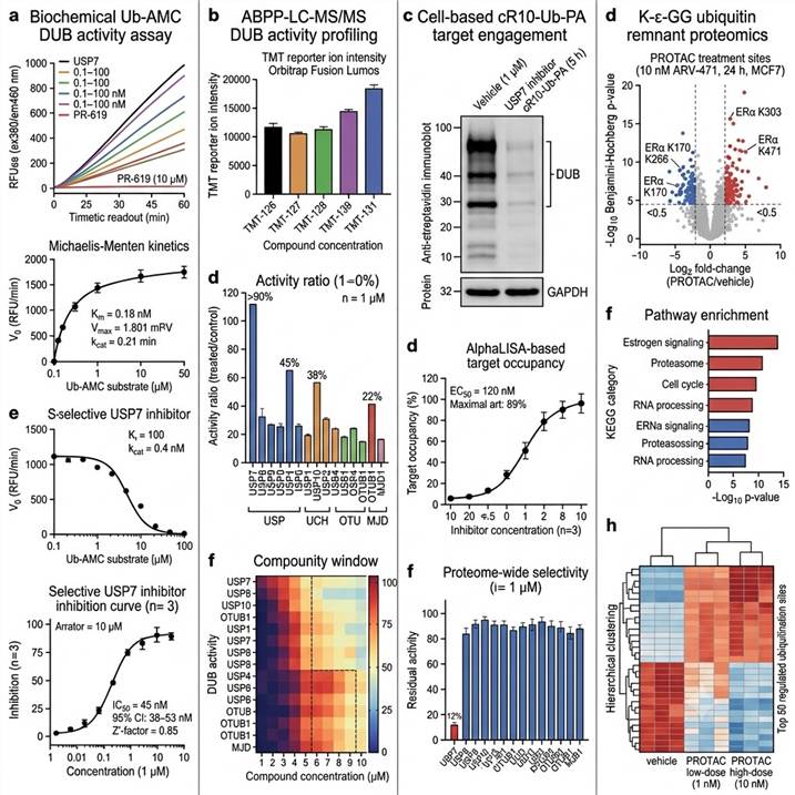
Representative data outputs from our DUB and Ubiquitin Enzyme Activity Assay platform. Left: Biochemical Ub-AMC kinetic assay with inhibitor IC₅₀. Center-left: ABPP-LC-MS/MS DUB family selectivity profiling. Center-right: Cell-based target engagement by cR10-Ub-PA probe. Right: K-ε-GG ubiquitin remnant proteomics in degrader-treated cells.
Key data deliverables included in every DUB and ubiquitin enzyme activity assay project:
- Biochemical activity assay data package — Raw fluorescence or AlphaLISA traces, V₀ values, IC₅₀ curves with 95% confidence intervals, Z′-factor quality metrics, and DUB panel selectivity ratios for each tested compound, with raw data files in standard plate-reader format
- ABPP-LC-MS/MS activity profiling data package — Complete list of all detected DUBs with MS/MS identification scores, TMT reporter ion intensities, activity ratios (treated/control) for each DUB at each compound concentration, selectivity heat maps, and raw MS data in mzML format
- Cell-based target engagement data package — Target occupancy percentages for each DUB at each compound concentration, proteome-wide selectivity profiles, pull-down immunoblot images, AlphaLISA dose-response curves, and EC₅₀ values with confidence intervals
- K-ε-GG ubiquitin remnant proteomics data package — Complete list of all identified and quantified ubiquitination sites with localization scores, differential ubiquitination analysis with statistical testing (FDR-corrected p-values), pathway enrichment results, PCA and volcano plots, and heat maps across all experimental groups
- Methods documentation — Complete protocols for enzyme preparations, probe synthesis or sourcing, assay conditions, LC-MS acquisition parameters, and data analysis methods, formatted for publication methods sections and regulatory reference
Related Services
Our DUB and ubiquitin enzyme activity assay platform is part of a comprehensive drug discovery service portfolio spanning activity-based profiling, mass spectrometry-based proteomics, and targeted protein degradation analysis for oncology, neurodegeneration, inflammation, and PROTAC development programs.
- Ubiquitylomics Analysis — Global K-ε-GG ubiquitin remnant LC-MS/MS profiling for comprehensive ubiquitination site identification and quantification in any biological system
- Protein Degrader Ubiquitination Efficacy — Targeted assessment of PROTAC-induced ubiquitination by MS-based ubiquitin remnant quantification and cellular ubiquitination assays
- Modificated Protein Degrader Proteomics — Multi-omics characterization of targeted protein degradation by PROTACs and molecular glues using quantitative proteomics
- AP-MS for Protein Degrader — Affinity purification-mass spectrometry for identifying degrader-induced protein interaction networks and ternary complex components
- PTM Enzyme Activity & Inhibitor Screening — Comprehensive PTM enzyme activity profiling and small-molecule inhibitor screening across kinase, DUB, HDAC, and other PTM enzyme classes
- Kinase Activity Profiling — Activity-based kinase profiling and inhibitor selectivity screening using multiplexed MS-based and biochemical assay platforms
- HDAC/HAT Activity Assays — Biochemical and cell-based activity assays for histone deacetylase and acetyltransferase characterization and inhibitor screening
- Ubiquitinated Peptide Enrichment — Immunoaffinity enrichment of ubiquitinated peptides (K-ε-GG, K-ε-LRGG) for comprehensive ubiquitination site mapping by LC-MS/MS
FAQs
What is the difference between DUB activity assays and ubiquitination proteomics?
DUB activity assays and ubiquitination proteomics answer distinct but complementary biological questions. DUB activity assays — whether biochemical (Ub-AMC, Ub-PL) or activity-based protein profiling (ABPP with Ub-VME/Ub-PA probes) — measure the functional catalytic activity of DUB enzymes: how much ubiquitin cleavage activity each DUB has in a sample, determined by the DUB's ability to process a ubiquitin-based substrate or react with an activity-based probe. Ubiquitination proteomics (K-ε-GG LC-MS/MS) measures the downstream product of ubiquitin enzyme activity: which proteins are ubiquitinated, on which lysine residues, and to what extent, at the time of sample collection. DUB activity data tell you what the enzymes are capable of doing; ubiquitination data tell you what has actually been done to the proteome. Together, they provide a complete picture: activity-based profiling reveals which DUBs are enzymatically active (and how active), while K-ε-GG proteomics reveals the ubiquitination state of the proteome — the net effect of all E3 ligase and DUB activities in the sample. Our service uniquely integrates both platforms, enabling researchers to directly correlate changes in DUB or E3 ligase activity with proteome-wide ubiquitination consequences.
What is the difference between biochemical activity assays and cell-based activity assays?
Biochemical DUB activity assays (Ub-AMC, Ub-PL, FRET-based) use purified recombinant DUB enzyme in a defined buffer system with a ubiquitin-based substrate, providing the most direct, controlled, and high-throughput measurement of DUB catalytic activity without the complexity of the cellular environment. Biochemical assays are ideal for compound IC₅₀ determination, structure-activity relationship (SAR) studies, mechanistic enzyme characterization, and high-throughput screening (HTS) campaigns where assay robustness and throughput (Z′ > 0.7, 50,000+ compounds/week) are paramount. Cell-based DUB activity assays (cell-permeable cR10-Ub-PA probes, competitive ABPP) measure DUB activity in the native cellular environment where the enzymes exist at endogenous expression levels, are subject to physiological regulation (phosphorylation, protein-protein interactions, subcellular localization), and compete with endogenous substrates and inhibitors. Cell-based assays provide target occupancy (the fraction of DUB active sites occupied by a test compound) and proteome-wide selectivity profiles in the relevant disease-relevant cellular context, but have lower throughput (20–40 samples/week) and greater variability than biochemical assays. Both formats are essential in drug discovery: biochemical assays for primary screening and lead optimization, cell-based assays for translational validation and PK-PD correlation.
What types of DUBs can be profiled by activity-based probes?
Our ABPP platform covers cysteine protease DUBs that use an active-site cysteine nucleophile for ubiquitin hydrolysis — including the USP (ubiquitin-specific protease), UCH (ubiquitin C-terminal hydrolase), OTU (ovarian tumor protease), and MJD (Machado-Joseph disease protease) subfamilies — which together comprise ~95% of all DUBs. The biotin-Ub-VME (vinyl methyl ester) and biotin-Ub-PA (propargyl amide) probes form an irreversible covalent bond with the active-site cysteine, enabling specific labeling of active enzymes regardless of subfamily. We have validated probe labeling and MS detection for 40+ DUBs including USP2/4/5/7/8/9x/10/11/14/15/16/20/21/22/24/25/28/30/34/36, UCHL1/3/5, OTUB1/2, OTUD1/2/3/4/5/6/7B, A20, Cezanne, ATXN3/3L, JOSD1/2, and BRCC36. The JAMM/MPN+ subfamily of metalloprotease DUBs (which use a zinc-coordinated catalytic water instead of a cysteine) cannot be labeled by cysteine-directed probes and require alternative activity-based approaches (substrate-based or metal-chelating probes) that can be developed on a project-specific basis.
What sample types are compatible with DUB activity assays?
Sample compatibility depends on the assay format. For biochemical Ub-AMC assays: purified recombinant DUB enzyme (10–100 ng per well in 384-well format), supplied as purified protein, bacterial lysate containing expressed DUB, or immunoprecipitated DUB from cellular lysates. For ABPP-LC-MS/MS DUB profiling: fresh or flash-frozen cell pellets (1–10 × 10⁶ cells per condition), tissue biopsies (10–50 mg), or tumor specimens, lysed in ABPP-compatible buffer (50 mM Tris pH 7.5, 150 mM NaCl, 0.5% NP-40, 1 mM DTT, protease inhibitors without NEM or iodoacetamide). For cell-based cR10-Ub-PA engagement: live adherent or suspension cells (> 90% viability, 2–5 × 10⁶ cells per condition), treated with probe in complete culture medium. For K-ε-GG ubiquitin remnant proteomics: cell pellets (5–20 × 10⁶ cells per IP), tissue (50–200 mg), or sorted cell populations (1 × 10⁶ minimum), flash-frozen and stored at −80°C. We strongly recommend including proteasome inhibitor treatment (MG-132, 10 μM, 4–6 hours) or DUB inhibitor treatment before harvesting for K-ε-GG proteomics to enrich ubiquitinated species.
How do you ensure specificity of DUB activity-based probes?
Activity-based probe specificity is ensured through a multi-layered validation strategy. First, all probes (biotin-Ub-VME, biotin-Ub-PA, Ub-AMC, Ub-PL) are tested against a panel of purified recombinant non-DUB cysteine proteases (caspase-3, cathepsin B, papain) and serine proteases (trypsin, chymotrypsin) to confirm the absence of cross-reactivity — the ubiquitin moiety itself provides substantial specificity since DUBs have evolved to specifically recognize ubiquitin, but we verify that non-DUB proteases with similar catalytic mechanisms do not process Ub-based probes. Second, probe specificity is confirmed in lysate ABPP experiments by pre-treatment with pan-DUB inhibitors (PR-619, N-ethylmaleimide) to demonstrate that probe labeling is DUB-dependent. Third, for Ub-AMC biochemical assays, we test each DUB enzyme preparation against the AMC substrate alone (without ubiquitin) to confirm that fluorescence release requires the ubiquitin moiety. Fourth, cell-based cR10-Ub-PA probes are validated in matched wild-type vs. DUB-knockout cell lines where available to confirm that the probe-labeled signal is specific to the target DUB. Fifth, for MS-based ABPP, TMT-based quantification ensures that probe-labeled DUBs meet minimum identification confidence thresholds (FDR < 1% at protein level, minimum 2 unique peptides per DUB), and the presence of the active-site peptide with the intact probe adduct in MS/MS spectra provides direct evidence of active-site labeling.
Can you assess DUB selectivity across the entire DUB family?
Yes — comprehensive DUB family selectivity profiling is a core capability of our ABPP-LC-MS/MS platform. In a single TMT-ABPP experiment, we can quantify the activity of 30–40+ DUBs (all DUBs that are expressed and active in the cell or tissue type of interest) in response to compound treatment, generating a quantitative selectivity matrix (compound concentration × DUB activity) that reveals the selectivity profile of any DUB inhibitor. Our standard DUB selectivity panel covers: USP subfamily (USP2, 4, 5, 7, 8, 9x, 10, 11, 14, 15, 16, 20, 21, 22, 24, 25, 28, 30, 34), UCH subfamily (UCHL1, UCHL3, UCHL5), OTU subfamily (OTUB1, OTUB2, OTUD1, OTUD2, OTUD3, OTUD6B, A20, Cezanne), and MJD subfamily (ATXN3, ATXN3L, JOSD1, JOSD2). The output is a heat map visualization showing the percentage of remaining DUB activity at each compound concentration, enabling rapid identification of the primary target (the DUB with the lowest activity ratio), secondary targets (DUBs showing > 50% inhibition at the test concentration), and the overall selectivity window. For DUB inhibitor discovery programs achieving selectivity against the broader DUB family is a critical requirement for minimizing mechanism-based toxicity. Cell-based competitive ABPP extends this selectivity assessment to the endogenous cellular environment, where probe labeling competes with cellular substrates and binding partners that may influence compound accessibility and target selectivity.
How do DUB and ubiquitin enzyme activity assays support PROTAC drug discovery programs?
DUB and ubiquitin enzyme activity assays support PROTAC drug discovery at multiple stages of the development pipeline. In target selection and E3 ligase feasibility assessment, E1/E2/E3 cascade assays establish whether a candidate E3 ligase (CRBN, VHL, MDM2, IAP, RNF4) has sufficient catalytic activity to support PROTAC-induced ubiquitination — if the E3 ligase has low intrinsic activity, it is unlikely to be effectively hijacked by PROTACs, regardless of ternary complex affinity. In PROTAC design and optimization, target-specific ubiquitination assays (recombinant target + E3 ligase + PROTAC + E1/E2/Ub) directly quantify compound-induced ubiquitination activity as a function of PROTAC concentration, linker length and composition, and warhead chemistry, enabling SAR optimization based on functional output (ubiquitination) rather than indirect measures (binding affinity or ternary complex formation). In lead characterization, cell-based target engagement assays using cell-permeable activity-based probes quantify PROTAC occupancy of the hijacked E3 ligase in live cells, providing cellular target engagement data that directly informs the PK-PD relationship. In selectivity assessment, K-ε-GG ubiquitin remnant proteomics maps the proteome-wide ubiquitination changes induced by PROTAC treatment, revealing off-target ubiquitination signatures that may indicate hijacking of unintended E3 ligases or promiscuous degradation of non-target proteins — a critical safety assessment. Throughout these stages, our integrated platform enables PROTAC programs to directly connect assay readouts (E3 activity, PROTAC-induced ubiquitination, cellular target engagement, proteome-wide ubiquitination) with compound structure and in vivo efficacy data, accelerating the discovery of selective, potent, and safe degrader therapeutics.
References
- van Tol BDM, et al. Neutron-encoded diubiquitins to profile linkage selectivity of deubiquitinating enzymes. Nature Communications. 2023;14:1661.
- Barroso-Gomila O, et al. BioE3 identifies specific substrates of ubiquitin E3 ligases. Nature Communications. 2023;14:1556.
- Liu Y, Yang J, Wang T, et al. Expanding PROTACtable genome universe of E3 ligases. Nature Communications. 2023;14:6509.
For research use only. Not for use in diagnostic procedures.