
Plasma Proteomics in Non-Model Species: Choosing Depletion, Discovery, and Targeted Validation
An HCP ELISA result and an LC-MS result can disagree without either method having failed. The two platforms observe different properties of a heterogeneous impurity population. ELISA integrates the response of the HCP species recognized by a polyclonal antibody reagent against an assay standard. LC-MS identifies peptide evidence from individual proteins that survive sample preparation, digestion, separation, ionization, acquisition, and data processing. Their reported values may carry the same unit while representing different measurement models.
For process development, the useful question is therefore not whether mass spectrometry should replace ELISA. It is which analytical evidence is needed to understand impurity clearance, assess ELISA coverage, explain a process excursion, or monitor a specific HCP of concern. A stage-appropriate strategy usually retains a suitable total-HCP ELISA and adds LC-MS where protein identity changes the decision.
USP General Chapter <1132> provides the broader framework for residual HCP measurement, while <1132.1> addresses residual HCP measurement in biopharmaceuticals by LC-MS. These chapters support orthogonal characterization; they do not make every discovery LC-MS workflow a validated lot-release method. The intended use, performance characteristics, standards, and validation state must remain explicit throughout the project.
Intended Use of Orthogonal HCP Analysis: The workflows discussed here support research, process development, clearance characterization, comparability, ELISA-reagent assessment, and root-cause investigation. A targeted LC-MS method may be developed toward quality-control use, but release testing requires product-specific method validation, specification justification, lifecycle controls, and appropriate regulatory acceptance.
Host Cell Protein Analysis in Biologics
Host cells release thousands of proteins into the culture environment through secretion, cell turnover, and lysis. Most are reduced by downstream purification, but a subset can persist because it is abundant, shares physicochemical properties with the product, interacts with the product, binds to chromatography media, or escapes a particular polishing step. The composition is specific to the host, cell line, culture conditions, product, and purification process.
The risk associated with an individual residual HCP is not determined by concentration alone. Relevant factors include persistence across process steps, enzymatic activity, potential effects on product or excipient stability, biological activity, immunogenicity, sequence homology to human proteins, route and frequency of dosing, and the amount of therapeutic product administered [1,2]. Identification by LC-MS enables that protein-specific assessment; it does not by itself establish clinical risk.
A total-HCP value remains valuable because it provides a sensitive, scalable view of overall process consistency. Protein-resolved LC-MS adds information that the aggregate result cannot supply. The methods are complementary because they answer different questions.
| Analytical route | Primary measurement | Best use | Main interpretive limitation |
|---|---|---|---|
| Multi-analyte HCP ELISA | Combined immunoreactivity relative to an HCP reference standard | Routine monitoring, process trending, and validated total-HCP measurement | Does not identify individual HCPs; result depends on antibody coverage and the relationship between sample and standard |
| Discovery DDA or DIA LC-MS | Peptide evidence assigned to individual HCPs, with relative or calibrated quantification | HCP identity, process-step profiling, ELISA coverage assessment, clearance mapping, and root-cause investigation | Sensitivity is constrained by product background, peptide observability, sample preparation, and data-processing thresholds |
| Targeted PRM/MRM or protein-specific immunoassay | Prespecified surrogate peptides or one defined protein | Sensitive follow-up of persistent or high-priority HCPs and assay development | Only measures the selected targets; specificity and calibration must be established for the intended matrix and use |
An orthogonal DIA-MS HCP profiling study is most informative when samples span the purification process and the analytical question is defined before acquisition. A single drug-substance result cannot reveal where an impurity entered, why it persisted, or whether the apparent level changed because of method response.
Why ELISA and LC-MS HCP Results Diverge
Discordance should be investigated mechanistically. Treating one platform as the universal reference risks hiding limitations in both.
HCP Antibody Coverage and Reference Standards
An HCP ELISA detects proteins for which the polyclonal reagent contains effective antibodies under the assay conditions. Coverage depends on the antigen preparation used for immunization, the host species and immune response, antibody affinity, epitope preservation, and the similarity between the assay standard and the actual process HCP population. Weakly immunogenic proteins, some low-molecular-weight proteins, and HCPs absent or underrepresented in the immunogen can receive little effective coverage [3].
The calibration standard also matters. ELISA reports an immunoreactive equivalent rather than a direct census of every protein molecule. A generic kit standard, a platform-specific standard, and a process-specific standard can yield different estimates for the same sample because their protein composition and antibody reactivity differ.
Coverage should therefore be investigated using a representative null-cell or process HCP antigen and a method that can identify which proteins are captured by the ELISA antibody reagent. Immunoaffinity capture followed by LC-MS is especially useful because it compares the HCP population before and after antibody capture at the protein level.
ELISA Hook Effect, Dilution Linearity, and Matrix Effects
Some in-process samples contain HCP concentrations high enough to exceed the useful range of a sandwich ELISA. Antigen excess can saturate capture and detection reagents and produce a hook effect or dilutional nonlinearity. This phenomenon is caused by analyte excess—not by the therapeutic antibody simply being abundant. It should be evaluated by serial dilution and definition of a minimum required dilution at which the corrected result is stable.
Product and buffer components can also alter recovery or antibody binding. A low result at one dilution and a higher corrected result after further dilution may indicate matrix interference or antigen excess rather than true biological change. Spike recovery and dilutional parallelism are therefore part of method suitability for the specific sample type.
LC-MS Dynamic Range and Peptide Detectability
In a purified monoclonal-antibody sample, the product can exceed an individual HCP by more than six orders of magnitude. The product digest dominates the peptide mixture, increasing competition during chromatography and ionization and reducing the sampling probability of trace HCP peptides [4]. Product depletion, HCP enrichment, offline separation, longer gradients, narrow-window DIA, or targeted acquisition can improve access, but each changes throughput and potential bias.
Absence from an LC-MS result does not prove absence from the sample. A protein may lack unique proteotypic peptides in the measurable mass range, digest inefficiently, contain unstable or heavily modified peptides, coelute with intense product peptides, or fail the peptide and protein inference criteria. Conversely, a single weak peptide-spectrum match should not be treated as sufficient evidence for a critical HCP.
An appropriately designed DIA quantitative proteomics method can improve consistency of fragment-ion acquisition across process samples, but sensitivity must be demonstrated in the relevant product matrix. Global identification count is not a substitute for recovery, precision, and lower-limit performance for the HCPs that matter to the decision.
Differences in HCP Quantitation by ELISA and LC-MS
Both ELISA and LC-MS may report ng HCP per mg drug substance, often expressed as ppm. The apparent agreement of units can obscure a difference in measurand.
- ELISA typically reports the total immunoreactive response converted through an HCP reference standard.
- Discovery LC-MS may report individual protein abundance by relative intensity, label-free estimation, or a semiquantitative model.
- Calibrated LC-MS can estimate individual HCP concentration using stable-isotope peptides, intact-protein standards, or another defined calibration strategy.
- Summing calibrated individual proteins can provide a total estimate, but that value reflects the proteins and peptides captured by the method.
Published comparisons have shown that ELISA and LC-MS can both report total HCP in ng/mg while producing different values because the underlying response mechanisms and standards differ [3]. The discrepancy should be reconciled through coverage, recovery, and protein-level evidence rather than forced numerical equivalence.
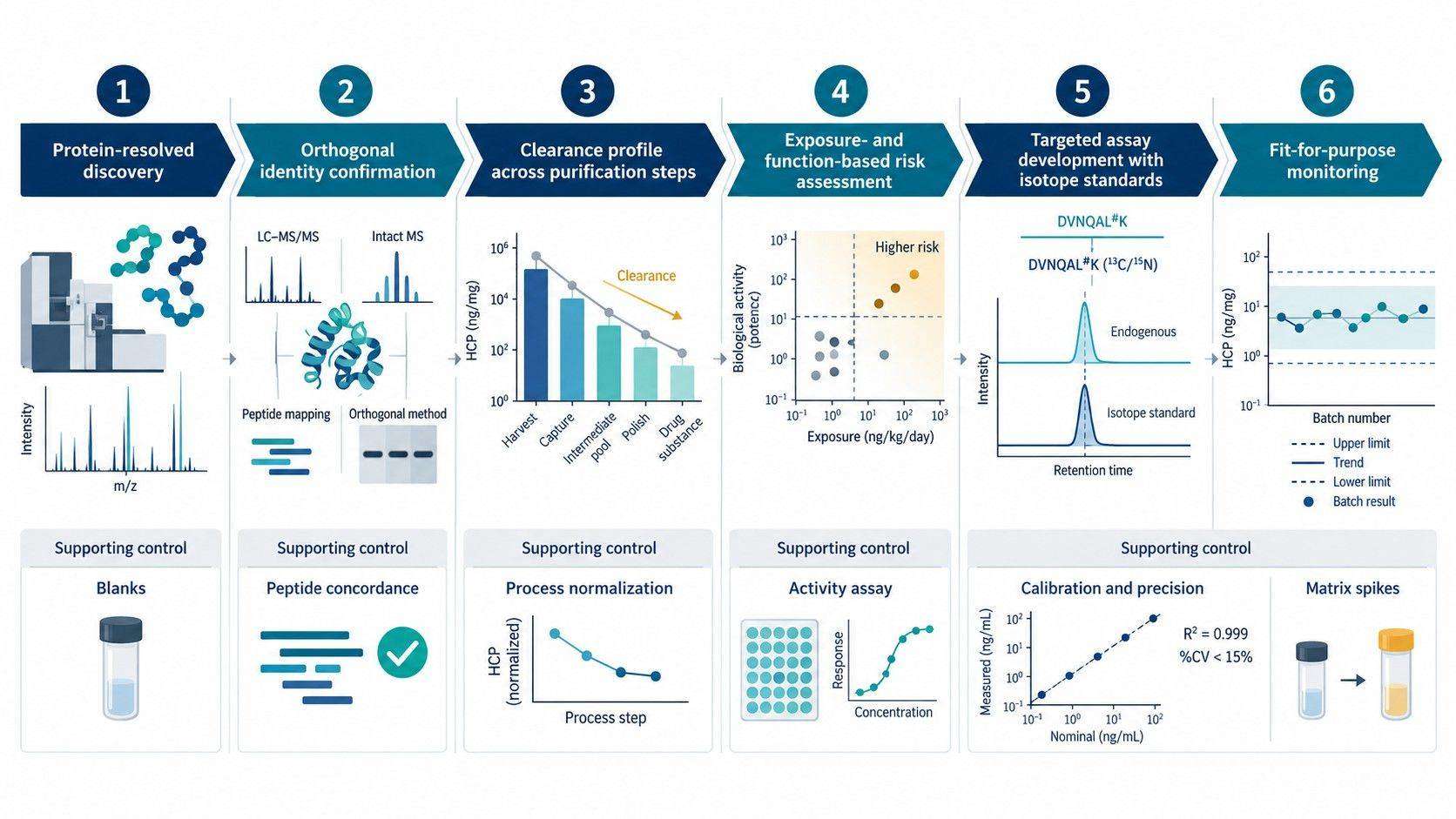 Figure 1. Distinct measurands and analytical blind spots of total-HCP ELISA, discovery DIA-MS, and targeted LC-MS.
HCP Method Selection by Bioprocess Development Stage
The analytical strategy should evolve with the process and the decision. Early development benefits from broad protein-level visibility. Later routine monitoring benefits from a robust, scalable assay. A specific persistent HCP may require a dedicated method that sits between those stages.
HCP Clearance Analysis During Process Development
For harvest, clarification, capture, intermediate purification, and polishing samples, broad LC-MS profiling can determine which HCPs are removed, which persist, and where the clearance slope changes. Samples should be collected from matched process runs, normalized to a defined product or sample basis, and analyzed with sufficient dilution or enrichment to manage the changing product-to-HCP ratio.
The objective is not merely to count proteins at each step. It is to create a clearance map for individual HCPs and distinguish three patterns:
- proteins that decrease consistently with bulk impurity clearance;
- hitchhiking proteins that co-purify with the product through multiple operations;
- proteins that appear to increase because the product is concentrated, the sample matrix changes, or measurement recovery differs between steps.
DIA can provide reproducible relative profiles across many process fractions. Where a key HCP requires absolute or lower-level quantification, stable-isotope dilution can be added for selected peptides. A dual strategy combining global DIA profiling with isotope-dilution quantification has demonstrated protein-specific measurements into the low-ppm range in process-development samples [5].
HCP ELISA Antibody Coverage Assessment
Coverage assessment asks whether the antibody reagent recognizes the HCP population relevant to the manufacturing process. It should use representative upstream or null-cell HCP material rather than only a generic reference mixture. Affinity extraction followed by LC-MS can identify proteins that are captured, incompletely captured, or missed.
A low coverage percentage should not be interpreted without considering abundance, protein identity, and the calculation method. Missing one persistent, biologically active protein may be more consequential than incomplete response to many low-priority proteins. Coverage should therefore be reported at the protein level and interpreted through a risk-based lens.
HCP Root-Cause Investigation
LC-MS becomes particularly valuable when total-HCP trends change unexpectedly, a polishing step loses clearance, a stability attribute deteriorates, or visible particles appear. The investigation should compare relevant process intermediates, lots, resin cycles, hold conditions, and formulation samples. Protein identity can connect the analytical signal to plausible mechanisms such as product binding, proteolysis, lipase activity, or polysorbate degradation.
For CHO-derived products, residual hydrolytic HCPs such as lipoprotein lipase (LPL) and lysosomal phospholipase A2 (LPLA2/PLA2G15) deserve specific attention when Polysorbate 20 or 80 loss, free-fatty-acid particles, or unexplained formulation instability is observed. LPLA2 has been linked experimentally to PS20 and PS80 hydrolysis in formulated antibodies, while broader CHO hydrolase studies identify LPL among the most active polysorbate-degrading enzymes [11,12]. Detection is still hypothesis-generating: enzyme activity, concentration, formulation conditions, and temporal association with degradation must be demonstrated before assigning causality.
The presence of a candidate enzyme is not sufficient to assign causality. The investigation may require activity assays, inhibitor experiments, spike studies, product-stability experiments, or cell-line/process modifications. LC-MS narrows the hypothesis; the root cause is established by converging evidence.
Targeted LC-MS Analysis of Known HCPs
Once a specific HCP is prioritized, PRM or SRM/MRM can focus acquisition on multiple unique surrogate peptides. Stable-isotope-labeled peptides support relative or absolute quantification, but peptide standards added after digestion do not correct for protein extraction and digestion recovery. Where those sources of error matter, an intact-protein or extended-peptide standard may be more appropriate.
Targeted method development should address peptide uniqueness, modifications, missed cleavages, matrix interference, carryover, calibration model, selectivity, precision, accuracy, recovery, dilution integrity, and stability. A transition from development support to release testing is a change in intended use and requires a corresponding validation and control strategy.
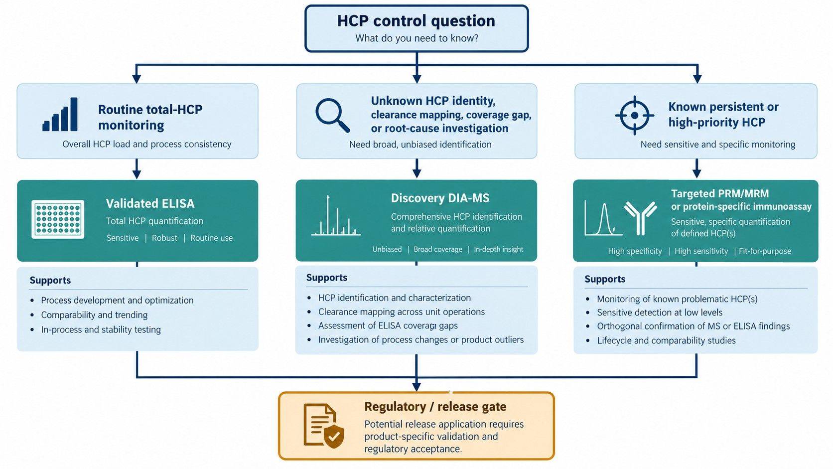 Figure 2. Stage-appropriate decision tree for selecting ELISA, discovery DIA-MS, and targeted PRM/MRM during HCP control.
Troubleshooting ELISA and LC-MS HCP Discrepancies
The pattern of disagreement often suggests the next experiment.
| Observed pattern | Plausible explanations | Most informative follow-up |
|---|---|---|
| High ELISA total HCP, few LC-MS identifications | Product-background suppression, insufficient enrichment, poor peptide observability, or broad immunoreactivity to proteins below the LC-MS limit | Matrix spike, product depletion or HCP enrichment, deeper DIA, and targeted analysis of expected HCPs |
| Low ELISA, multiple LC-MS HCPs | Antibody coverage gap, nonrepresentative standard, low-affinity antibodies, or ELISA matrix interference | Antibody-affinity extraction MS, dilutional parallelism, spike recovery, and protein-specific review |
| Stable ELISA total, changing LC-MS composition | Different HCPs contribute to a similar aggregate response | Protein-level clearance map and risk ranking of newly persistent species |
| ELISA increases after dilution | Antigen excess or matrix interference | Dilution series, minimum required dilution, and parallelism assessment |
| One LC-MS HCP persists while total ELISA decreases | Product association or poor clearance of a specific protein | Targeted quantification across process steps, product-interaction testing, and process-parameter investigation |
| Targeted LC-MS and discovery DIA differ | Different calibration, integration, peptide choice, or interference handling | Review peptide concordance, standard addition, chromatograms, transition ratios, and digestion controls |
The comparison should be made at matched sampling points and on a consistent basis. Changing from volume-normalized upstream samples to product-mass-normalized downstream samples without documenting the basis can create an artificial clearance trend.
Risk Assessment for Persistent and High-Risk HCPs
A protein should not be labeled high risk solely because it was detected. Risk ranking should integrate analytical persistence with biological and product context.
HCP Identification Confidence
Require multiple unique peptides where feasible, consistent retention and fragment-ion evidence, control of peptide- and protein-level FDR, and review of sequence homology. For low-level critical findings, confirmation using synthetic standards or a targeted method is preferable to relying on a discovery score alone.
HCP Clearance and Patient Exposure
Track the protein from harvest through the final relevant process stage. Estimate concentration relative to drug substance and translate that value into a potential administered amount using the therapeutic dose and regimen. A protein at the same ppm level can imply different exposure for a low-dose and a high-dose product.
Product Quality and Biological Activity
Consider whether the HCP is an enzyme, cytokine, chaperone, protease, lipase, or another protein with plausible activity in the product or patient context. Some HCPs can degrade product or excipients at concentrations below those that dominate the total HCP result. Activity should be tested experimentally when it drives the risk hypothesis.
Prior Knowledge and Product-Specific Risk
Known examples such as CHO phospholipase B-like 2 illustrate that HCP-product interactions and immunogenicity can be product and process specific [6,7]. Clusterin and other proteins have been observed to persist in Protein A eluates for multiple antibodies, but recurrence does not make every detection equally consequential [8]. The evidence must be interpreted for the actual molecule, process, and dose.
Quality Controls and Documentation for Orthogonal HCP Analysis
The study design should allow the result to be reconstructed and compared across platforms. Essential elements include:
- host organism, cell line, product construct, and process-stage definitions;
- matched sample collection, storage, and preparation records;
- drug-substance concentration and the normalization basis used for each result;
- ELISA kit or reagent lot, standard type, dilution range, parallelism, and spike recovery;
- LC-MS sample preparation, depletion or enrichment, digestion, acquisition, database, and FDR settings;
- peptide-to-protein inference rules and treatment of shared peptides;
- quantitative standards, where added, and the process step at which they were introduced;
- blank, matrix, carryover, and system-suitability controls;
- protein-level evidence for any HCP used to support a process or risk decision.
The targeted proteomics stage should inherit the identity evidence and process context established during discovery. It should not begin with a peptide panel selected solely from database annotations without confirming that the corresponding proteins and proteotypic peptides are observable in the relevant product matrix.
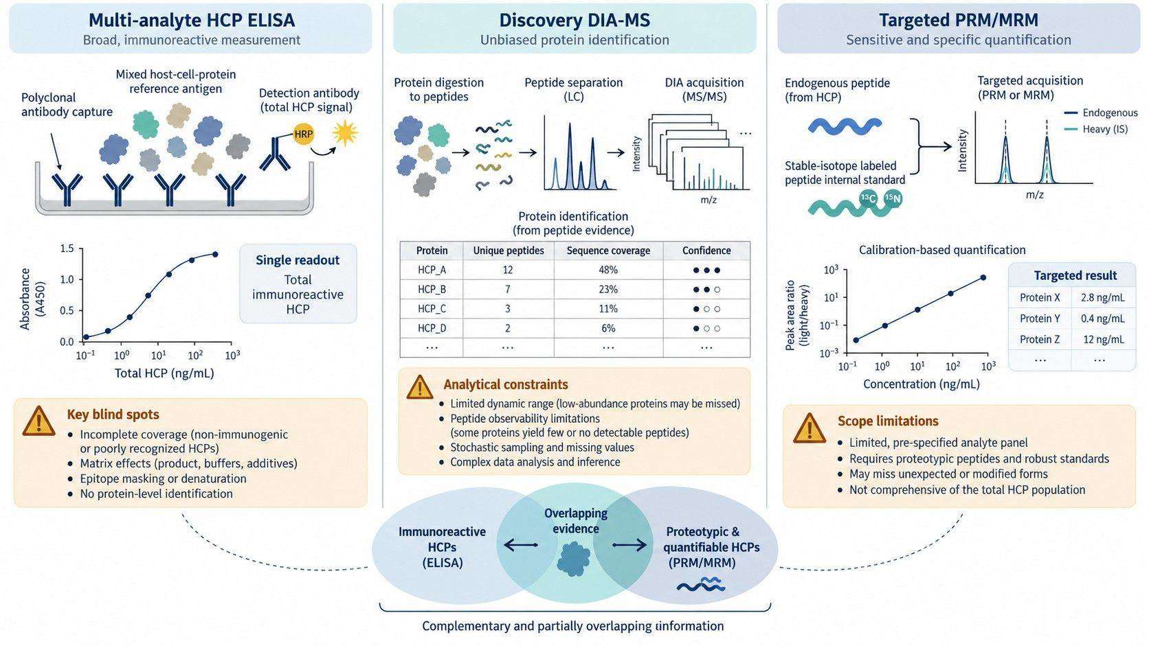 Figure 3. Evidence chain for advancing an HCP from discovery identification to process tracking, risk assessment, and fit-for-purpose targeted measurement.
Orthogonal HCP Analysis Workflow: ELISA, DIA-MS, and Targeted LC-MS
An orthogonal HCP program assigns each platform a defined analytical role and establishes how evidence moves from one method to the next.
| Method | Primary role | Evidence handed to the next stage |
|---|---|---|
| Total-HCP ELISA | Scalable monitoring of aggregate immunoreactivity across process samples and routine lots | Dilutional behavior, recovery, process trend, and antibody-coverage questions that may require protein-level investigation |
| Discovery DIA-MS | Protein-resolved clearance mapping, coverage-gap assessment, and root-cause investigation | Confirmed protein identities, proteotypic peptides, process-stage behavior, and a risk-ranked list for focused measurement |
| Targeted PRM/MRM | Fit-for-purpose monitoring or quantification of selected persistent or high-priority HCPs | Peptide-specific performance, calibration and precision evidence, and longitudinal data for the intended decision |
The methods should be connected through matched samples, traceable standards, and a declared intended use. Discordance is not resolved by selecting whichever result appears more favorable. It should trigger a mechanism-specific investigation of antibody coverage, matrix recovery, product interference, peptide observability, calibration basis, and changes in the composition of the residual HCP population.
This integrated interpretation keeps aggregate process monitoring separate from protein-specific risk assessment while allowing both to support the same control strategy. Teams evaluating an ELISA coverage gap, an unexpected clearance profile, or a difficult-to-remove protein can use DIA-MS HCP profiling to define the protein-level evidence needed before advancing selected targets to PRM or MRM.
References:
- de Zafra CLZ, et al. Host cell proteins in biotechnology-derived products: a risk assessment framework. Biotechnology and Bioengineering. 2015. PMID: 26010760.
- Coye L, et al. Host Cell Protein Clinical Safety Risk Assessment—An Updated Industry Review. Biotechnology and Bioengineering. 2025.
- Pilely K, Johansen MR, Lund RR, et al. Monitoring process-related impurities in biologics—host cell protein analysis. Analytical and Bioanalytical Chemistry. 2022;414:747–758. DOI: 10.1007/s00216-021-03648-2.
- Guo J, Kufer R, Li D, et al. Technical advancement and practical considerations of LC-MS/MS-based methods for host cell protein identification and quantitation to support process development. mAbs. 2023;15:2213365. DOI: 10.1080/19420862.2023.2213365.
- Husson G, Delangle A, O'Hara J, et al. Dual Data-Independent Acquisition Approach Combining Global HCP Profiling and Absolute Quantification of Key Impurities during Bioprocess Development. Analytical Chemistry. 2018;90(2):1241–1247. DOI: 10.1021/acs.analchem.7b03965.
- Vanderlaan M, Zhu-Shimoni J, Lin S, Gunawan F, Waerner T, Van Cott KE. Experience with host cell protein impurities in biopharmaceuticals. Biotechnology Progress. 2018;34(4):828–837. DOI: 10.1002/btpr.2640.
- Tran B, et al. Investigating interactions between phospholipase B-like 2 and antibodies during Protein A chromatography. Journal of Chromatography A. 2016. PMID: 26896920.
- Zhang Q, Goetze AM, Cui H, Wylie J, Trimble S, Hewig A, Flynn GC. Comprehensive tracking of host cell proteins during monoclonal antibody purifications using mass spectrometry. mAbs. 2014;6(3):659–670. DOI: 10.4161/mabs.28120.
- United States Pharmacopeia. General Chapter
<1132>Residual Host Cell Protein Measurement in Biopharmaceuticals. - United States Pharmacopeia. General Chapter
<1132.1>Residual Host Cell Protein Measurement in Biopharmaceuticals by Liquid Chromatography–Mass Spectrometry. - Hall T, Sandefur SL, Frye CC, Tuley TL, Huang L. Polysorbates 20 and 80 degradation by group XV lysosomal phospholipase A2 isomer X1 in monoclonal antibody formulations. Journal of Pharmaceutical Sciences. 2016;105(5):1633–1642. DOI: 10.1016/j.xphs.2016.02.022.
- Maier M, Weiß L, Zeh N, et al. Illuminating a biologics development challenge: systematic characterization of CHO cell-derived hydrolases identified in monoclonal antibody formulations. mAbs. 2024;16(1):2375798. DOI: 10.1080/19420862.2024.2375798.
 4D Proteomics with Data-Independent Acquisition (DIA)