
What Is Matrisome and ECM Proteomics?
Matrisome and extracellular matrix (ECM) proteomics applies specialized sample preparation and LC-MS/MS to quantitatively characterize structural ECM proteins and matrix-associated regulators. It is designed for fibrotic tissue, tumor stroma, and decellularized scaffolds, where crosslinked or glycosylated matrix proteins can be under-represented by conventional workflows.
Our DIA quantitative proteomics workflow integrates sample-appropriate fractionation, solubilization, and, where appropriate, deglycosylation to improve recovery of collagens, glycoproteins, proteoglycans, and ECM regulators. Results are delivered as relative-abundance and fraction-specific matrisome profiles to support mechanism-focused studies in fibrosis, oncology, and regenerative medicine.
Content Guide
- Matrisome Classification
- Sequential Extraction SOP
- Disease Applications
- Workflow
- Platform Selection
- Sample Submission
- Deliverables
Matrisome Classification: Core Matrisome vs. Matrisome-Associated Regulators
Proteins are mapped directly against the In Silico Matrisome Project framework:

1. Collagens (Core Matrisome)
All 44 human collagen chains: fibrillar types (COL1A1/A2, COL3A1, COL5A1), basement membrane networks (COL4A1–A6), and microfibrillar collagens (COL6A1–A3).

2. ECM Glycoproteins (Core Matrisome)
Multi-domain adhesive proteins: Fibronectin (FN1), Laminins (LAMA/B/C), Tenascins (TNC, TNXB), Fibrillins (FBN1/2), Periostin (POSTN), and Vitronectin (VTN).

3. Proteoglycans (Core Matrisome)
Compressive and hydrating matrix components: Aggrecan (ACAN), Versican (VCAN), Decorin (DCN), Biglycan (BGN), Lumican (LUM), and Perlecan (HSPG2).

4. ECM Regulators & Growth Factors
Crosslinkers (LOX, LOXL1–4, TGM2), proteases (MMP1/2/9/14, ADAMTS), inhibitors (TIMP1–4), and sequestered ligands (TGF-β, BMPs, VEGFs).
Pre-Analytical Protocol: 4-Step Sequential Chemical Extraction
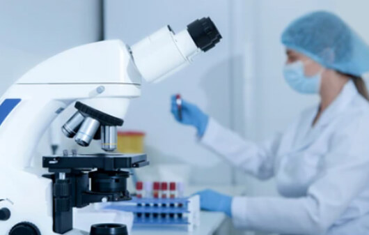
To enrich insoluble matrix components while reducing cellular protein masking, we use a sequential fractionation workflow that is selected during project feasibility review:
- Fraction 1 (Cytosolic Depletion): Hypotonic buffer (0.5 M NaCl + 0.1% Triton X-100) washes away soluble intracellular proteins while preserving the intact matrix scaffold.
- Fraction 2 (Membrane Clearance): Mild 0.1% SDS incubation clears organellar debris and cellular chaperones, yielding an enriched decellularized matrix pellet.
- Fraction 3 (Chaotropic Extraction): 4M Guanidine-HCl + reducing agents solubilizes non-covalently crosslinked glycoproteins and matrisome-associated factors.
- Fraction 4 (Core Pellet Solubilization & Deglycosylation): The insoluble crosslinked pellet is treated with PNGase F and Chondroitinase ABC to strip GAG chains, followed by high-temperature S-Trap enzymatic digestion into tryptic peptides.
Translational Disease Applications: Fibrosis, Desmoplasia, and Biomaterials
| Application | Model Systems | Analytical Goal | Key Molecular Readouts |
|---|---|---|---|
| Fibrotic Disease Profiling | MASH/NASH liver, IPF lung, renal interstitial fibrosis. | Quantify collagen accumulation and relative abundance patterns of crosslinking regulators. | COL1A1/COL3A1 ratios, LOXL2, Periostin, Tenascin-C, Fibronectin ED-A/B. |
| Tumor Stroma & Desmoplasia | Pancreatic ductal adenocarcinoma (PDAC), TNBC, CAF co-cultures. | Characterize ECM features associated with T-cell exclusion and reduced therapeutic penetration. | Versican (VCAN), basement membrane remodelers (COL4/COL6), MMP/TIMP ratios. |
| Decellularized Scaffolds (dECM) | Tissue engineering matrices, hydrogels, cardiac patches. | Verify cellular antigen clearance while confirming matrix protein retention. | Relative depletion of cellular marker proteins alongside retention profiles for structural laminins and collagens. |
| Anti-Fibrotic Drug Screening | Fibrotic tissues treated with LOX inhibitors, TGF-β blockers, Pirfenidone. | Measure treatment-associated changes in matrix deposition and crosslinking-regulator abundance. | Dose-associated changes in LOX/TGM2 abundance and ECM remodeling markers. |
Step-by-Step Matrisome Proteomics Workflow
Define tissue cohorts (healthy vs. fibrotic, stroma vs. tumor core) and anti-fibrotic treatment arms.
Execute hypotonic cellular depletion, 0.1% SDS wash, 4M Guanidine-HCl extraction, and core pellet recovery.
Apply selected deglycosylation steps when compatible with the sample type, followed by S-Trap trypsin/Lys-C digestion.
High-depth single-shot DIA on Orbitrap Astral or 4D-DIA (dia-PASeF on timsTOF Pro 2) with pooled QC monitoring.
Automated classification into Core Matrisome (Collagens, Glycoproteins, Proteoglycans) vs. Associated factors.
Matrix subcategory breakdown, crosslinking network mapping, GSEA fibrosis scoring, and PRM validation shortlists.
- Insoluble matrix recovery: 4-step chemical extraction captures crosslinked collagens without pellet loss
- Matrisome Project classification: direct mapping of Core Matrisome and Matrisome-Associated factors
- Enzymatic deglycosylation: PNGase F and Chondroitinase ABC pre-treatment ensures deep proteoglycan coverage
- Decellularization validation: quantitative confirmation of cellular antigen removal for biomaterial scaffolds
Matrisome Proteomics Analytical Platform Decision Guide
| Study Goal | Strategy | Primary Platform | Deliverable Scope |
|---|---|---|---|
| Bulk Matrisome Discovery (Fibrotic Liver / Lung / Stroma) |
Discovery DIA Proteomics | Orbitrap Astral / Exploris 480 / timsTOF Pro 2 | Fit-for-purpose DIA depth and quantitative precision for qualified tissue cohorts; expected coverage and CV are confirmed during feasibility review. |
| Microscale & Needle Biopsies (Core Biopsies / LCM Stroma) |
High-Sensitivity 4D-DIA | timsTOF Pro 2 (dia-PASeF) / timsTOF Ultra | TIMS ion mobility supports limited tissue input and microdissected stroma projects; expected coverage depends on tissue region, extraction yield, and matrix composition. |
| Decellularized Scaffolds (dECM) (Hydrogels / Biomaterials) |
dECM Quality & Purity DIA | Orbitrap Exploris 480 / timsTOF Pro 2 | Profiles structural matrisome retention and relative depletion of cellular protein markers; pair with DNA and histology controls when qualifying decellularization. |
| Mechanosensing Signaling (Integrin-FAK-YAP Kinetics) |
4D Phosphoproteomics | timsTOF Pro 2 / Orbitrap Exploris 480 | Provides high-coverage phosphoproteomics for integrin-FAK-YAP pathway investigation; achievable phosphosite coverage depends on input and enrichment performance. |
| Targeted Fibrosis Biomarkers (LOX, MMPs, Collagen Turnover) |
Targeted PRM Panels | Orbitrap PRM / Triple Quadrupole | Absolute quantification can be configured for selected matrisome biomarkers when suitable internal standards and assay performance criteria are established. |
Sample Submission Requirements and Guidelines
| Sample Type | Recommended Input | Minimum Input | Handling & Shipping |
|---|---|---|---|
| Fresh-Frozen Fibrotic Tissue | 30–50 mg wet weight | 10 mg wet weight | Dissect rapidly; rinse excess blood in cold saline; snap-freeze in liquid N2. Ship on dry ice (-80°C). |
| Tumor Stroma / Margin Tissue | 30–50 mg wet weight | 10 mg wet weight | Dissect tumor vs. margin; snap-freeze immediately in liquid N2. Ship on dry ice. |
| Decellularized Scaffolds (dECM) | 10–20 mg dry / wet weight | 5 mg wet weight | Rinse thoroughly with sterile deionized water to remove detergents; snap-freeze. Ship on dry ice. |
| 3D Organoid / Hydrogel Cultures | 50–100 organoids in dome | 20 organoids | Recover domes with cell recovery solution or freeze intact dome in liquid N2. Ship on dry ice. |
| Pre-Extracted Fractions | 20–50 μL at 1–2 mg/mL | 10 μg total protein | Document extraction buffer composition (Guanidine/Urea). Ship on dry ice. |
Deliverables and Decision-Ready Outputs
Quantitative matrisome matrices, Matrisome Project classifications, and fibrosis scorecards
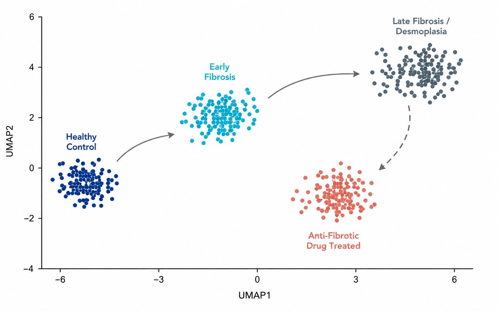
Sample-level PCA evaluates whether matrisome composition clusters strictly by disease stage (early vs. late fibrosis).
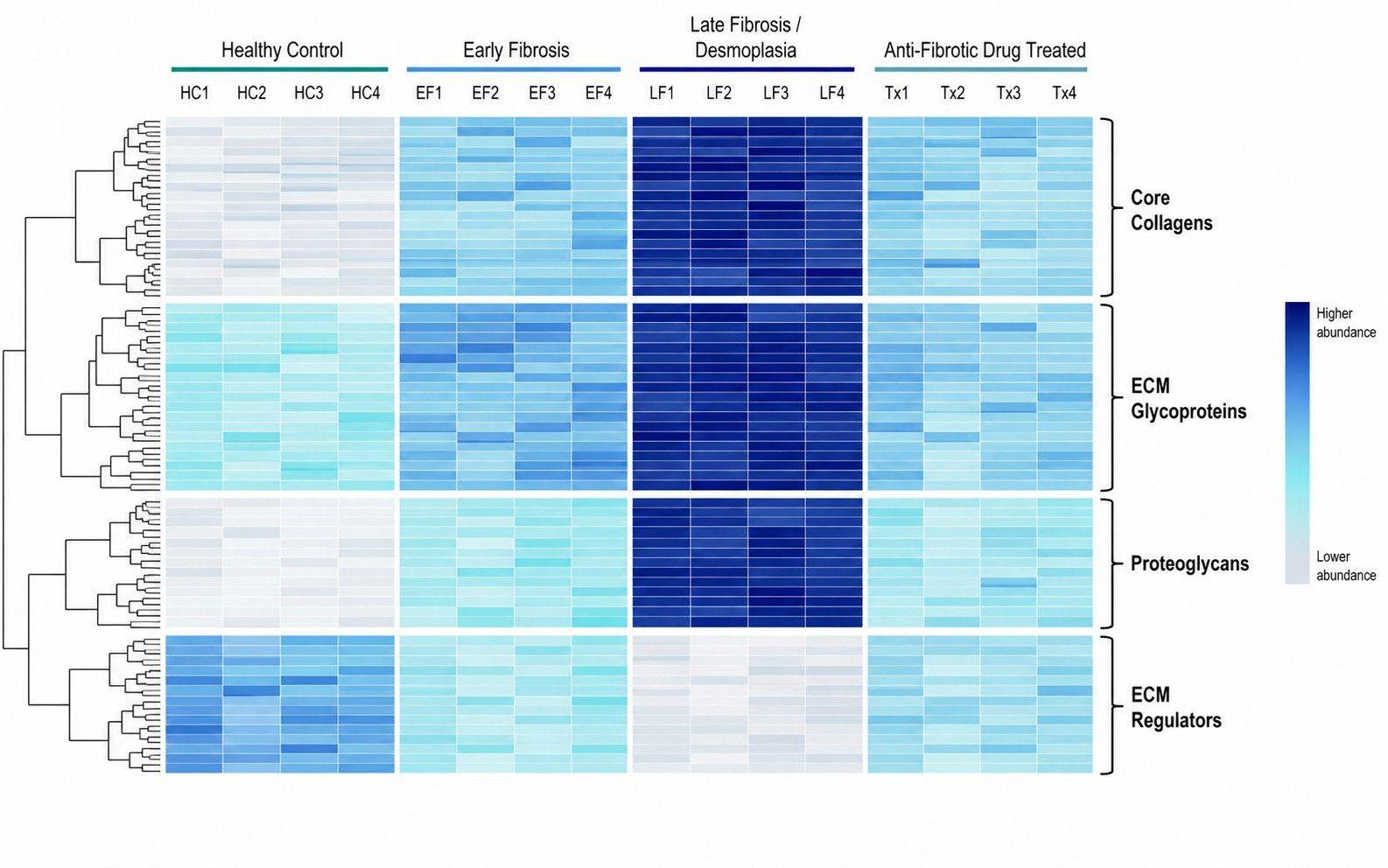
Hierarchical clustering resolves distinct matrix subcategories (collagens, proteoglycans, glycoproteins) across groups.
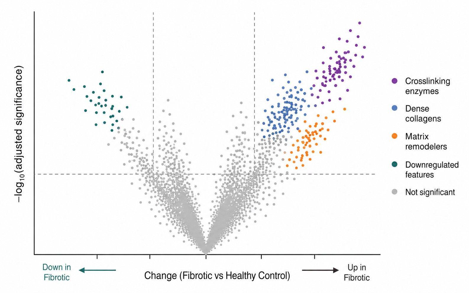
Differential volcano plots prioritize statistically significant matrix crosslinking enzymes (LOX/LOXL2), MMPs, and collagen chains.
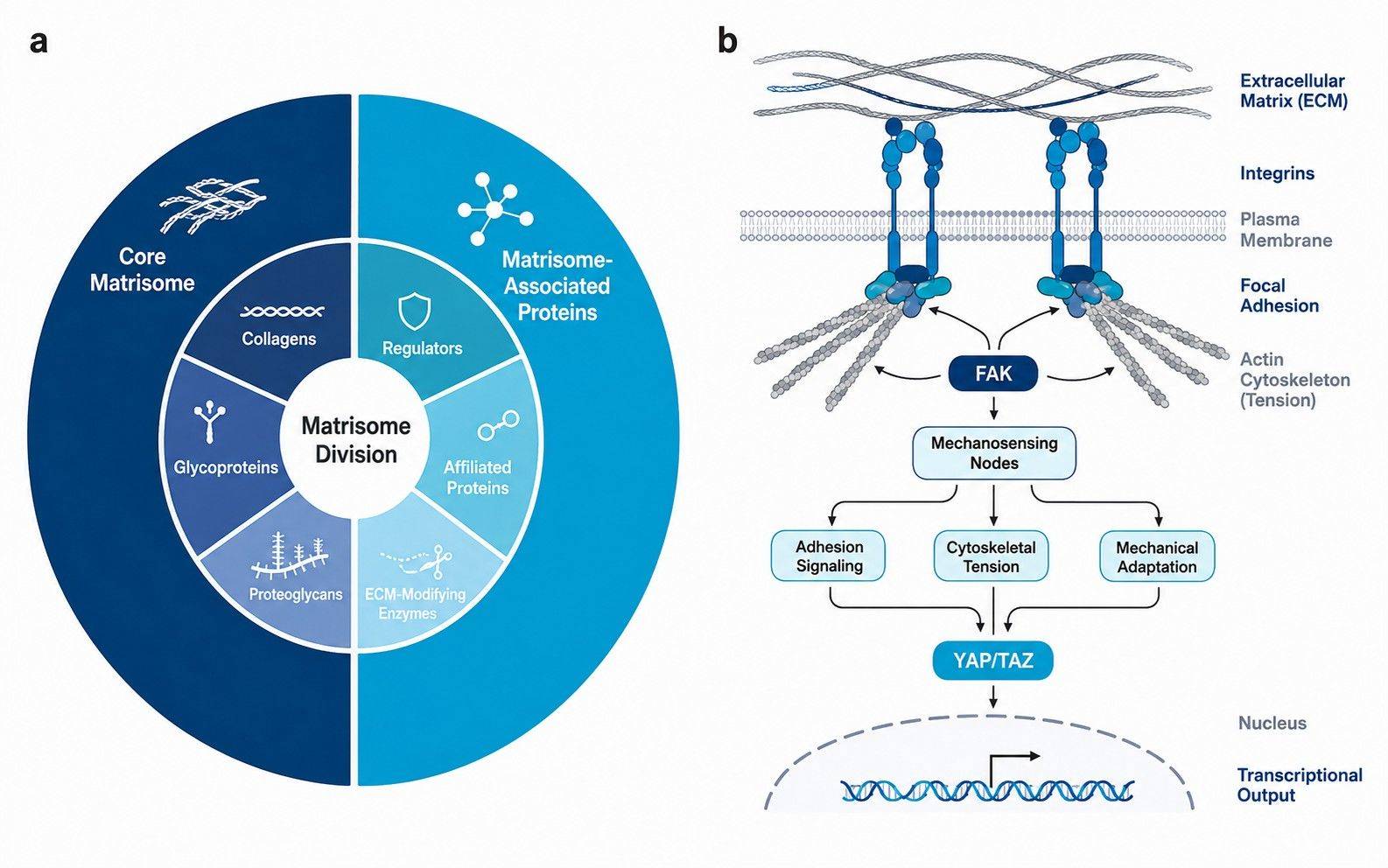
Matrisome division breakdown and integrin-FAK mechanotransduction networks map biomechanical remodeling pathways.

Classified Matrisome Intensity Tables
- Normalized protein- and peptide-level matrices annotated by Matrisome Project divisions (Core vs. Associated).

Quality & Decellularization Summary
- Extraction efficiency metrics, digestion completeness, pooled QC CV distributions, and intracellular protein clearance reports.

Core-to-Associated Ratio Metrics
- Quantitative ratios of fibrillar collagens (COL1/COL3), crosslinking-regulator abundance patterns, and fraction-specific matrix remodeling profiles.

Pathway & Mechanotransduction Networks
- GSEA fibrosis pathway enrichment, Integrin-FAK-YAP/TAZ mechanotransduction signaling, and pre-metastatic niche scorecards.

Comprehensive Final Report
- Publication-ready figures, auditable methods documentation, and candidate shortlists for targeted PRM validation.