
What Is Host–Pathogen Quantitative Proteomics?
Host–pathogen quantitative proteomics is the simultaneous mass spectrometry-based profiling of host cellular defense networks and pathogen virulence factors within infected systems. Powered by high-depth DIA quantitative proteomics and 4D ion-mobility mass spectrometry, it resolves dynamic pathogen subversion and host defense kinetics across time courses and antimicrobial interventions.
While RNA-seq and antibody panels capture static transcriptional activation or isolated surface antigens, they fail to detect viral host shutoff, pathogen-mediated proteolysis, or pre-formed bacterial effector injection. Direct dual-species quantitative LC-MS/MS resolves the true functional proteome by quantifying thousands of host defense regulators alongside scarce pathogen effectors from a single infection lysate.
Primary Study Scenarios & Decision Nodes:
- Viral Pathogenesis & Host Shutoff: Map viral non-structural proteins (e.g., RdRp, proteases) alongside host interferon cascades (cGAS-STING, ISG15, IFIT1–3) and translational arrest kinetics (0.5–24 h).
- Intracellular Bacterial Vacuolar Escape: Quantify Type III/IV/VI secretion system effectors (T3SS/T4SS) and host immunometabolic switches (ACOD1/itaconate, Rab GTPases) across infected macrophages.
- Virulence Attenuation & Knockout Profiling: Benchmark wild-type pathogens against isogenic Δvirulence or Δtoxin mutants to pinpoint direct cytotoxicity mechanisms and Caspase-1/Gasdermin D pyroptosis.
- Antimicrobial & Antiviral Drug Deconvolution: Track dose-dependent suppression of pathogen structural complexes alongside the resolution of host pro-inflammatory cytokine storms.
Content Guide
- Dual-Species Dynamic Range
- Host vs. Pathogen Modules
- Research Applications
- Step-by-Step Workflow
- Platform Decision Guide
- Sample Submission
- Deliverables & Outputs
Overcoming the Asymmetric Dynamic Range in Dual-Species Proteomics
The primary analytical bottleneck in host–pathogen proteomics is severe biomass asymmetry: host proteins frequently account for >95–99% of total sample protein, while pathogen proteins represent less than 1–5% of the total mass spectrometry signal. Creative Proteomics deploys a multi-tiered analytical strategy to maximize pathogen peptide recovery without sacrificing host depth:

- Subcellular & Phagosomal Enrichment: For intracellular pathogens, sucrose gradient fractionation isolates intact phagosomes and host vacuolar membranes, enriching pathogen-associated factors by 10- to 30-fold before lysis.
- Selective Microbial Pellet Recovery: Differential centrifugation combined with mild non-ionic detergent washes selectively lyses host plasma membranes while preserving intact bacterial cell walls for enriched microbial profiling.
- High-Capacity 4D-MS Separation: Trapped ion mobility spectrometry (TIMS) on the timsTOF Pro 2 separates co-eluting, high-abundance host peptides from low-abundance pathogen ions in the collision cross-section (CCS) dimension, drastically increasing spectral purity.
- Concatenated Dual-Species Database Searching: Joint search of host and pathogen FASTA databases with independent target-decoy calculation and strict false discovery rate (FDR ≤ 1%) controls eliminates cross-species mismatch artifacts.
Dual-Layer Functional Modules: Host Defense vs. Pathogen Virulence
Proteins quantified across infection cohorts are systematically clustered into coordinated host-response and pathogen-effector modules:

1. Host Innate Immunity & PRRs
Pattern recognition receptors (TLR2/4, cGAS-STING, RIG-I), adaptors (MyD88, TRIF, MAVS), and classical interferon-stimulated factors (ISG15, IFIT1–3, MX1, OAS1–3). Explore pathogen-mediated Ubiquitination Rewiring.

2. Host Stress & Immunometabolism
ER stress/UPR effectors (BiP, CHOP), glycolytic switches (GLUT1, HK2, LDHA), and antibacterial metabolic enzymes (ACOD1/IRG1 itaconate biosynthesis).

3. Pathogen Virulence Machinery
Bacterial secretion system effectors (Type III, IV, VI effectors, e.g., Inv/Prg/Spi), pore-forming toxins, and viral replication-transcription complexes. Map surface Glycoprotein Interactions.

4. Pathogen Defense & Counter-Measures
Antioxidant enzymes (catalases, SodA/KatG), bacterial heat-shock chaperones (DnaK, GroEL), multi-drug efflux pumps, and host-targeted proteases.
Translational Research Applications: Virology, Bacteriology, and Drug Discovery
| Application | Model Systems | Analytical Objective | Key Molecular Readouts |
|---|---|---|---|
| Viral Infection & Host Shutoff | SARS-CoV-2, Influenza A, Dengue/Zika, RSV in primary epithelial cells. | Map viral protein accumulation and host translational arrest. | Viral RdRp/nucleoproteins, ribosomal subunits, ISGs (MX1, IFIT1), phosphorylated IRF3. |
| Bacterial Vacuolar Escape | M. tuberculosis, Salmonella, Listeria in Primary Macrophages. | Track phagosomal maturation arrest and metabolic reprogramming. | Bacterial T3SS/T4SS effectors, Rab5/Rab7, LAMP1, ACOD1/itaconate enzymes, v-ATPase. |
| Virulence Factor Perturbation | Wild-type vs. Δvirulence mutant pathogens in co-culture models. | Identify direct cytotoxicity and host immune evasion pathways. | Caspase-1/3/8 activation, Gasdermin D cleavage, NF-κB target cytokines, pathogen fitness. |
| Antimicrobial Drug Screening | Infected in vitro / in vivo models treated with antibiotics or antivirals. | Measure pathogen clearance and Drug Mechanism of Action regarding inflammatory cytokine storms. | Pathogen structural protein degradation, reduction of pro-inflammatory cytokines, tissue repair. |
| Fungal Dimorphism & Invasion | Candida albicans, Aspergillus co-cultured with host endothelial cells. | Quantify adhesins driving yeast-to-hyphae morphological transition. | Hyphal adhesins (Als3, Hwp1), secreted aspartyl proteases (Saps), host tight-junction claudins. |
Step-by-Step Host–Pathogen Proteomics Workflow
Define biological cohorts (Mock vs. infected, time series 0–48 h, MOI titrations) with matched uninfected controls and ≥4 biological replicates.
Execute verified chaotropic lysis (4% SDS / 8M Urea) and heat inactivation (95°C, 10–15 min) ensuring complete non-infectivity for BSL-2 compatibility.
Automated S-Trap micro-column processing for total detergent clearance, reduction/alkylation, and rapid high-efficiency tryptic digestion.
Single-shot DIA on Orbitrap Astral or 4D-DIA (dia-PASeF on timsTOF Pro 2) with interleaved pooled QC injections to ensure quantitative stability.
Search concatenated host and pathogen FASTA databases with independent target-decoy calculation and strict species-specific FDR ≤ 1% filtering.
Deliver normalized dual-species expression matrices, host defense pathway enrichment, pathogen virulence kinetics, and targeted PRM validation lists.
- Simultaneous host & pathogen resolution: captures both defense cascades and virulence effectors in one run
- Joint concatenated database searching: independent target-decoy calculation prevents cross-species FDR inflation
- High-capacity 4D-MS ion mobility: separates co-eluting host peptides from low-abundance microbial ions
- Standardized biosafety workflows: validated chemical and thermal inactivation protocols compliant with BSL-2 standards
Host–Pathogen Proteomics Analytical Platform Decision Guide
| Study Goal | Strategy | Primary Platform | Deliverable Scope |
|---|---|---|---|
| Global Infection Discovery (Bulk Cell Lysates / In Vivo Tissues) |
Discovery DIA Proteomics | Orbitrap Astral / Exploris 480 / timsTOF Pro 2 | Deepest proteome depth (>6,000–8,500 host proteins; hundreds of microbial pathogen proteins), CV < 15%, missing value rates < 5%. |
| Microscale & Sorted Pathogen Samples (FACS-Sorted Infected Cells / Organoid Foci) |
High-Sensitivity 4D-DIA | timsTOF Pro 2 / timsTOF Ultra (dia-PASeF) | TIMS ion mobility separates scarce pathogen peptides from abundant host ions; sensitive down to 50,000 sorted cells. |
| Immediate PRR & Kinase Signaling (cGAS-STING, TBK1, NF-κB, 15–120 min) |
4D Phosphoproteomics | timsTOF Pro 2 / Orbitrap Exploris 480 | Captures >15,000–25,000+ phosphosites; KSEA maps immediate kinase activation cascades triggered by pathogen contact. |
| Targeted Virulence & Toxin Panels (T3SS Effectors, Viral Antigens, Toxins) |
Targeted PRM Panels | Orbitrap PRM / Triple Quadrupole (TSQ Altis) | Absolute quantification of 20–50 curated virulence targets across drug cohorts with zero missing values using SIL peptides. |
| Polymicrobial & Microbiomes (Gut, Stool, Mucosal Flora) | 4D Metaproteomics Services | timsTOF Pro 2 / Orbitrap Astral | Taxonomic classification and functional pathway profiling across complex uncultivated multi-species microbial communities without host reference dependencies. |
| Host Antigen Presentation (MHC-I / MHC-II Epitopes) | Immunopeptidome Profiling Service | Orbitrap Eclipse / Exploris 480 / timsTOF Ultra | Direct immunoaffinity isolation and mass spectrometry sequencing of pathogen-derived peptides physically presented by host HLA complexes. |
Sample Submission Requirements and Guidelines
| Sample Type | Recommended Input | Minimum Input | Inactivation & Shipping Guidelines |
|---|---|---|---|
| Infected Adherent Cells | 2–5 × 10⁶ cells / pellet | 1 × 10⁶ cells | Wash 2× with cold PBS; lyse in 4% SDS or 8M Urea; heat inactivate (95°C, 10 min); ship on dry ice (-80°C). |
| Infected Suspension / Sorting Pellets | 2–5 × 10⁶ cells / pellet | 5 × 10⁵ cells | Pellet cells (300 × g, 5 min); rinse with cold PBS; lyse in chaotropic buffer; heat inactivate; ship on dry ice. |
| Infected Animal Tissues (Lung, Liver) | 30–50 mg wet weight | 15 mg wet weight | Dissect rapidly; bead-beat in SDS lysis buffer in BSL-2 hood; heat inactivate before packaging; ship on dry ice. |
| Conditioned Media (Secretomes) | 5–10 mL culture supernatant | 2 mL | Clarify (1,000 × g, 10 min); filter via 0.22 µm membrane; concentrate via 3 kDa filter; ship on dry ice. |
| Pre-Extracted & Inactivated Lysates | 50–100 µg total protein | 20 µg total protein | Document buffer formulation and sterility verification in submission documentation; ship on dry ice. |
Deliverables and Decision-Ready Outputs
Dual-Species Expression Matrices, Infection Kinetics, and Virulence Factor Scorecards
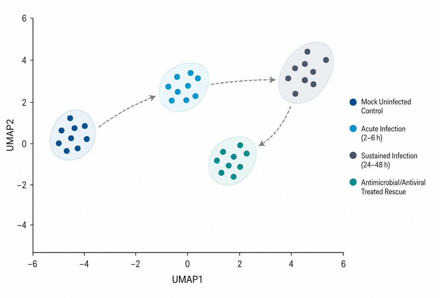
Sample-level PCA evaluates biological cohort separation across mock, acute, and sustained infection stages.
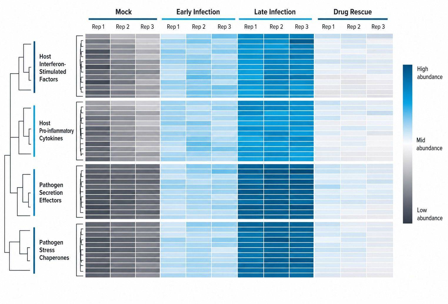
Hierarchical clustering resolves coordinated host defense induction versus pathogen virulence factor kinetics.
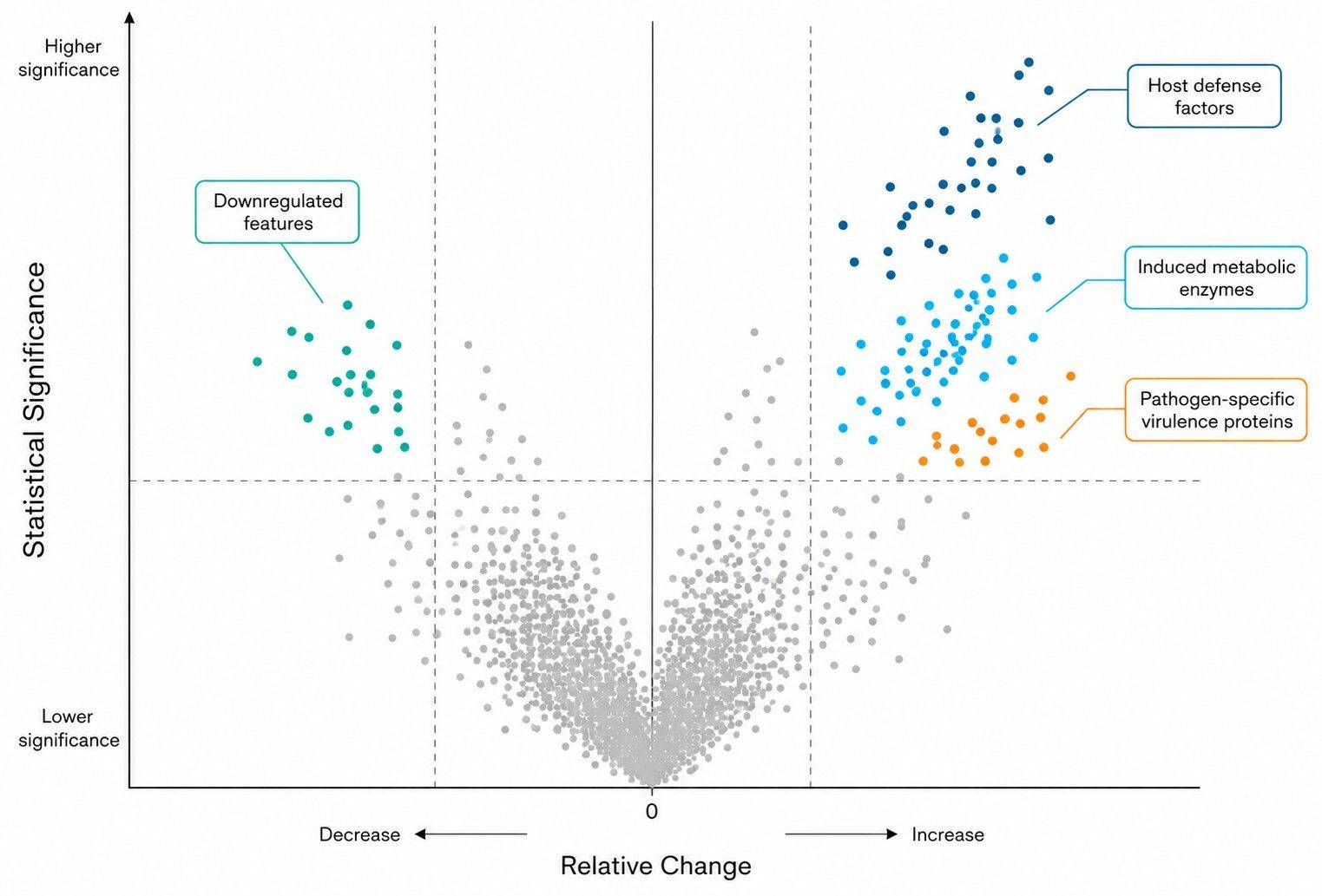
Dual-species volcano plots identify statistically significant host effectors and targeted pathogen virulence factors.
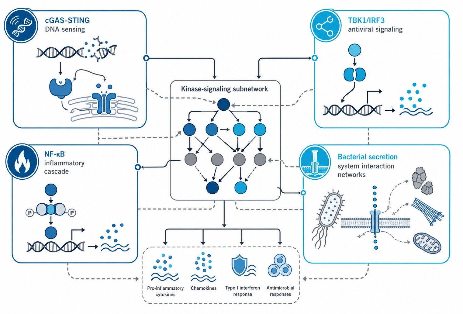
Integrated signaling networks map cGAS-STING, NF-κB, and microbial secretion effector interactions.

Dual-Species Normalized Quantitative Matrices
- Log2-transformed expression tables containing separate and concatenated host and pathogen abundance metrics with species-specific FDR ≤ 1% filtering.

Kinetic Clustering & Virulence Profiling
- Dynamic trajectory clustering of secretion system effectors, pore-forming toxins, and host antiviral/antibacterial regulators across infection time courses.

Statistical Contrast & Volcano Filtering
- Pairwise significance testing (adjusted p-value ≤ 0.05, fold change ≤ 2.0) comparing Mock vs. Infected, mutant strains, and antimicrobial rescue cohorts.

Innate Immunity Pathway & GSEA Reports
- Gene Set Enrichment Analysis (GSEA) mapping cGAS-STING, Type I interferon, NF-κB, inflammasome activation, and immunometabolic rewiring cascades.