
Organoid Proteomics Workflow and Study Design Strategies
Organoid proteomics is a specialized bioanalytical approach that combines 3D cell culture systems with high-resolution liquid chromatography-tandem mass spectrometry (LC-MS/MS) to systematically identify and quantify the global proteome, post-translational modifications, and signaling pathways within organotypic models. By preserving physiological cell-cell contacts, nutrient gradients, and differentiation states, organoid proteomics provides a high-fidelity molecular readout for oncology drug discovery, biomarker identification, and mechanistic biology.
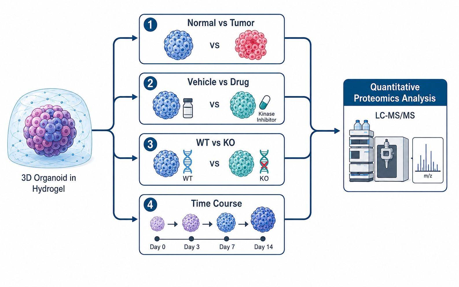
Unlike standard monolayer cultures, 3D organoids are grown embedded within complex extracellular hydrogels and exhibit multi-scale biological variance. Successful quantitative proteomics requires defining the exact biological comparison and unit of replication prior to harvest—whether contrasting patient-matched normal vs. tumor organoids, vehicle vs. drug treatments, drug-sensitive vs. resistant lines, or longitudinal differentiation trajectories.
Content Guide
- Project Strategy
- Common Challenges
- ECM Depletion & Prep
- Sample Submission Guide
- Replicate Design & Normalization
- DIA & 4D-DIA Platforms
- Research Applications
- Project Workflow
- Pre-Analytical & MS QC
- Bioinformatics & Deliverables
- Complementary Modalities
| Study Model / Contrast | Core Biological Question | Recommended Proteomic Readout |
|---|---|---|
| Normal vs. Tumor Organoids (PDTOs) | Identify dysregulated oncogenic driver pathways, aberrant surface targets, and tumor-specific metabolic adaptations. | Global DIA / 4D-DIA profiling with deep protein coverage (6,000–9,000+ protein groups). |
| Vehicle vs. Drug-Treated Organoids | Map acute and adaptive response signatures, on-target inhibition, pathway rewiring, and secondary resistance mechanisms. | Paired DIA global profiling and phosphoproteomic quantification for signaling network kinetics. |
| Responder vs. Non-Responder Biobanks | Correlate baseline ex vivo molecular phenotypes with patient clinical outcomes and pharmacological IC50 profiles. | Cohort-scale DIA quantitative proteomics with cross-batch pooled QC alignment. |
| Parental vs. Gene-Edited 3D Models | Determine downstream proteomic consequences, compensatory networks, and phenotypic shifts following CRISPR KO/knockdown. | High-precision label-free DIA with statistical fold-change and pathway enrichment. |
| Differentiation / Maturation Time Course | Track lineage commitment, functional polarization, and architectural maturation across developmental time points. | Longitudinal DIA quantitative series with cluster trajectory modeling. |
Define the Biological Unit Before Sample Pooling
Because organoid structures are small, researchers frequently face the decision of whether to pool material across wells. While pooling increases total protein yield, it masks intra-well and structure-to-structure heterogeneity. A robust project design must explicitly define whether the biological replicate represents an independent donor, an established clonal line, a discrete culture well, or a specific passage number.
- Maintain Donor Autonomy: Keep patient-derived lines and distinct donor specimens unpooled to preserve between-subject variance.
- Balance Culture Batches: Distribute control and treated organoid wells evenly across culture plates and harvesting dates to avoid confounding batch effects.
- Record Full Metadata: Systematically track passage number, dome count, seeding density, culture medium lot, and harvest timing alongside MS runs.
- Include Negative Matrix Blanks: Harvest cell-free Matrigel/BME domes processed under identical conditions to enable accurate background filtering.
Common Organoid Proteomics Challenges
Overcoming extracellular matrix carryover, low starting material, and nested cohort variance in 3D culture models.

Heavy ECM / Matrigel Background
Residual basement membrane hydrogels (laminin, collagen IV, entactin) suppress electrospray ionization and obscure low-abundance cellular proteins.

Microscale Sample Inputs
Precious 3D patient specimens yield microgram-level protein amounts, requiring ultra-sensitive microscale digestion and nanoLC-MS/MS platforms.

Multi-Tiered Nested Replicates
Donor-to-donor differences, passage drift, and intra-well dome size variability complicate statistical normalization and differential analysis.

Signaling Network Rewiring
Kinase inhibitor screening in PDTOs often drives rapid phosphosignaling rewiring before total protein abundance shifts occur.

Multi-Plate Cohort Batch Drift
Longitudinal organoid culture across multiple 24/48-well plates risks analytical drift, requiring periodic pooled QC and retention-time alignment.

Cell-Type Heterogeneity Boundaries
Bulk profiling reports ensemble average proteomes, requiring clear boundary identification when spatial or single-cell resolution is necessary.
Organoid Harvesting, ECM Depletion, and Sample Preparation Protocols
The primary pre-analytical challenge in 3D culture proteomics is the presence of protein-rich basement membrane hydrogels (such as Matrigel, Cultrex BME, or Geltrex). These matrices contain massive amounts of murine structural proteins—principally laminin subunits (α1, β1, γ1), collagen IV (α1, α2), nidogen-1 (entactin), and perlecan. Residual matrix proteins suppress electrospray ionization, mask low-abundance intracellular proteins, and skew quantitative normalization.
Pre-Analytical ECM Removal & Lysis Protocols
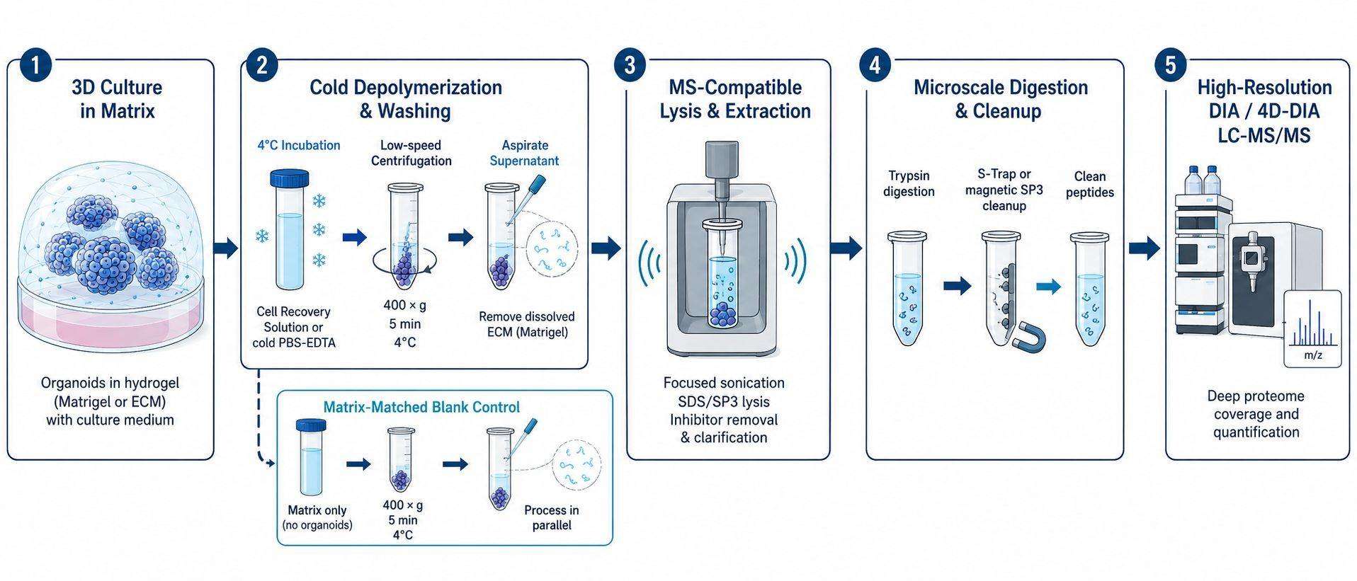
- Non-Enzymatic Cold Depolymerization: Incubation in ice-cold Cell Recovery Solution or ice-cold PBS-EDTA (4°C for 30–60 min) depolymerizes the hydrogel network via thermal transition without lysing organoid cell membranes or cleaving surface epitopes.
- Controlled Enzymatic Recovery: For dense or collagen-rich matrices, mild dispase or collagenase digestion (10–15 min at 37°C) followed by immediate cold quenching effectively releases organoid bodies.
- Low-Speed Differential Centrifugation: Centrifugation at 300–800 × g at 4°C pellets intact organoid structures while maintaining solubilized ECM proteins in the supernatant for complete aspiration.
- High-Efficiency Detergent Lysis: Recovered pellets are lysed using MS-compatible lysis buffers (e.g., S-Trap SDS lysis, SP3-compatible buffer, or RapiGest) coupled with focused ultrasonication for complete extraction of nuclear, membrane, and cytosolic proteins.
Sample Requirements and Submission Guidelines
To ensure high proteomic depth and quantitative accuracy, we provide flexible submission formats tailored for both standard research studies and microscale low-input 3D specimens:
| Sample Category | Recommended Input | Minimum Feasibility | Harvesting & Shipping Guidelines |
|---|---|---|---|
| Standard Organoid Pellets (ECM-Depleted) | 3–5 Matrigel domes (~150 μL gel volume) (10–20 μg total protein) |
1–2 domes (≥ 2 μg protein) |
Depolymerize ECM at 4°C, wash pellet with ice-cold PBS 3×, remove supernatant, flash-freeze in liquid N2. Ship on dry ice (-80°C). |
| Microscale / Low-Input Organoids | 1–2 small domes (~50 μL) (1–5 μg total protein) |
500 ng total protein (~10,000 cells) | Harvest into low-binding microcentrifuge tubes; direct lysis in 4D-DIA compatible lysis buffer; flash-freeze. Ship on dry ice. |
| Intact Organoids in Gel (Lab Recovery) | 3–6 intact domes in culture plates or cryovials | 2 intact domes | Aspirate media, add cold preservation reagent/PBS, freeze immediately. Creative Proteomics performs standardized on-site matrix depletion. |
| Organoid Phosphoproteomics | 15–30 Matrigel domes (100–200 μg total protein) |
50 μg protein | Lysis with phosphatase inhibitors (PhosSTOP, sodium orthovanadate); immediate flash-freezing in liquid N2. Ship on dry ice. |
| Conditioned Media (Secretome / EVs) | 2–5 mL conditioned medium per biological replicate | 1 mL medium | Culture in serum-free / defined media for 24–48 h; clarify at 2,000 × g to remove debris; flash-freeze supernatant. Ship on dry ice. |
Statistical Replicate Design and Data Normalization for 3D Cultures
Organoid experiments introduce nested hierarchies of variation: donor background, clonal line establishment, passage history, culture well, dome structure size, and MS analytical batches. Distinguishing genuine biological effects from technical variance requires a rigorous experimental design and appropriate normalization strategy:
Biological Replicates
Utilize ≥3–5 independent donor lines or independently established culture wells per experimental condition to empower robust statistical inference (FDR ≤ 0.05).
Contaminant Filtering
Subtract murine ECM peptides (laminin, collagen, nidogen) and culture supplement proteins (BSA, insulin, transferrin) using project-matched blank controls.
Robust Normalization
Apply median-polish, total-proteome intensity (TPA), or cyclic LOESS normalization across endogenous organoid proteins, excluding residual ECM spikes.
High-Throughput DIA and 4D-DIA Quantitative Proteomics for 3D Cultures
Selecting the optimal mass spectrometry strategy depends on starting material constraints, ECM complexity, and cohort scale. Data-Independent Acquisition (DIA) and ion-mobility-enhanced 4D-DIA (dia-PASeF) provide deep proteome coverage, high quantitative completeness, and reproducible quantification across multi-plate 3D studies:
Label-Free DIA Proteomics
Designed for multi-sample 3D cohorts, standard DIA quantitative proteomics provides systematic MS2 fragmentation across predefined isolation windows, capturing 6,000–9,000+ protein groups with high quantitative reproducibility and minimal missing data.
4D-DIA (dia-PASeF)
For precious or microscale organoids (500 ng – 2 μg), 4D-DIA quantitative proteomics introduces trapped ion mobility spectrometry (TIMS) to add collision cross-section (CCS) alignment, resolving co-eluting peptides and maximizing proteome depth from minimal dome inputs.
4D Phosphoproteomics
To resolve kinase activation cascades and drug resistance mechanisms in PDTOs, 4D phosphoproteomics couples microscale Fe-NTA/Ti-IMAC enrichment with TIMS-DIA to quantify 15,000–30,000+ phosphosites from as little as 50–100 μg total protein.
Research Applications: PDTO Drug Screening, Disease Modeling, and PTMs
| Application Domain | Scientific Value & Experimental Design |
|---|---|
| Patient-Derived Tumor Organoids (PDTOs) | Establish patient-specific proteomic profiles, compare primary vs. metastatic organoids, and identify predictive biomarkers associated with clinical therapy response. |
| High-Throughput Drug Screening & IC50 Correlation | Quantify proteome-wide and phosphoproteome shifts induced by kinase inhibitors, targeted therapeutics, or chemotherapeutics to resolve mechanism-of-action (MoA). |
| Stem-Cell-Derived Organoid Maturation | Track developmental proteome dynamics and structural polarization in cerebral, intestinal, hepatic, cardiac, or renal organoids during differentiation protocols. |
| CRISPR Gene Perturbation in 3D | Map functional downstream proteome remodeling, target degradation kinetics, and pathway compensation in knockout (KO), knockdown, or knock-in organoid lines. |
| Secretome and Extracellular Vesicle (EV) Profiling | Analyze paracrine factors, cytokines, and exosome cargo secreted by organoids into conditioned media to investigate cell-cell communication and microenvironment signaling. |
Organoid Proteomics Project Workflow
The workflow integrates biological scoping, ECM depletion, microscale digestion, high-depth mass spectrometry, and systems-level bioinformatics into a unified, traceable pipeline:
Define biological contrasts (PDTOs, drug treatment, CRISPR KO, maturation time course). Review hydrogel matrix type, donor metadata, dome counts, and replicate structure before harvest planning.
4°C non-enzymatic cold depolymerization (or controlled dispase) with differential centrifugation (400 × g) removes >95% of hydrogel mass without cell lysis. Parallel matrix blanks are collected.
Automated microscale S-Trap/SP3 digestion followed by nanoLC-MS/MS acquisition on timsTOF Pro 2 (dia-PASeF) or high-field Orbitrap instruments for deep single-shot proteome profiling.
Bioinformatic filtering of murine ECM contaminants (laminin, collagen, nidogen) using matrix blanks, followed by median-polish/TPA normalization and pooled QC reproducibility checks (CV < 15%).
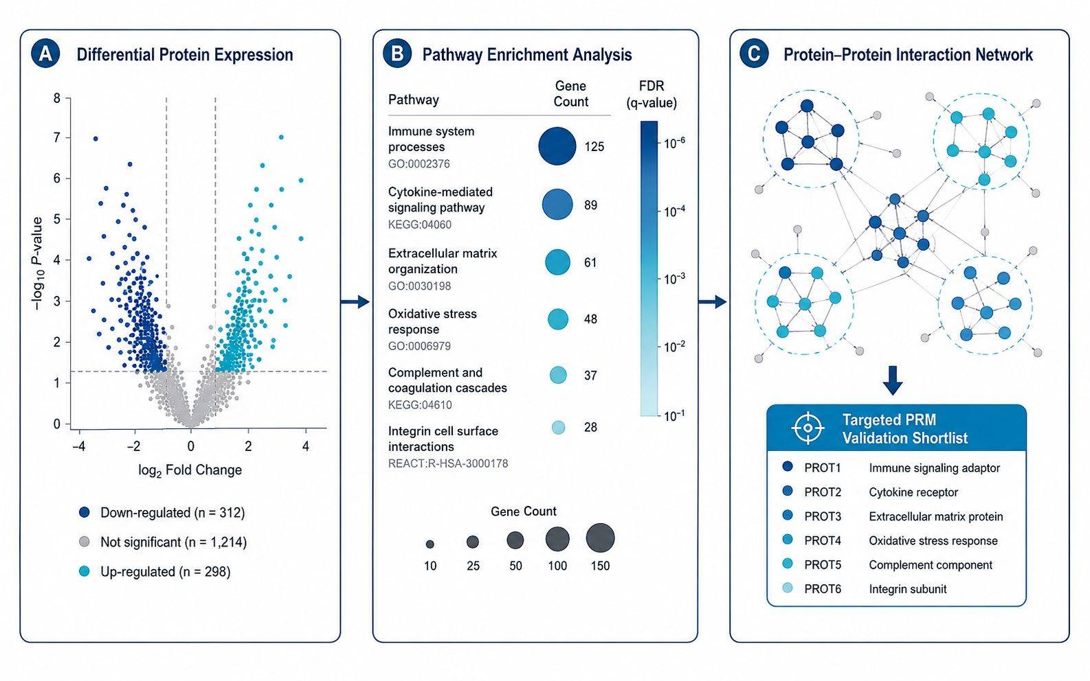
Volcano plots, GSEA/KEGG/Reactome pathway enrichment, kinase-substrate phosphorylation networks, and ranking of prioritized candidates for targeted PRM orthogonal validation.
Pre-Analytical and Analytical Quality Control for 3D Models
Rigorous quality control spans the entire lifecycle of an organoid proteomics project to ensure that proteomic variations reflect true biology rather than culture artifacts or analytical drift:
ECM Depletion Verification
Evaluate residual matrix protein abundance (laminin, collagen, nidogen) against total proteome intensity; verify background is below 5% of total signal.
Pooled QC Stability
Inject pooled organoid reference samples periodically throughout the LC-MS batch to track retention time stability, peak width, and quantitative CV (<15%).
Cohort Metadata Tracking
Document passage number, donor ID, harvest batch, and lysis yield alongside MS raw files to control for biological and technical confounding factors.
Data Deliverables, Bioinformatics, and Candidate Prioritization
We deliver publication-ready, fully annotated bioinformatics data packages designed to translate massive mass spectrometry datasets into actionable biological hypotheses:
- Normalized Quantitative Matrices: Filtered protein and peptide expression matrices with complete sample metadata and missing value annotations.
- Comprehensive QC Report: Assessment of ECM contamination levels, peptide digestion efficiency, pooled QC reproducibility, and run-order drift.
- Differential Expression Analysis: Volcano plots, statistical fold-change tables, and Benjamini-Hochberg false discovery rate (FDR) statistics.
- Pathway & Network Enrichment: Gene Ontology (GO), KEGG, Reactome, and Gene Set Enrichment Analysis (GSEA) identifying activated or repressed biological pathways.
- Kinase-Substrate & PPI Networks: Phosphorylation motif analysis and protein-protein interaction (STRING/Cytoscape) mapping for signaling networks.
- Validation Candidate Shortlist: Prioritized protein candidates ranked for orthogonal validation via targeted PRM proteomics or Western blotting.
Information That Helps Us Scope Your Organoid Study
- Organoid lineage, disease model, donor background, and culture matrix type (e.g., Matrigel, BME)
- Culture format, passage number, dome count, and available sample input levels
- Experimental conditions: vehicle vs. drug treatments, CRISPR edits, or time-course series
- Primary research objective: global profiling, phosphoproteomics, or targeted candidate validation
When Bulk Organoid Proteomics Requires Complementary Modalities
Bulk organoid proteomics measures the ensemble average protein abundance across all cell types present in the 3D structure. When your experimental question requires resolving spatial architecture or distinct rare cell lineages (such as stem cells, enterocytes, or neuroendocrine cells within an intestinal organoid), spatial proteomics or single-cell proteomics may provide critical orthogonal resolution. Furthermore, when investigating dynamic metabolic pathway turnover, substrate utilization, or energy flux rather than protein abundance, our dedicated organoid metabolomics service provides the ideal primary functional readout.
Selected Scientific References
- Zhang Y, et al. In-Depth Comparison of Matrigel Dissolving Methods on Proteomic Profiling of Organoids. Molecular & Cellular Proteomics. 2022;21(1):100181. doi:10.1016/j.mcpro.2021.100181.
- Proteomic profiling of brain organoids and extracellular vesicles identifies early Alzheimer's disease biomarkers and drug response heterogeneity. Cell Death & Disease. 2024;15:130. PMC13058922.
- Pharmaco-proteogenomic characterization of liver cancer organoids for precision oncology. Nature Communications. 2024;15:2314. doi:10.1038/s41467-024-46542-8.