

Extracellular vesicles (EVs)—including exosomes and other small EV subtypes—have become a core analytical target in translational research and biopharma R&D. Their cargo (proteins, lipids, and nucleic acids) can reflect cell state and mechanism, and EVs themselves can modulate intercellular signaling. But EV biology is only as trustworthy as the isolation workflow that precedes your downstream assays.
This resource explains how ultracentrifugation isolates EVs, which parameters actually matter for reproducibility, and when ultracentrifugation is the right choice compared with size exclusion chromatography (SEC), precipitation, tangential flow filtration (TFF), and emerging microfluidic approaches. The emphasis is practical: how to balance purity versus yield, how to troubleshoot low recovery, and how to validate EV integrity so proteomics (and other omics) readouts stay interpretable.
Why Ultracentrifugation is the Gold Standard for EV Isolation
Exosomes (often discussed within the broader EV umbrella) are typically nanoscale vesicles released through the endosomal pathway, while larger EV populations can bud directly from the plasma membrane. In real-world samples, you rarely get a single EV “type”—you get a continuum of particles and non-vesicular species that overlap in size and density. That’s the central challenge: EV isolation is a separation problem with imperfect boundaries.
Ultracentrifugation has earned “gold standard” status largely because it is reagent-free, broadly applicable across matrices, and historically well-characterized. Differential ultracentrifugation (dUC) can process substantial sample volumes and produces EV-enriched pellets with predictable failure modes. Density gradient ultracentrifugation (dgUC) adds an additional physical dimension—buoyant density—to improve purity and reduce co-isolated contaminants.
For biopharma teams, ultracentrifugation is often the method you use when you need (1) transparent physics, (2) controllable wash steps, and (3) isolation conditions that won’t introduce polymer carryover or column-derived artifacts that complicate sensitive downstream readouts.
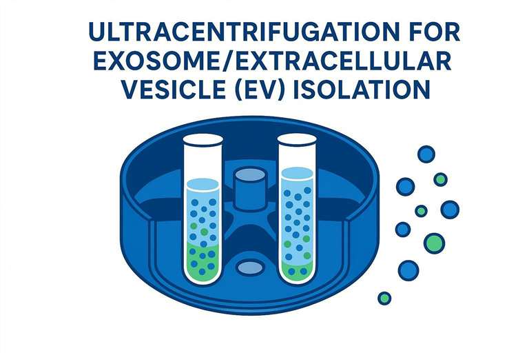
Principles of Ultracentrifugation for EV Isolation (Ultracentrifugation for Exosome Isolation)
At a high level, ultracentrifugation separates particles based on how quickly they migrate in a centrifugal field. EV sedimentation depends on a combination of particle size, density relative to the medium, rotor geometry, viscosity, and run time. This is why two protocols that both claim “100,000×g” can yield meaningfully different EV populations.
How ultracentrifugation works: separating EVs based on size and density
- Differential ultracentrifugation uses sequential spins to progressively remove larger components (cells, debris, apoptotic bodies, large microvesicles) before pelleting smaller EVs at high g-force.
- Density gradient ultracentrifugation separates particles until they reach a layer where buoyant density matches the surrounding medium—helping resolve EVs from similarly sized non-vesicular contaminants.
In practice, differential spins are more “throughput-friendly” and easier to standardize within a lab. Gradients are more purity-oriented but require more hands-on time and careful fraction handling.
Key physics concepts: centrifugal force (RCF vs RPM) and calculation methods
The most common source of protocol drift is confusing RPM (rotations per minute) with RCF (relative centrifugal force, reported as ×g). RPM is a rotor speed. RCF is the force experienced by the sample—and it changes with rotor radius.
The practical takeaway for reproducibility is simple: when you report or replicate an EV protocol, include RCF, rotor type, and the effective radius (or at least the manufacturer’s rotor specs). A theoretical analysis of differential centrifugation showed that rotor geometry can change sedimentation behavior in ways that cannot be “fixed” by naïvely scaling time using K-factors when switching to fixed-angle rotors (see Isolation of exosomes by differential centrifugation: Theoretical analysis of a commonly used protocol).
Pro Tip: When comparing SOPs across sites, standardize by RCF at the relevant radius and document the rotor model. “Same RPM” across rotors is not a controlled condition.
Differences between differential ultracentrifugation and density gradient ultracentrifugation
Differential ultracentrifugation is fundamentally a rate-based enrichment: you pellet what sediments within a defined time window. In contrast, density gradient ultracentrifugation exosomes workflows are closer to equilibrium separation: particles migrate until density matches the gradient layer, which helps separate EVs from soluble protein complexes and some lipoprotein fractions that can be problematic in plasma/serum.
Because many contaminants overlap with EVs in size, gradients are often the step that turns an “EV-enriched pellet” into an “EV preparation you can defend” in downstream proteomics or biomarker work.
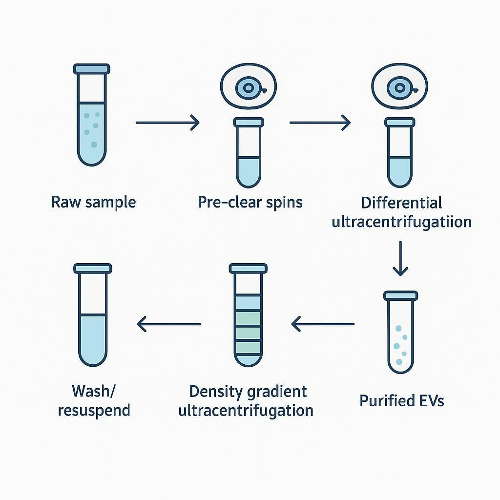
Ultracentrifugation Workflow and Key Parameters for EV Isolation
Sample preparation: removal of debris and larger particles
Before you ultracentrifuge, you’re really doing risk management. Poor pre-clearance makes every later step harder.
A typical pre-clear sequence is designed to remove:
- intact cells and large debris
- apoptotic bodies and large vesicles
- aggregates introduced by freeze–thaw or harsh homogenization
For viscous or protein-rich matrices (plasma/serum), pre-clearance also reduces fouling and makes downstream purity more achievable.
Step-by-step differential ultracentrifugation procedure: optimizing time, g-force, and rotor type
A practical differential ultracentrifugation protocol for EVs can be described as a staged enrichment, with each stage having a specific “removal target.” Exact parameters vary by matrix, rotor, and tube format, but the logic is consistent.
| Stage | Goal | Typical considerations (matrix-dependent) |
|---|---|---|
| Low-speed spins | Remove cells + large debris | Gentle handling to avoid generating debris; keep cold |
| Medium-speed spin | Reduce large vesicles + organelle fragments | Helps prevent overloading the high-speed step |
| High-speed UC | Pellet small EV-enriched fraction | RCF, time, and rotor geometry strongly affect recovery |
| Wash UC | Reduce soluble proteins | Improves purity but can reduce yield if pellet handling is rough |
Two points matter more than any single “published number” (and they’re why fixed-angle vs swinging-bucket rotor choice can change what you recover):
- Pellet handling is part of the protocol. EV pellets can be nearly invisible. Aggressive aspiration, incomplete resuspension, or harsh pipetting can cost you more yield than changing g-force by a few percent.
- Rotor choice changes the sedimentation path. Fixed-angle and swinging-bucket rotors do not behave identically for small particles. If you’re harmonizing across sites, align the rotor type where possible—and if not, harmonize by RCF/radius and validate outputs (NTA/TEM/WB) rather than assuming equivalence.
Density gradient ultracentrifugation: media selection and fraction collection
Density gradients typically use sucrose or iodixanol (often preferred for EV work due to gentler osmotic properties and practical fractionation). The purpose is not to “get more EVs” but to remove what looks like EVs to your downstream assay—for example, lipoproteins and protein complexes that can inflate particle counts or confound proteomics.
Fraction collection is where gradients succeed or fail:
- use consistent fraction volumes
- record fraction density (or at minimum, fraction order and gradient composition)
- avoid cross-contamination between layers during aspiration
When plasma/serum is the input, gradients are often the most defensible way to reduce non-vesicular carryover before sensitive downstream analyses.
Tips for optimizing key parameters: g-force, centrifugation time, rotor selection, temperature control
Optimization isn’t about chasing a maximum yield number. It’s about defining a stable operating window that preserves EV integrity and produces reproducible QC metrics.
g-force and time control the cut-off of what sediments. Longer or harder spins can increase recovery and increase co-precipitation. A methods overview notes that extended ultracentrifugation can increase non-EV protein contamination and that rotor type and viscosity influence isolation efficiency (as summarized in Isolation of Extracellular Vesicles: General Methodologies and Considerations, 2018).
Rotor selection should be driven by:
- sample volume needs
- whether you’re running gradients (often more convenient with swinging-bucket)
- your tolerance for protocol transfer risk across sites
Temperature control (commonly 4°C) helps reduce enzymatic activity and can protect labile cargo. Just remember: colder temperatures can increase viscosity, which can change sedimentation behavior. That makes reporting (and consistent practice) even more important.
Advantages and Limitations of Ultracentrifugation
Ultracentrifugation is powerful because it’s simple in principle—but it’s not “easy.” The advantages and limitations are both real, and biopharma teams should treat them as design constraints rather than surprises.
Key advantages
Ultracentrifugation remains attractive because it:
- Avoids chemical reagents that can co-precipitate or carry over into downstream assays
- Scales to larger volumes than many kit-based approaches
- Allows controlled wash steps to manage soluble protein contamination
- Is broadly interoperable with downstream characterization (NTA, TEM, immunoblotting) and omics when the workflow is well-controlled
Limitations
High equipment costs and operational complexity. Ultracentrifuges, rotors, and compatible consumables are expensive, and small deviations (tube fill volume, balancing, rotor specs) can affect outcomes.
Impact of high g-force on EV integrity and downstream analysis. High forces can compact pellets, promote aggregation, and make resuspension harsh. Those effects can distort NTA distributions and complicate interpretation.
Balancing yield versus purity in EV isolation. The same conditions that pellet more material also tend to pellet more contaminants. For clinical matrices, that trade-off becomes central: what looks like “higher yield” might be higher non-vesicular particle carryover.
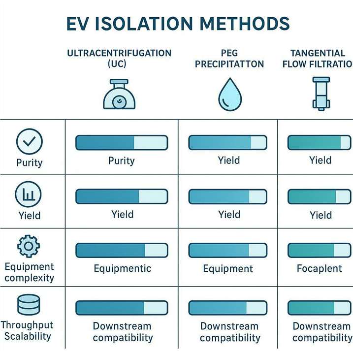
Ultracentrifugation vs Other EV Isolation Methods
Most method debates can be reduced to two questions:
- What contamination can your downstream readout tolerate?
- What throughput and reproducibility do you need across sites and studies?
Ultracentrifugation vs Size Exclusion Chromatography (SEC)
SEC is gentle and often produces EV fractions with reduced soluble protein carryover compared with crude pellets, but columns can be sensitive to overload and require careful fraction selection. SEC can be an excellent “polishing” step after concentration, but it becomes operationally heavier when sample volume is large.
Ultracentrifugation vs PEG/precipitation methods
Precipitation is operationally simple and can give high apparent recovery, but co-precipitation of non-EV material is a known limitation—especially problematic when you need EV preparations that stand up to downstream proteomics or biomarker discovery. A protocol comparison discusses trade-offs between ultracentrifugation and precipitation workflows (see Exosome Isolation by Ultracentrifugation and Precipitation).
Ultracentrifugation vs Tangential Flow Filtration (TFF) and microfluidic technologies
TFF is often chosen for large-volume processing and concentration, with the added benefit of being relatively gentle when run at appropriate shear conditions. Microfluidic methods can provide high selectivity, but many teams treat them as niche tools unless the workflow is already validated for their sample type and endpoints.
Decision guide on choosing the best method based on experimental goals and sample types
Below is a practical decision matrix that maps common biopharma goals to a “first-pass” method choice. In practice, hybrid workflows (e.g., concentration + polishing) are common.
| Your primary goal | Typical priority | Often a strong starting point | Notes |
|---|---|---|---|
| Proteomics-ready EV prep from plasma | Purity > yield | dgUC (often after pre-clearance) | Helps reduce co-isolated lipoproteins/protein complexes |
| High-throughput screening from conditioned media | Throughput + reproducibility | dUC or TFF → optional SEC | Consider how you’ll normalize input and validate across batches |
| Low-input matrices (CSF) | Recovery + integrity | dUC with careful handling; consider polishing only if feasible | Losses during extra steps can dominate |
| Tissue homogenate EVs | Deconvolution of debris/aggregates | strong pre-clearance + dUC; consider dgUC for purity | Tissue prep quality drives everything |
| RNA-focused discovery | Integrity + inhibitor control | method depends on contaminants; validate inhibitors early | Avoid assuming particle count equals usable RNA |
Common Issues and Optimization in Ultracentrifugation for EV Isolation
Low yield issues: common causes and troubleshooting strategies
Low yield is often not “insufficient g-force.” It’s usually one (or several) controllable process failures:
- Pellet loss during aspiration: EV pellets can be translucent. Leave a small volume above the pellet and standardize aspiration technique.
- Inadequate pre-clearance: Debris overload can trap EVs or create aggregates that behave unpredictably.
- Matrix viscosity (notably plasma/serum): sedimentation slows as viscosity rises; harmonize conditions and validate rather than copying a culture-media SOP.
- Rotor mismatch across sites: identical RPM does not guarantee identical RCF; document rotor radius and report RCF.
If you’re debugging across labs, treat the isolation as an assay: fix the input definition (volume, storage history, hemolysis status for blood), then fix the reporting (RCF/radius/rotor), then fix the handling.
Addressing EV degradation or inactivation during ultracentrifugation
EV integrity can be compromised by:
- repeated freeze–thaw
- prolonged room-temperature handling
- harsh resuspension of compacted pellets
Standardize cold-chain handling, minimize processing delays, and use gentle resuspension to reduce shear. If your readout is functional (uptake assays, bioactivity), consider running an integrity checkpoint before committing to large batches.
Contaminant co-precipitation: identification and strategies for minimizing contamination
For plasma/serum, co-isolated species can include lipoproteins and abundant soluble proteins; for urine, uromodulin complexes are a known issue. A methods overview notes common contamination and marker considerations across matrices, and highlights the need for multi-modal characterization (Isolation of Extracellular Vesicles: General Methodologies and Considerations, 2018).
Practical strategies include:
- using density gradients when purity is critical
- adding a wash UC step (accepting some yield loss)
- avoiding over-spinning that increases non-EV co-pelleting
- validating contaminants with negative markers (not just positive EV markers)
⚠️ Warning: If your NTA particle concentration increases after a protocol change, that’s not automatically “better.” It may be increased non-vesicular particles. Always pair particle counts with protein metrics and marker profiling.
Downstream Compatibility and EV Validation Metrics
A credible EV isolation workflow ends with validation that matches your downstream question. For biopharma R&D, the goal is usually not “I have EVs.” It’s “I have a preparation whose composition is stable enough that my biomarker or mechanistic signal is interpretable.”
Key purity assessment metrics
NTA (Nanoparticle Tracking Analysis) helps estimate particle size distribution and concentration. It is sensitive to sample dilution, refractive index, and non-vesicular particles, so interpret it alongside orthogonal metrics.
TEM (Transmission Electron Microscopy) provides morphological confirmation (vesicular structures, integrity) and can reveal aggregation or debris.
Western blot (or orthogonal immunoassays) confirm presence of EV-associated markers and help track contaminants. A methods overview lists commonly used EV markers such as CD9, CD63, CD81, TSG101, and Alix, and notes contamination pitfalls (Isolation of Extracellular Vesicles: General Methodologies and Considerations, 2018).
A practical marker panel often includes:
- positive markers (e.g., tetraspanins and endosomal-associated proteins)
- negative/contaminant markers relevant to your matrix (e.g., abundant plasma proteins; matrix-specific contaminants)
For urine-derived preparations, document how you handled urine-specific confounders and verify with orthogonal readouts—because EV isolation from urine can look “high-yield” on particle counts while still being contaminant-heavy.
Preparing EVs for downstream applications: proteomics, lipidomics, and metabolomics
Downstream omics compatibility is where isolation trade-offs become expensive:
- Proteomics is particularly sensitive to co-isolated abundant proteins (plasma) and to polymer carryover (precipitation). Purity-oriented workflows (often dgUC and/or SEC polishing) may be preferable.
- Lipidomics benefits from clean separation from lipoproteins, especially in blood-derived matrices.
- Metabolomics requires tight control of background small molecules and buffers; extra washes and careful buffer selection matter.
If your goal is an omics-grade EV preparation with reporting-ready QC, you can also consider outsourcing portions of the workflow—especially when you need consistency across programs. Creative Proteomics provides Exosome Proteomics Services and Extracellular Vesicle Metabolomics Service that can be used to extend a validated isolation into downstream multi-omics readouts.
Validating EV integrity for reliable experimental results
A minimal “biopharma defensibility” standard is:
- at least two orthogonal characterization modalities (e.g., NTA + TEM)
- marker-based confirmation (including at least one negative/control marker)
- documented protocol parameters (RCF, rotor, time, temperature, wash steps)
If you’re moving toward regulated contexts, consider upgrading this into a formal QC panel and acceptance criteria specific to your sample type and endpoint.
Decision Tree for EV Isolation Method Selection
The most robust method is the one that matches your constraints. Use this decision logic to choose a starting workflow, then validate and iterate.
Decision logic: considerations for sample volume, equipment, purity vs. yield priorities
- Is your matrix high-risk for co-isolates (plasma/serum, some tissue homogenates)?
- If yes and downstream is proteomics/biomarker discovery → prioritize dgUC (and/or SEC polishing) over “single-step pelleting.”
- Is your volume large (conditioned media at scale) and throughput is limiting?
- If yes → consider TFF for concentration, then choose a polishing step (SEC or dgUC) based on purity needs.
- Is your input low (CSF) where each extra step can destroy recovery?
- If yes → minimize step count; optimize handling and validate with sensitive assays rather than adding multiple purification stages.
- Do you need method transferability across sites?
- If yes → standardize reporting (RCF/radius/rotor) and define QC acceptance metrics, not just “follow this RPM.”
Summary table of when to choose ultracentrifugation, SEC, precipitation, or TFF based on experimental needs
| Method | Choose when… | Watch-outs |
|---|---|---|
| Ultracentrifugation (dUC) | You need a reagent-free baseline workflow and can control reporting + handling | Co-pelleting contaminants; pellet loss; rotor-to-rotor variability |
| Density gradient UC (dgUC) | Purity is critical (plasma/serum proteomics; mechanistic claims) | Hands-on complexity; EV loss in gradients |
| SEC | Gentle polishing and reduced soluble protein carryover matter | Column overload; fraction selection impacts yield |
| PEG precipitation | Operational simplicity matters more than purity (early exploratory work) | Polymer/protein co-precipitation; downstream interference |
| TFF | Large volume concentration and scalability are needed | Membrane selection; shear/processing conditions; requires validation |
FAQs
Protocol parameters (RCF, rotors, and run settings)
Q: What’s the difference between RPM and RCF in ultracentrifugation for exosome isolation?
RCF (×g) is the effective force applied to the sample, while RPM is the rotor speed. Because RCF depends on rotor radius, the same RPM can generate different forces in different rotors, which changes EV sedimentation and reproducibility.
Q: How do I convert RPM to RCF for an EV ultracentrifugation protocol?
Use the rotor’s effective radius and the standard conversion equation provided by the centrifuge manufacturer. For method transfer across sites, document rotor model and radius; reporting only RPM is not sufficient for reproducible EV isolation.
Q: Does rotor type (fixed-angle vs swinging-bucket) affect EV isolation results?
Yes. Rotor geometry changes sedimentation path length and pelleting efficiency, which can alter yield and co-isolated material. If you switch rotors, validate the output (NTA/TEM/marker panel) rather than assuming time scaling produces equivalent EV populations.
Purity and contamination (especially for plasma/serum)
Q: How can I reduce lipoprotein contamination when isolating EVs from plasma or serum?
Use a purification step that separates by density (e.g., density gradient ultracentrifugation) and confirm improvement with contaminant-aware markers, not particle count alone. For plasma-derived EV proteomics, treat lipoprotein removal as a design requirement rather than a “nice-to-have.”
Q: Why does my ultracentrifugation EV pellet have high protein but weak EV marker signals?
Co-pelleted soluble proteins and protein complexes can dominate the pellet, especially in protein-rich matrices or when spins are extended. Adding a wash step and/or moving to a density gradient can improve specificity, but you should expect some yield loss and plan accordingly.
Validation and downstream readiness
Q: What are the minimum validation steps after ultracentrifugation-based EV isolation?
Use at least two orthogonal methods (commonly NTA for size/concentration and TEM for morphology) plus a marker-based assay (e.g., western blot) that includes both EV-associated markers and at least one contaminant/negative marker appropriate to your sample type.
Q: Which is better for EV validation: NTA or TEM?
They answer different questions. NTA is best for particle size distribution and concentration trends across batches, while TEM is best for confirming vesicular morphology and spotting aggregation/debris. Using both provides much stronger evidence than either alone.
Q: How should I prepare EVs for downstream proteomics?
Prioritize purity controls that reduce abundant co-isolated proteins and document QC metrics before committing to large studies. For plasma/serum, consider density-based purification and validate that protein contamination is not driving apparent biomarker signals.
Sample-type nuances
Q: Why is EV isolation from CSF so challenging with ultracentrifugation? CSF is often low-input for EVs, so each additional handling step can reduce recovery. Minimize unnecessary purification steps, focus on gentle pellet handling, and rely on sensitive validation assays to confirm that the isolated material matches your intended EV population.
Q: What’s the biggest mistake when isolating EVs from tissue homogenate? Treating tissue homogenate like conditioned media. Tissue prep can generate debris, aggregates, and organelle fragments that overload downstream separation; rigorous pre-clearance and controlled homogenization are usually more important than increasing the ultracentrifugation force.
Next steps
If you need an EV isolation workflow that’s optimized for your sample type and validated with fit-for-purpose QC (especially for plasma/serum or omics-ready readouts), Creative Proteomics’ Exosome Isolation and Purification Service and Exosome Analysis Services can support method selection, purification, and characterization as part of an end-to-end study design.
References
- Isolation of exosomes by differential centrifugation: Theoretical analysis of a commonly used protocol
- Isolation of Extracellular Vesicles: General Methodologies and Considerations
- Exosome Isolation by Ultracentrifugation and Precipitation
- Framework for rapid comparison of extracellular vesicle isolation methods
- Comparison of extracellular vesicle isolation processes for proteomic analysis of plasma
* For Research Use Only. Not for use in diagnostic procedures.
