We configure identification, quantitative comparison, post-translational modification analysis, and targeted verification around the starting material, study design, and evidence required from the protein data.
| Service | Use This Service When You Need To | Main Results | Project Entry Point |
|---|---|---|---|
| Exosome protein identification | Determine which proteins are detected in an exosome or small extracellular vesicle preparation | Confidence-controlled peptide and protein identification tables with functional and vesicle-related annotations | Pre-isolated vesicles, protein lysate, prepared peptides, or source material with upstream preparation |
| Quantitative exosome proteomics | Compare protein abundance across treatments, models, time points, batches, or research groups | Quantitative protein matrix, replicate assessment, differential analysis, and functional interpretation | Matched groups processed through a consistent preparation and LC-MS/MS workflow |
| Exosome membrane and surface protein-focused analysis | Prioritize membrane-associated or surface-accessible protein candidates | Enriched or prioritized membrane-associated protein results with topology and annotation review | Intact or appropriately prepared vesicles; workflow selected according to the surface-accessibility question |
| Exosome protein PTM analysis | Investigate a defined modification class such as phosphorylation, glycosylation, or ubiquitin-related changes | Enriched modified peptides, candidate sites, localization confidence, and comparative results | Sufficient protein input and a modification-specific enrichment plan |
| Targeted PRM or SRM/MRM verification | Measure a predefined set of candidate proteins or peptides after discovery | Target-focused peptide measurements with analyte-level quality-control information | Candidate list, suitable proteotypic peptides, and defined comparison groups |
| Integrated exosome preparation and characterization | Begin with plasma, serum, urine, cerebrospinal fluid, conditioned medium, or another source material | A coordinated route from vesicle enrichment and characterization to protein analysis | Source material requiring an exosome isolation and purification service before proteomics |
If vesicles have already been isolated, we review the preparation method, buffer composition, storage history, available particle or marker data, and remaining sample amount before selecting the downstream route. When the material has not yet been characterized, exosome identification services can be incorporated before or alongside proteomic analysis.
For projects centered on microvesicles, larger vesicles, apoptotic vesicles, or mixed EV populations rather than exosome- or small-EV-enriched preparations, see our extracellular vesicle proteomics services or the broader extracellular vesicle analysis services.
We use exosome proteomics to identify proteins in EV-enriched preparations, compare defined groups, prioritize candidate markers, assess perturbation responses, and select targets for orthogonal verification.
Our interpretation accounts for proteins from the vesicle membrane, vesicle lumen, protein corona, co-isolated lipoproteins, soluble protein complexes, cellular debris, and preparation reagents. We review the isolation method, sample matrix, controls, and characterization data with the protein results because proteomics alone cannot establish vesicle biogenesis, intravesicular localization, or functional causality.
| Research Question | Recommended Analysis | Results Provided | Study Design Notes |
|---|---|---|---|
| Which proteins are present in my preparation? | Identification-focused LC-MS/MS | Peptide and protein identifications with confidence metrics and annotations | Detection depends on abundance, extraction, digestion, preparation background, and acquisition depth |
| Which proteins differ between groups? | DIA, label-free, or TMT quantitative proteomics | Quantitative matrix, replicate QC, statistical comparison, and differential proteins | Group differences can reflect inconsistent vesicle recovery or contaminant carryover if upstream processing is not controlled |
| Are candidate proteins reproducibly measurable? | Targeted PRM or SRM/MRM | Targeted peptide measurements and analyte-level QC | Target proteins and suitable proteotypic peptides are reviewed before assay development |
| Are protein modifications changing? | Modification-specific enrichment followed by LC-MS/MS | Modified peptides, candidate sites, and quantitative comparison | Enrichment efficiency and site localization require separate quality assessment |
| Is the protein profile associated with a specific vesicle subpopulation? | Marker-directed capture or single-particle phenotyping followed by a compatible protein-analysis plan | Subpopulation-focused results | Affinity capture enriches marker-defined subsets and does not represent all vesicles |
| Does an exosomal protein change alter recipient-cell behavior? | Proteomics followed by functional or uptake assays | Protein candidates combined with orthogonal biological results | Protein abundance alone does not establish transfer, activity, or causality |
Current extracellular-vesicle guidance emphasizes transparent nomenclature, separation from nonvesicular extracellular particles, and characterization using complementary methods. We use “exosome” when it matches the project definition and available evidence. Where biogenesis has not been established, results can be reported more conservatively as extracellular vesicle or small-EV-enriched preparations.
We select discovery, quantitative, multiplexed, modification, or targeted workflows according to sample amount, matrix background, group structure, expected missingness, and downstream use.
| Analysis Option | Recommended For | Quantitative Approach | Study Design Notes | Results Provided |
|---|---|---|---|---|
| DDA discovery proteomics | Initial protein identification, exploratory studies, or spectral-library generation | Broad discovery through data-dependent precursor selection | Stochastic precursor selection can increase missing values across larger sample sets | Protein identifications, peptide evidence, and relative abundance estimates |
| Label-free quantitative proteomics | Flexible group comparisons without chemical labeling | Accommodates varied sample numbers and preserves per-run data | Run order, instrument drift, batch balance, normalization, and missing-data handling require control | Normalized protein matrix, QC, and differential analysis |
| DIA quantitative proteomics | Cohort studies or comparisons that prioritize consistency across runs | Systematic acquisition can improve quantitative completeness and reproducibility | Window scheme, chromatographic stability, library strategy, and batch structure affect performance | Quantitative matrix with replicate and group-comparison results |
| TMT quantitative proteomics | Multiplexed designs requiring several samples to be analyzed together | Reduces run-to-run missingness within a multiplex and supports efficient group comparison | We review channel balance, reference-channel design, batch bridging, and ratio compression | Reporter-ion-based relative quantification and differential analysis |
| PTM proteomics | Defined phosphorylation, glycosylation, ubiquitin-related, or other modification questions | Enrichment increases access to low-stoichiometry modified peptides | Requires modification-specific enrichment, controls, and site-localization assessment | Modified-peptide and site-level tables with quantitative results when included |
| PRM or SRM/MRM | Verification of a predefined candidate list | Focused acquisition for selected peptides | We review protein selection, proteotypic peptides, interferences, and the reference-material strategy before acquisition | Targeted peptide measurements and analyte-level QC |
No single method is universally best. DDA can support discovery and library generation. DIA is often selected for reproducible comparison across a larger sample series. TMT can suit multiplexed designs when channel allocation and batch bridging are planned in advance. Targeted MS is used after the target list is sufficiently defined; it is not a substitute for unbiased discovery when the relevant proteins are unknown.
Isolation directly affects vesicle recovery, protein background, lipoprotein carryover, protein-corona composition, and vesicle subpopulations. All study samples should therefore follow a consistent preparation protocol unless method comparison is the objective.
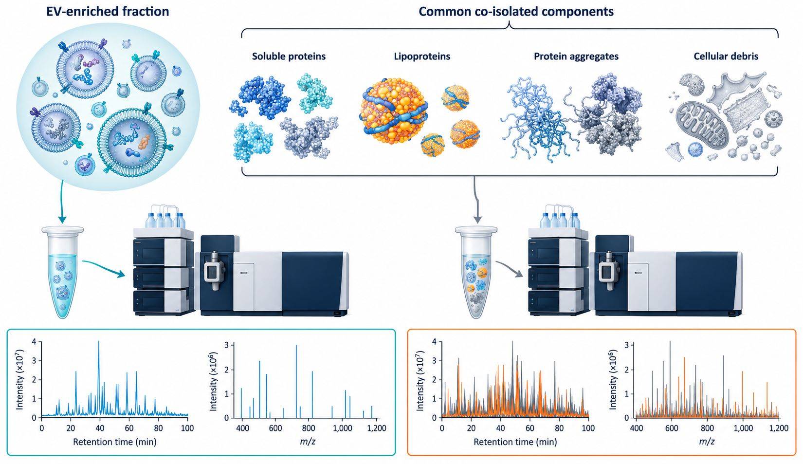
Exosome proteomics interpretation should account for vesicles, soluble proteins, lipoproteins, protein aggregates, and preparation-related carryover.
| Preparation Approach | Useful When | Proteomics Considerations |
|---|---|---|
| Size-exclusion chromatography | Reducing soluble protein background while maintaining intact vesicles | Fraction selection and concentration must be standardized; lipoproteins and other particles may remain |
| Differential or ultracentrifugation-based enrichment | Processing conditioned medium or larger source volumes with an established protocol | Recovery, aggregation, operator variability, and co-pelleting of nonvesicular material should be monitored |
| Density-gradient separation | Increasing separation by buoyant density when purity is prioritized | Additional handling can reduce recovery; gradient material must be removed before protein preparation |
| Immunoaffinity capture | Studying a marker-defined vesicle subset | Capture biology and antibody specificity bias the recovered population; bead and isotype controls may be required |
| Polymer precipitation | Rapid enrichment when yield is prioritized | Co-precipitated proteins and polymers can interfere with digestion and LC-MS/MS unless additional cleanup is performed |
| Submitted pre-isolated preparation | The client has an established isolation workflow | Preparation history, buffer, storage, characterization, and contamination risk are reviewed before analysis |
Particle size or concentration alone does not establish vesicle identity or purity. Depending on the project and submitted material, the proteomics plan can incorporate complementary characterization services before sample processing.
| Supporting Service | Role Before or Alongside Proteomics | Typical Evidence |
|---|---|---|
| NTA-based exosome identification | Assess particle concentration and size distribution and compare recovery across preparations | Particle concentration, size-distribution metrics, and optional fluorescence-based measurements |
| TEM-based exosome identification | Document vesicle morphology and evaluate visible nonvesicular material | Electron-microscopy images and morphology observations |
| Western blot exosome verification | Evaluate selected positive and negative protein markers in the preparation | Marker-specific band evidence with project-appropriate controls |
| Exosome marker analysis | Assess targeted marker panels when a broader marker context is required | Marker-focused results for vesicle characterization or candidate follow-up |
| Single-particle ExoView profiling | Resolve marker-defined subpopulations and co-expression patterns beyond bulk measurements | Captured-particle counts, marker-positive fractions, and colocalization results |
For quantitative projects, characterization is not only a publication requirement. It helps determine whether an apparent protein difference is consistent with biology or could reflect variation in vesicle recovery, plasma-protein contamination, lipoprotein abundance, cell death, culture-medium composition, or sample handling.
The amounts below are planning targets for standard protein identification, label-free quantification, or DIA-based analysis. They are not universal acceptance limits. Final requirements are confirmed after review of the matrix, preparation status, expected protein yield, study design, and requested quality controls.
| Starting Material | Typical Planning Input | Limited-Input Review | Submission Notes |
|---|---|---|---|
| Plasma or serum | 1 mL per sample | 0.5 to <1 mL | Submit 2–3 mL when vesicle characterization, repeat preparation, or reserve material is also required |
| Cell-culture conditioned medium | 50 mL per sample | 10 to <50 mL | Serum-free or EV-depleted medium is preferred; low-secretion cultures may require 100–200 mL |
| Urine | 50 mL per sample | 10 to <50 mL | Use consistent collection, clarification, storage, and normalization procedures across the study |
| Cerebrospinal fluid | 2 mL per sample when available | Less than 2 mL | Limited volumes are reviewed according to the analytical endpoint, preparation losses, and available reserve |
| Pre-isolated exosomes or EVs | 50 µg total protein | 10 to <50 µg | Provide protein concentration, isolation method, buffer, and particle or characterization data when available |
| Exosome or EV protein lysate | 20 µg total protein | 5 to <20 µg | Approximately 50 µg is preferred when repeat preparation or additional quality control is planned |
| Prepared peptides | 5–10 µg | Reviewed individually | Provide digestion, cleanup, concentration, and solvent information |
Particle count is useful supporting information but is not used alone to accept or reject a preparation. Particle measurements can include nonvesicular material, and protein recovery varies with the source matrix and isolation method. Projects that include NTA, TEM, marker analysis, technical repeats, TMT labeling, extensive fractionation, or modification-specific enrichment may require additional material. Phosphoproteomics, glycoproteomics, and other PTM workflows are reviewed separately before shipment.
| Starting Material | Information We Review | Frequent Risk Factors |
|---|---|---|
| Cell-culture conditioned medium | Cell type, culture conditions, confluence, collection interval, medium composition, serum or EV-depletion strategy, viability, clarification steps, and storage | Serum-derived vesicles and proteins, cell-death products, inconsistent conditioning time, low volume, and repeated freeze-thaw cycles |
| Plasma or serum | Species, collection tube or anticoagulant, processing interval, centrifugation history, hemolysis or lipemia observations, storage, and freeze-thaw history | Abundant plasma proteins, lipoproteins, platelets, hemolysis, clotting-related differences, and inconsistent preprocessing |
| Urine | Collection format, timing, normalization plan, clarification, pH handling, protease-control strategy, storage, and freeze-thaw history | Variable concentration, precipitation, cellular debris, bacterial contamination, and collection-time effects |
| Cerebrospinal fluid or another limited biofluid | Collection and processing history, available volume, storage, study groups, and required analytical endpoint | Low vesicle and protein input, adsorption losses, limited repeat analysis, and contamination from blood |
| Pre-isolated vesicles | Source matrix, isolation method, fraction or pellet description, buffer, storage, particle data, marker results, morphology data, and available material | Incompatible detergents or polymers, insufficient cleanup, unknown recovery, high background, and incomplete characterization |
| Protein lysate or prepared peptides | Lysis or digestion method, detergent and salt composition, reduction and alkylation conditions, cleanup, concentration, and storage | MS-incompatible buffer components, keratin, overprocessing, peptide loss, and inability to perform particle-level characterization |
Store samples in single-use aliquots at −80 °C, ship on dry ice, and avoid repeated freeze-thaw cycles. If the available amount falls within or below a limited-input review range, send the sample details before shipment so that analytical depth, repeat capacity, and appropriate controls can be evaluated.
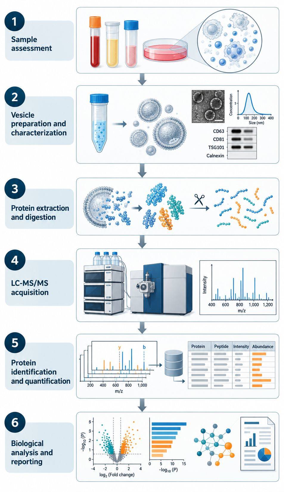
Integrated workflow for exosome preparation, characterization, protein processing, LC-MS/MS, data analysis, and reporting.
We review the biological question, sample source, preparation status, study groups, replicate structure, available material, target proteins or pathways, and the intended downstream use of the results.
Source material can undergo project-appropriate vesicle isolation, enrichment, purification, concentration, and selected characterization. Submitted preparations are reviewed for compatibility with protein extraction and LC-MS/MS.
Proteins are recovered using a workflow appropriate for membrane-rich, low-input material. Reduction, alkylation, enzymatic digestion, cleanup, and optional fractionation or modification enrichment are configured around the selected analysis.
Prepared peptides are analyzed using the selected DDA, label-free, DIA, TMT, PTM-focused, or targeted acquisition route. Samples are ordered and balanced to reduce avoidable batch confounding.
Data processing covers peptide-spectrum or precursor-level assessment, protein inference, quantitative normalization, replicate consistency, missingness, batch effects, and comparison-specific quality checks. Search settings and protein databases are matched to the species and experimental design.
We organize the results into protein and peptide tables, quantitative matrices, QC summaries, differential comparisons, annotations, functional analyses, and candidate lists that match the project scope. Follow-up recommendations are limited to technically appropriate next steps supported by the data.
The report distinguishes measured proteins from biological interpretation because an EV-enriched preparation does not establish exclusive vesicle origin for every identification.
A protein list should not be interpreted as a list of proteins uniquely located inside exosomes. Candidate selection considers the sample matrix, isolation route, known abundant contaminants, marker context, peptide-level support, and consistency across biological replicates. When appropriate, selected discoveries can move into targeted PRM or SRM/MRM, western blotting, immunoassays, or functional experiments.
| Research Area | How Exosome Proteomics Is Applied | Typical Results |
|---|---|---|
| Biomarker research | Compare vesicle-enriched protein profiles across defined research cohorts or experimental groups | Differential proteins and prioritized candidates for independent verification |
| Exosome function research | Identify protein cargo associated with donor-cell state, stimulation, stress, or environmental change | Protein modules and pathways that support mechanistic hypotheses |
| Drug-response and mechanism studies | Measure treatment-associated changes in extracellular-vesicle protein cargo | Regulated proteins, pathway changes, and candidates for matched cellular or functional assays |
| Engineered exosome research | Compare cargo or surface-protein profiles across constructs, loading conditions, production batches, or labeling and tracking strategies | Relative cargo profiles and candidates for orthogonal characterization |
| Neurological and other limited-biofluid studies | Profile proteins from cerebrospinal fluid or other low-input matrices after feasibility assessment | Detectable protein cargo and candidate differences with matrix-specific limitations documented |
| Multi-omics studies | Combine protein results with exosome metabolomics, exosome lipidomics, or other molecular measurements | Cross-omic pathway hypotheses and coordinated candidate prioritization |
When protein, metabolite, and lipid changes need to be evaluated together, the proteomics workflow can be coordinated with exosome metabolomics and lipidomics services. Experimental design, sample allocation, and normalization should be planned before sample processing so that the datasets remain comparable.
Projects can begin with source material, pre-isolated vesicles, protein lysates, or peptides. Preparation, characterization, protein processing, LC-MS/MS, data analysis, and reporting are planned as one connected workflow.
We consider the isolation method, sample matrix, marker information, likely co-isolated components, and replicate behavior when interpreting the protein profile. This reduces the risk of treating plasma proteins, serum-derived material, lipoproteins, or preparation artifacts as exosome-specific biology.
DDA, label-free, DIA, TMT, PTM-focused, and targeted approaches answer different questions. We select the analytical route according to the comparison structure, sample availability, expected targets, and required output.
Discovery results can be prioritized using peptide evidence, quantitative consistency, annotation, and biological context. Suitable candidates can then move into targeted mass spectrometry or another orthogonal method without presenting the discovery list as validated biomarkers.
Share your sample source, isolation status, comparison groups, available characterization data, and whether you need protein discovery, quantitative comparison, PTM analysis, or targeted verification. Our scientists will recommend a workflow suited to the material and research objective.
Can I submit pre-isolated exosomes or extracellular vesicles?
Yes. Please provide the source matrix, isolation or enrichment method, buffer composition, storage history, available amount, and any NTA, TEM, western blot, ExoView, or other characterization results. We review the preparation for LC-MS/MS compatibility and determine whether additional cleanup or characterization is advisable.
Can the project start with plasma, serum, urine, cerebrospinal fluid, or conditioned medium?
Yes. A project can start with source material when vesicle isolation or enrichment is needed before proteomics. The preparation route is selected according to the matrix, study objective, available material, required purity, and downstream analysis. All comparison samples should be collected and processed consistently.
How do I choose between DDA, DIA, TMT, and label-free quantification?
DDA is useful for discovery and library generation. DIA often suits studies that prioritize quantitative consistency across a sample series. TMT supports multiplexed comparisons when channel allocation and batch bridging are planned carefully. Label-free analysis provides flexible sample numbers without chemical labeling. We recommend the route after reviewing the group design, sample amount, batch structure, and expected output.
How many proteins will be identified?
Protein coverage depends on the source matrix, vesicle recovery and purity, available protein, extraction and digestion efficiency, chromatographic performance, acquisition method, database, and protein abundance distribution. We do not use a fixed protein count as a universal expectation. A low-abundance or highly hydrophobic protein may remain undetected even when it is biologically present.
Do I need NTA, TEM, and marker analysis before proteomics?
The appropriate characterization package depends on the material and the claims the study needs to support. Particle analysis, morphology, positive and negative markers, and subpopulation profiling provide complementary information. No single method proves vesicle identity or purity. Existing characterization can be reviewed, or selected assays can be added to the project.
How can plasma proteins and lipoproteins affect the results?
Plasma and serum contain highly abundant soluble proteins and lipoprotein particles that can co-isolate with extracellular vesicles. These components can dominate peptide signal, suppress lower-abundance vesicle proteins, and create apparent group differences if preparation quality varies. Consistent preprocessing, an appropriate isolation strategy, negative controls, and purity-aware data review are therefore important.
Can exosome surface proteins be analyzed?
Surface-focused projects are possible, but the workflow must distinguish general membrane association from experimentally supported surface accessibility. Intact-vesicle labeling or capture strategies, topology annotation, and orthogonal antibody-based confirmation may be required. Standard whole-vesicle proteomics alone does not prove that a detected protein is exposed on the outer surface.
Can discovery candidates be verified in the same program?
Suitable candidates can be transferred to PRM or SRM/MRM after proteotypic peptides, expected abundance, interferences, and sample availability are reviewed. Western blotting, immunoassays, or functional experiments may be more appropriate for some proteins. The verification method is selected according to the biological question and the evidence required.
For Research Use Only. Not for use in diagnostic procedures.

Hi there - let me know if you have any questions.
×