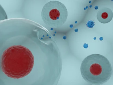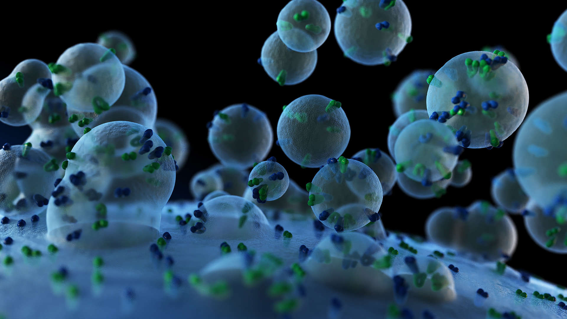
Secretomics is the large-scale study of proteins actively secreted by cells into the extracellular environment. These include cytokines, growth factors, enzymes, and other signaling molecules that regulate immune response, cell communication, and tissue remodeling.
Unlike intracellular proteins, secreted proteins are often present at low abundance and highly sensitive to cellular states or environmental changes. Their profiles vary across cell types, time points, and experimental conditions—making them valuable indicators of dynamic biological processes.
Due to dilution in media and interference from background proteins, secretome analysis requires sensitive instrumentation and optimized enrichment. Creative Proteomics offers a specialized platform to capture and quantify secreted proteins with precision, delivering data you can trust for mechanism studies, biomarker discovery, and functional pathway analysis.

Quantitative identification of soluble secreted proteins from cell culture media
Supports label-free and TMT-based multiplexing
Compatible with conditioned media, explants, organoids

Removal of extracellular vesicles (EVs) for soluble-only protein profiling
Enrichment of true soluble secreted factors without exosomal content

Dual workflow to compare exosome-bound and soluble fractions
Ideal for studying EV-mediated and classical secretion pathways

Predicts secretion route: SignalP (classical), SecretomeP (non-classical), GO term mapping
Tiered secretion confidence scoring included in report

Statistical comparison across treatment groups, time points, or conditions
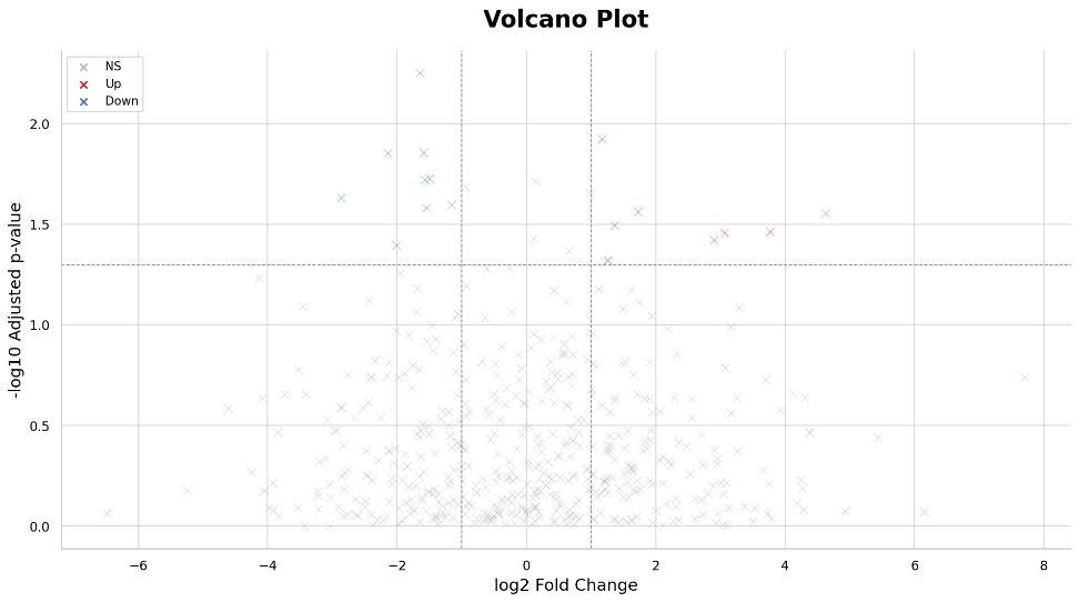
Volcano plots, hierarchical clustering, and enrichment analysis provided

Integration with transcriptomics, phosphoproteomics, cytokine panels, or other datasets
Supports co-analysis and pathway alignment

Secreted proteins are often present at very low concentrations.
Our solution:
Serum-free culture, protein enrichment, and ultra-sensitive LC-MS/MS enable reliable detection.

Culture media and lysed cell debris may interfere with secretome purity.
Our solution:
Sample cleanup, filtration, and secretion-specific annotations (e.g., SignalP, SecretomeP) reduce false positives.

Secretome composition changes across cell states, time points, and treatments.
Our solution:
We support replicates, time-course designs, and statistical comparisons to ensure confident biological interpretation.

Identification alone does not explain function or relevance.
Our solution:
Integrated pathway enrichment (GO, KEGG, Reactome) reveals functional insights behind protein shifts.

Secretome data often lacks context without integration.
Our solution:
We support transcriptomics, exosome proteomics, and phosphoproteomics co-analysis for multi-layer insight.
Our streamlined secretomics workflow ensures sample integrity, reproducibility, and biological relevance at every step.
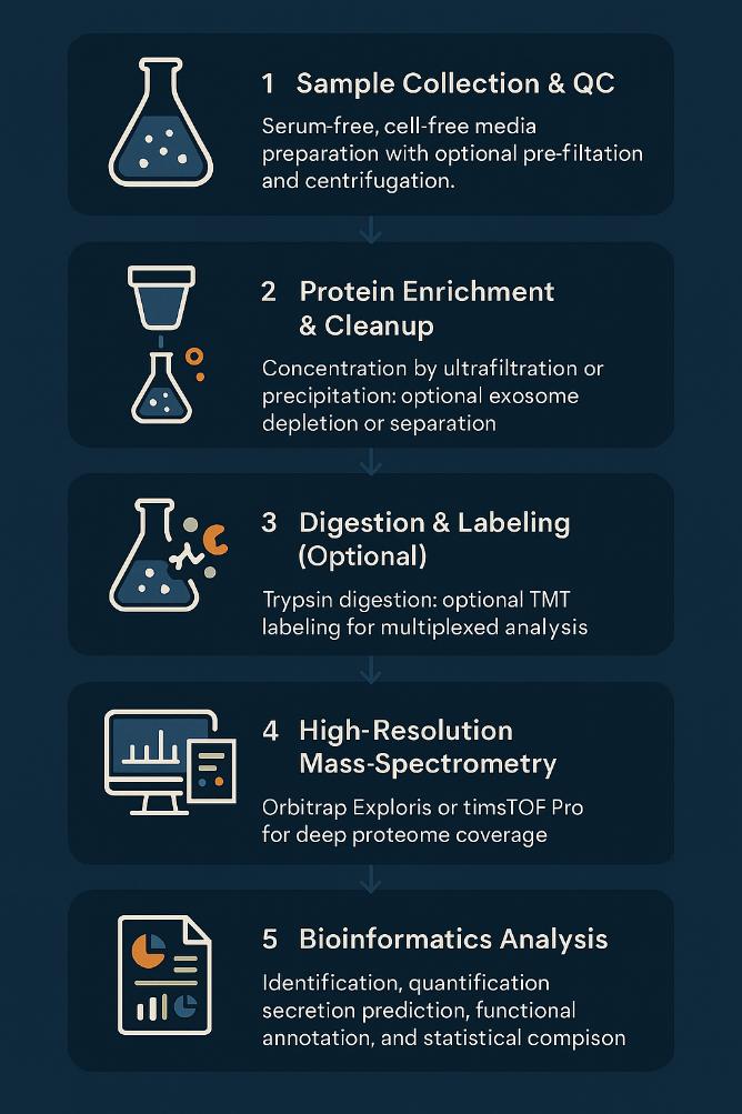
Sample Collection & QC
Serum-free, cell-free media preparation with optional pre-filtration and centrifugation.
Protein Enrichment & Cleanup
Concentration by ultrafiltration or precipitation; optional exosome depletion or separation.
Digestion & Labeling (Optional)
Trypsin digestion; optional TMT labeling for multiplexed analysis.
High-Resolution Mass Spectrometry
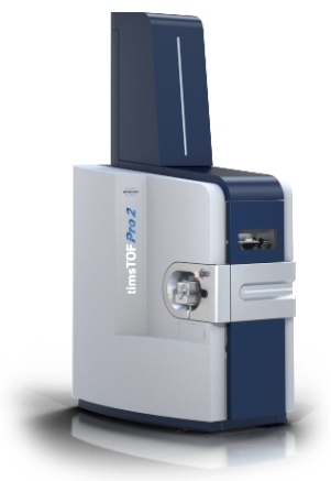
Orbitrap Exploris or timsTOF Pro for deep proteome coverage.
Bioinformatics Analysis
Identification, quantification, secretion prediction, functional annotation, and statistical comparison.
Results Delivery
Annotated data tables, visual analytics, QC metrics, and pathway insights.
Supports both label-free and TMT-labeled experiments.
Optional parallel workflows available for soluble and exosomal fractions.
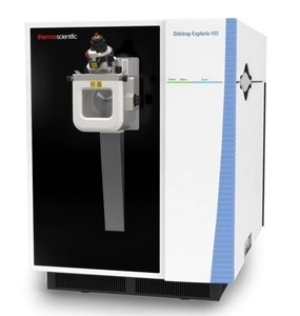
Orbitrap Exploris™ 480
Bruker timsTOF Pro 2
nanoElute® UHPLC (Bruker)

Ultra-Sensitive Detection
Detects secreted proteins at picogram-level concentrations, enabling confident quantification of low-abundance cytokines and regulatory peptides.

Deep Protein Coverage
Typical samples yield >1500 proteins identified, with up to 50% classified as secreted based on SignalP, SecretomeP, and GO enrichment.

High Quantitative Accuracy
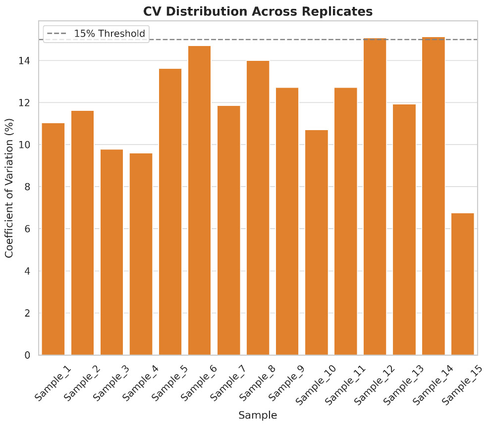
Achieves <15% CV in label-free quantification and R² ≥ 0.99 for TMT-based workflows, validated across internal standard replicates.

Comprehensive Secretion Annotation
Dual-layer secretion prediction with SignalP & SecretomeP, plus GO mapping of extracellular proteins. Includes confidence scoring for secretion pathways.

Biological Pathway Resolution
Each project includes enrichment of 50–200+ biological processes, covering immune modulation, signal transduction, and extracellular matrix remodeling.

Scalable Multi-Group Design
Supports 2–6 biological conditions (standard) or TMT 16-plex for expanded comparisons across treatments, time points, or co-cultures.

Reduced Background Interference
Optimized workflows—including serum-free media and EV depletion—cut contamination from non-secreted proteins by >70%, improving signal clarity.
Every secretome profiling project at Creative Proteomics incorporates rigorous, built-in quality control measures to ensure consistent performance, low variation, and high data confidence. These controls are embedded throughout the LC-MS/MS workflow, from sample injection to final bioinformatics reporting.
Our Quality Assurance Measures Include:
To distinguish true secreted proteins from background or lysed-content artifacts, Creative Proteomics applies a multi-layered annotation strategy that integrates computational predictions and curated biological ontologies.
Our Secretome Annotation Pipeline Includes:
The final output includes secretion-classified protein lists with pathway tags and prediction scores, ready for direct functional interpretation or follow-up screening.
Raw Data Files (.raw, .d, .mzML)
Full-resolution MS spectra from Orbitrap or timsTOF platforms.
Protein ID & Quantification Tables (Excel)
Normalized protein intensities with group-based comparisons.
Secretion Annotations
SignalP, SecretomeP, and GO-based classification with confidence scoring.
Statistical Visualization
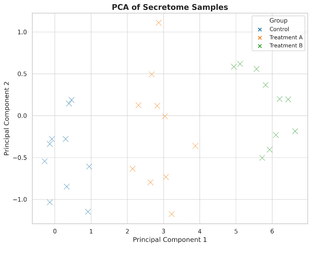
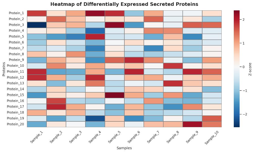
Volcano plots, heatmaps, and PCA to highlight group differences and sample quality.
Functional Enrichment Maps
KEGG, Reactome, and STRING pathway mappings with clustering options.
Quality Control Reports
Metrics on ID rate, RT shift, signal consistency, and CV distribution.
Optional Bioinformatics Add-ons
Multi-omics integration, custom filtering, or cross-species pathway alignment.
| Category | Instruction |
|---|---|
| Accepted Sample Types | Cell culture supernatants (serum-free), organoid-conditioned media, plasma (EDTA/heparin), cerebrospinal fluid (CSF) |
| Recommended Volume | ≥ 1 mL per condition (for cell supernatants); ≥ 200 µL (for biofluids) |
| Culture Conditions | Use serum-free medium or dialyzed FBS (if essential); collect after 24–48 h conditioning |
| Pre-processing | Centrifuge at 2,000 × g for 10 min; filter through 0.22 μm if needed |
| Protein Input Target | ≥ 10 µg total protein per sample; BCA or Bradford quantification recommended |
| Storage | Snap-freeze immediately after collection; store at -80 °C |
| Shipping | Ship on dry ice with clearly labeled tubes and completed submission form |
What is the minimum number of biological replicates recommended for secretome analysis?
To ensure statistical robustness, we typically recommend at least three biological replicates per condition. This allows for reliable detection of differentially secreted proteins and supports downstream statistical modeling (e.g., PCA, volcano plots).
How do you control for background noise from serum or additives in the culture medium?
We use serum-free media or dialyzed FBS during sample preparation and apply advanced bioinformatics filtering to exclude contaminating proteins from serum, plasticware, or cell lysis. Additionally, background reference databases (e.g., CRAPome) can be incorporated.
Can this service differentiate between classical and non-classical secretion pathways?
Yes. We incorporate tools like SignalP (for classical secretion) and SecretomeP (for non-classical secretion), along with GO and KEGG annotations, to classify secretion modes and help interpret vesicle-based or leaderless export mechanisms.
Is it possible to compare multiple experimental groups or time points in one analysis?
Absolutely. Our analysis pipeline supports multi-group comparisons and longitudinal profiling, allowing clients to track dynamic changes in secreted protein expression across treatments, cell types, or temporal stages.
How do you ensure the secretome data are not confounded by intracellular proteins released from cell death?
We advise clients to monitor cell viability before collection and apply preprocessing steps such as low-speed centrifugation and 0.22 μm filtration. In silico QC metrics are also used to flag potential contamination from apoptotic or lysed cells.
Can I integrate my secretome results with proteome, transcriptome, or metabolome data?
Yes. We offer optional multi-omics integration to align secretome data with proteomics, RNA-seq, or metabolomics datasets, enabling network-based interpretation of secretion events within a broader biological context.
What types of enrichment or fractionation methods are used before MS analysis?
We typically apply protein concentration steps (e.g., ultrafiltration, TCA precipitation) and optionally glycoprotein or exosome enrichment depending on the project scope. These methods help increase signal specificity and boost detection of low-abundance secreted factors.
How are post-translational modifications (PTMs) in secreted proteins handled?
Upon request, our workflow can include PTM-aware database searching (e.g., N-glycosylation, phosphorylation), and we can annotate modification sites within the secreted protein pool to support biomarker discovery or mechanistic studies.

Hi there - let me know if you have any questions.
×