

Building Reviewer-Ready EV Studies with MISEV2023
In this article, "MISEV2023 reporting requirements for extracellular vesicles" refers to the minimum practical information needed for reviewers to assess EV identity, purity, quantitation limits, and functional claims without relying on assumptions.
In extracellular vesicle (EV) research, strong experimental design alone may not be enough to move a manuscript through editorial triage. Increasingly, reviewers also expect studies to be reported in a way that allows reproducibility, interpretability, and technical limitations to be evaluated without guesswork.
That's why the International Society for Extracellular Vesicles (ISEV) updated its consensus reporting guidance with MISEV2023. The primary guideline—Minimal information for studies of extracellular vesicles (MISEV2023): from basic to advanced approaches—builds on MISEV2018 but expands expectations in the places peer review most often breaks: pre-analytics, quantitative reporting limits, and functional claims (see the full text on PMC: Minimal information for studies of extracellular vesicles (MISEV2023)).
Three conceptual shifts drive most "major revision" requests under MISEV2023:
- Pre-analytical constraints must be documented, not implied. Reviewers increasingly expect you to state what your workflow can and cannot control at the sample stage, and what the practical limits are for interpreting changes.
- Quantitative claims must acknowledge method limits (LOD/LOQ). If you present particle counts, fluorescence signals, or MS-based quantitation, you must show what's above noise and what isn't.
- Nomenclature and functional claims are held to a higher bar. MISEV2023 pushes researchers toward operational terms (based on physical properties and method context) and requires tighter controls for uptake/release assays so artifacts don't masquerade as biology.
Used correctly, a compliance checklist is not extra paperwork. It helps prevent common causes of editorial rejection and major revision by making the study's foundational measurements transparent and auditable.
Key Takeaway: MISEV2023 compliance is a peer-review strategy as much as a reporting standard—your goal is to make every key claim auditable from the Methods and QC.
The Pre-Analytical Checklist: Source Metadata and Limitations
Many EV papers fail before isolation is even discussed, because the manuscript doesn't let a reviewer interpret whether the starting material was comparable across conditions. MISEV2023 elevates pre-analytical variables from "nice to have" to a central part of experimental meaning.
Donor and Sample Metadata: what to report (and why reviewers care)
Pre-analytics is where hidden variability creeps in: hemolysis, platelet activation, temperature drift, inconsistent processing delays, and storage differences that change particle background. Even in cell culture, serum-derived particles and handling steps can dominate what you later call "EV cargo."
Use the checklist below as a reviewer-facing way to ensure you've declared the conditions that bound your conclusions.
| What reviewers expect you to report | Examples of reportable details (adapt to your system) | Why it matters under MISEV2023 |
|---|---|---|
| Collection method and materials | Collection tube type, needle gauge, anticoagulant (for plasma), urine preservatives (if used) | Source handling changes background particles and co-isolates before EV isolation begins. |
| Time-to-processing and pre-clearing | Delay from draw/collection to processing; centrifugation steps to remove cells/debris; filtration pore sizes | Reduces ambiguity about cell/platelet carryover and later "contamination" claims. |
| Storage conditions | Temperature, storage duration, number of freeze–thaw cycles; flash freezing vs slow freezing | Freeze–thaw and storage can change EV integrity and co-isolate profiles. |
| Input quantity | Biofluid volume; tissue mass; cell number and culture volume | Needed for normalized yields and for comparing across studies. |
| Known limitations you cannot control | Clinical heterogeneity, variable diet/medications, sample transport constraints | Reviewers look for transparent boundaries, not "perfect" stories. |
A practical way to write this in a Methods section is to treat pre-analytics like instrument settings: concise, explicit, and reproducible.
EV-Depleted Inputs: Validating Background Particle Control
If you use serum (e.g., FBS) or other complex supplements in culture, MISEV2023 is clear about the risk: serum-derived particles can be carried through isolation and appear as "cell-derived EVs." That can compromise both characterization (e.g., particle counts) and downstream functional readouts.
MISEV2023 therefore expects you to validate and report the efficacy of your EV-depletion approach when you claim "EV-depleted serum." This EV-depleted serum validation language matters because reviewers will otherwise assume background contamination could explain your particle counts, proteomics hits, or phenotypes.For studies that include membrane or protein marker readouts, an LC–MS/MS-based workflow such as Mass Spectrometry Based Proteomics can help document residual background proteins after depletion and pre-clearance. At minimum, your manuscript should make it possible to answer:
- How was depletion performed (e.g., ultracentrifugation, filtration, SEC, commercial depletion)?
- How was depletion validated (same characterization method you use later, applied to the depleted input)?
- What negative controls were run to ensure background particles don't explain the phenotype?
If your manuscript includes culture-derived EV function or cargo claims, this step often determines whether reviewers view your work as interpretable.
Isolation and Characterization: Technique Limits and LOD/LOQ
If you're looking for an EV study methods reporting checklist you can actually apply during experiments (not just at manuscript submission), treat this section as the point where "how we isolated" and "what we can reliably quantify" become inseparable.
A paper that says "EVs were isolated by ultracentrifugation" is no longer sufficient. MISEV2023 pushes toward method-specific parameter reporting so other labs can reproduce the separation window.
If you need a standardized route from isolation to orthogonal characterization (so the Methods section stays consistent), Exosomes Identification is a relevant service page to reference.
Isolation reporting is not the method name—it's the method parameters
Below is a manuscript-ready checklist for two common workflows.
| Isolation approach | Parameters that should be explicitly reported | Common reviewer concern it resolves |
|---|---|---|
| Ultracentrifugation (UC) | Rotor type; rotor k-factor; g-force and duration; temperature; braking settings; wash steps; tube type | "100,000g" alone is not comparable across rotors and protocols; k-factor clarifies sedimentation performance. |
| Size-exclusion chromatography (SEC) | Resin type; column bed volume; fraction collection scheme; how EV-enriched fractions were defined; buffer composition | Without bed volume and fraction logic, readers can't interpret whether proteins/lipoproteins likely co-eluted. |
When your Methods section is this explicit, it becomes easier to defend two things reviewers care about most: (1) why you believe your preparation contains vesicles, and (2) what non-vesicular material is plausibly present.
This becomes especially important when downstream analyses include proteomics. In that setting, isolation reporting should be detailed enough for reviewers to determine whether an apparent marker shift may reflect isolation bias rather than biological change.
For teams managing multi-cohort or repeat EV studies, a defined isolation protocol supported by downstream characterization can improve reporting consistency across projects. Key parameters such as UC rotor k-factor, SEC bed volume, and fraction-selection criteria should be documented in a standardized format.
LOD/LOQ Reporting: Defining the Limits of Quantitative EV Claims
One of the most practical updates in MISEV2023 is the expectation to report limits of detection (LOD) and, when relevant, limits of quantitation (LOQ) for characterization techniques when you present quantitative metrics.
This matters because EV characterization often lives near measurement boundaries:
- NTA can register background particles and noise as "vesicles" if thresholds aren't justified.
- High-sensitivity flow methods can confuse swarm effects, antibody aggregates, or background fluorescence with EV events.
- Fluorescence-based uptake studies can be dominated by free dye, dye micelles, or label transfer rather than vesicle internalization.
A reviewer-friendly way to present this is to state, for each quantitative platform:
- What the working range is for your assay under your exact settings
- What background looks like in buffer-only and process controls
- What you treat as below-LOQ and therefore do not interpret quantitatively
MISEV2023's larger point is not that everyone must use the same instrument—it's that quantitative claims must be tethered to what the method can actually resolve.
Updated Protein Marker Requirements for EV Purity
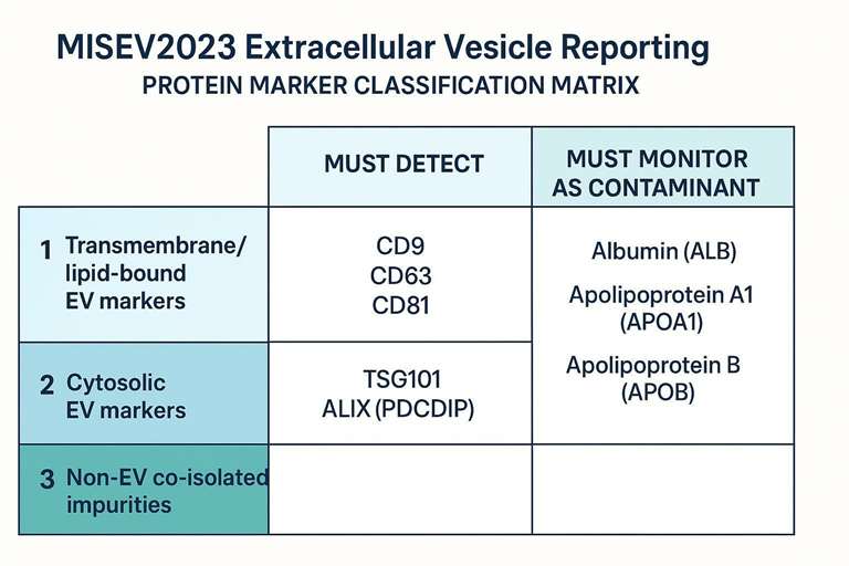
MISEV2023 keeps the concept that you should not "prove EVs" with a single marker. It formalizes marker reporting into categories that help authors document both EV-associated features and co-isolated non-EV components. This structure makes Category 1, 2, and 3 marker reporting clearer and less prone to overinterpretation.
A strong manuscript makes it easy for reviewers to see two truths at once:
- "Yes, this preparation contains lipid-bilayer particles consistent with EVs."
- "Here's what non-EV material is still present, and how we measured it."
Demonstrating EV Presence (Categories 1 & 2)
Category 1 markers support the presence of lipid-bilayer vesicles by detecting at least one transmembrane or lipid-bound EV-associated protein. The classic examples are tetraspanins (CD9, CD63, CD81), but your marker choice should fit your source and claim.
Category 2 markers provide evidence of internal EV-associated components, commonly cytosolic proteins enriched in EV preparations (e.g., TSG101, ALIX/PDCD6IP). Together with Category 1, they help reviewers see that you are not only detecting "stuff in the sample," but components consistent with intact vesicles.
A practical reporting tip: if you use Western blotting, specify antibody details and loading logic (what input amount, what normalization). If you use MS-based profiling, specify search parameters and FDR logic (and later, deposit raw data).
Assessing Co-Isolation and Impurities (Category 3)
MISEV2023 is unusually explicit about something many papers try to minimize: co-isolation is expected, and you must report it. Category 3 reporting provides a structured way to communicate the extent of co-isolated non-EV material.
In plasma-derived EV work, reviewers often want to see whether lipoproteins and abundant soluble proteins were monitored. Practical examples include:
- Apolipoproteins (e.g., APOA1, APOB) that track lipoprotein carryover
- Albumin as a high-abundance protein that can dominate low-input preparations
This is where high-resolution LC–MS/MS is particularly useful: rather than testing only a few proteins, it can map a broader impurity profile and show relative abundance patterns that help interpret functional or biomarker claims. In this context, LC–MS/MS can support more disciplined interpretation of low-abundance signals by helping distinguish true EV-associated proteins from background proteins and co-isolated contaminants.
For studies that need global marker coverage and impurity mapping aligned to MISEV categories, untargeted LC–MS/MS profiling can generate a defensible marker/contaminant profile and support transparent reporting. (If you also need orthogonal, non-MS characterization for the same isolate, pair proteomics with particle-level sizing and imaging so morphology and readouts can be reported alongside marker categories.)
Functional Assays: Validating Release and Uptake
MISEV2023 places much more weight on functional claims than many authors expect, especially when those claims involve EV uptake. In practice, functional sections trigger intense review because they are highly susceptible to artifacts.
Release Design and Functional Dose
Functional experiments often fail review not because the phenotype is uninteresting, but because the dose and normalization make it impossible to interpret whether the effect is plausible or comparable.
MISEV2023-aligned reporting for release models should make these points explicit:
- What release system was used (cell type, culture conditions, stimulation, and any stressors)
- How EV release was normalized, such as normalization to live cell count or another justified output measure
- How the "functional dose" was defined, and why that definition is appropriate for your experimental question
A reviewer is likely to ask: is the functional effect supported by a dose-response, or is it a single high-dose condition that could reflect non-physiological exposure?
The safest reporting pattern is:
- define dose in operational units (particle number, protein amount, or EV-to-cell ratio)
- state the rationale for that unit
- show at least a minimal dose-response or otherwise justify why one dose is interpretable
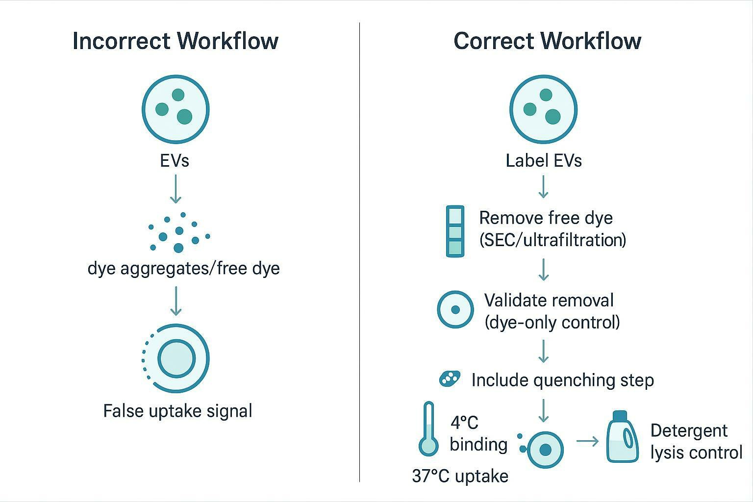
Uptake Labeling and Artifact Controls
Fluorescent labeling is one of the most scrutinized EV experiments in peer review because several common workflows can produce convincing-looking uptake "signals" that are not EV uptake.
MISEV2023 pushes authors toward controls that make the source of signal falsifiable:
- Free-dye removal validation: demonstrate that the label is not carried through as soluble dye or dye aggregates (this is the core "free dye removal" control reviewers look for).
- Dye-only controls: run the exact labeling and cleanup process without EVs; process the control identically.
- Temperature-shift assays: compare uptake at 4°C vs 37°C to help distinguish surface binding/passive association from energy-dependent internalization.
- Quenching controls: use an appropriate approach to distinguish surface-bound fluorescence from internal signal when the assay allows it.
Recent literature on EV dye artifacts and labeling strategies highlights the importance of these controls; for example, see Factors to Consider Before Choosing an EV Labeling Method for Fluorescence-Based Analysis (2024). You can reinforce this control logic by reporting uptake readouts alongside orthogonal characterization outputs (e.g., particle-level sizing and morphology), so reviewers see both particle-level evidence and functional controls in the same narrative.
Data Transparency and Omics Reporting
If your EV study includes LC–MS/MS, RNA-seq, or other omics, MISEV2023 compliance includes an unglamorous requirement that can make or break trust: data transparency.
Raw data deposition is part of the Methods
For proteomics, raw data deposition isn't an optional add-on—it's how reviewers and readers can audit identifications, filters, and potential batch effects.
A practical manuscript checklist for proteomics transparency:
| If you ran LC–MS/MS, report… | Why reviewers ask | Where to point readers |
|---|---|---|
| Dataset accession and repository | Enables reanalysis and method auditing | PRIDE submission guidance |
| Sample prep + LC–MS/MS acquisition details | Determines comparability across cohorts | Methods section (concise but complete) |
| Search/ID filters (e.g., FDR logic) | Prevents over-claiming IDs | Methods + deposited analysis files |
EV-TRACK: reduce reviewer ambiguity with EV-METRIC
MISEV2023 also encourages open, structured reporting. EV-TRACK is a community platform designed to improve transparency in EV research. Registering your study helps ensure that key methodological fields are captured consistently, and EV-TRACK provides an EV-METRIC score that reflects completeness of reporting.
For background and rationale, see Is your article EV-TRACKed? (2017) and the primary EV-TRACK paper on PubMed: EV-TRACK: transparent reporting and centralizing knowledge in extracellular vesicle research (2017).
Supporting Reviewer-Ready EV Characterization and Proteomics
If your team needs EV characterization workflows aligned with MISEV2023 expectations, Creative Proteomics can support studies through integrated particle-level characterization, morphology assessment, marker/contaminant profiling, and LC–MS/MS-based proteomics via Extracellular Vesicles Proteomics Services.
Frequently Asked Questions
What is the most significant change from MISEV2018 to MISEV2023?
MISEV2023 shifts from "check the box" reporting to context-dependent transparency, with stronger expectations for pre-analytical variables, method-specific parameter reporting, and acknowledging quantitative method limits like LOD/LOQ. It also raises the bar on functional claims by requiring EV uptake assay controls that make uptake/release conclusions reproducible and artifact-resistant.
Does MISEV2023 still require Electron Microscopy (EM) for all EV publications?
High-resolution imaging remains a cornerstone expectation when morphology is used to support EV claims, but the key is that imaging must be appropriate to the claim and reported transparently. If you present EM-based evidence, report acquisition conditions and sample preparation clearly so readers can judge whether structures are consistent with lipid-bilayer particles rather than preparation artifacts.
Can I still use the term "exosome" in my manuscript?
You can, but MISEV2023 strongly discourages using biogenesis-implied terms unless you have direct evidence supporting that biogenesis pathway. In most manuscripts, operational terms like "small EVs" (e.g., <200 nm) are safer and more defensible because they describe what was measured and isolated rather than what is assumed.
How do I report LOD/LOQ for EV characterization without overstating instrument performance?
Start by defining what you treat as background using buffer-only and process controls, then specify the validated working range of your method under your exact settings. Report results below LOQ as "detected but not quantifiable" (or equivalent) rather than turning noise into a concentration claim.
What controls are most important for EV uptake assays using fluorescent dyes?
Dye artifacts are a leading cause of disputed uptake claims, so the minimum defensible set is: dye-only controls processed identically, validated free-dye removal, and a temperature-shift comparison (4°C vs 37°C) to separate binding from internalization. Add quenching or detergent-sensitivity controls when your assay format allows you to distinguish surface-associated signal from true uptake.
Do I need to register my EV study in EV-TRACK to be considered MISEV2023 compliant?
Not always, but EV-TRACK registration is an efficient way to demonstrate reporting completeness and reduce reviewer uncertainty about missing metadata. If your manuscript is likely to face heavy methods scrutiny (clinical biofluids, multi-omics, or functional claims), an EV-METRIC-backed reporting record can strengthen transparency.
How can proteomics help fulfill MISEV2023 Category 1, 2, and 3 marker requirements?
Untargeted LC–MS/MS can assess multiple marker categories in one dataset, supporting EV-associated markers (Categories 1–2) while also quantifying common co-isolated impurities (Category 3). Compared with a few targeted blots, this broader coverage makes it easier to report both EV identity evidence and contamination context in a way reviewers can audit.
References
- Minimal information for studies of extracellular vesicles (MISEV2023): from basic to advanced approaches
- EV-TRACK: transparent reporting and centralizing knowledge in extracellular vesicle research
- Factors to consider before choosing EV labeling method for fluorescence-based analysis
* For Research Use Only. Not for use in diagnostic procedures.
