

Parkinson's disease (PD) is often described through its most recognizable cellular hallmark: Lewy pathology inside vulnerable dopaminergic neurons. For peripheral biomarker development, however, the key question is not only what occurs inside vulnerable neurons, but how PD-related pathology moves through neural networks and appears in measurable biofluid compartments. It's how pathology moves through connected neural networks, and what fragments of that process escape into biofluids in a measurable, interpretable form.
Exosomes and other extracellular vesicles (EVs) sit in the middle of that problem. They can carry misfolded proteins, enzymes tied to lysosomal failure, and inflammatory mediators. They also create a practical opportunity: if you enrich the right EV subpopulation and quantify the right proteoforms, it becomes possible to reduce the matrix-related ambiguity that limits many total-protein plasma biomarker approaches.
Key Takeaway: In PD, the most informative EV biomarkers are often not "more alpha-synuclein," but which alpha-synuclein proteoform (and which cell-type EV) you can quantify after rigorous enrichment and QC.
EV-Mediated Alpha-Synuclein Propagation in Parkinson's Disease
Parkinson's disease is driven by progressive accumulation of misfolded alpha-synuclein (α-syn) into aggregates that ultimately form Lewy bodies and Lewy neurites in susceptible neuronal populations. The problem is not just aggregation. It's distribution. Pathology appears to move across anatomically connected regions over time, tracking with symptom evolution.
The prion-like spreading hypothesis
The "prion-like" spreading hypothesis proposes that misfolded α-syn can seed conformational change of native α-syn in recipient cells, amplifying pathology as it spreads. This framework is often discussed alongside Braak staging, where PD-related pathology is described as advancing from lower brainstem regions toward cortical areas as disease progresses.
In practice, this model has two implications for translational researchers:
- You're not tracking a single static lesion; you're tracking a dynamic propagation process.
- A biomarker that reflects "propagation-ready" species (or the cellular stress states that promote their release) is more likely to align with disease trajectory than a bulk abundance measure.
Neuron-derived extracellular vesicles as transport vectors
Neuron-derived extracellular vesicles (NDEVs), including exosomes, are plausible transport vectors for α-syn because they are produced through regulated intracellular trafficking pathways and can be taken up by recipient cells. EVs are not passive debris. They can be actively loaded with cargo and secreted.
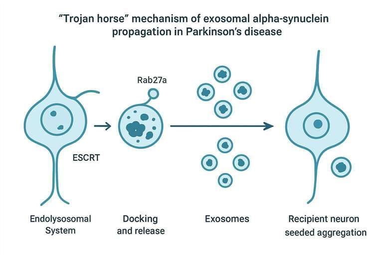
A mechanistic thread that repeatedly emerges in PD EV biology is the endolysosomal system. When proteostasis is stressed and lysosomal degradation is compromised, cells can increase EV-mediated export as a pressure-release route. In a PD context, that "relief valve" can become a propagation route. For example, Alvarez-Erviti and colleagues reported that lysosomal dysfunction increased exosome-mediated α-syn release and transmission in a cellular model (Brain, 2011; PMID: 21303699).
Mechanistically, EV biogenesis is tied to endosomal maturation into multivesicular bodies (MVBs), with cargo sorting influenced by endolysosomal and ESCRT-associated machinery. Rab GTPases, including Rab27a, regulate docking and release of vesicles at the plasma membrane. When neurons package aggregated α-syn into intraluminal vesicles and release them as exosomes, they may reduce intracellular burden while inadvertently delivering seeding-competent species to connected cells.
A practical note for assay design: mechanistic features such as ESCRT-associated sorting and Rab27a-regulated release are directly relevant to assay design. They explain why EV cargo changes under lysosomal stress, which is exactly when PD-relevant signals can become measurable.
If you need a service framework to experimentally interrogate EV-mediated transfer (for example, donor/recipient co-culture designs, uptake assays, and EV cargo readouts), Exosome Transfer Research is a relevant entry point for structuring those studies.
Overcoming the Analytical Bottleneck: The Proteoform and Matrix Challenge
Most PD biomarker programs encounter a key analytical challenge early: alpha-synuclein is not rare in blood. It is abundant in peripheral compartments, and the parts that are most relevant to PD are often present at low abundance, in specific modified or aggregated states.
That creates two coupled bottlenecks:
- Matrix confounding (especially red blood cell contamination and hemolysis)
- Proteoform ambiguity (total α-syn is not the same as pathogenic α-syn)
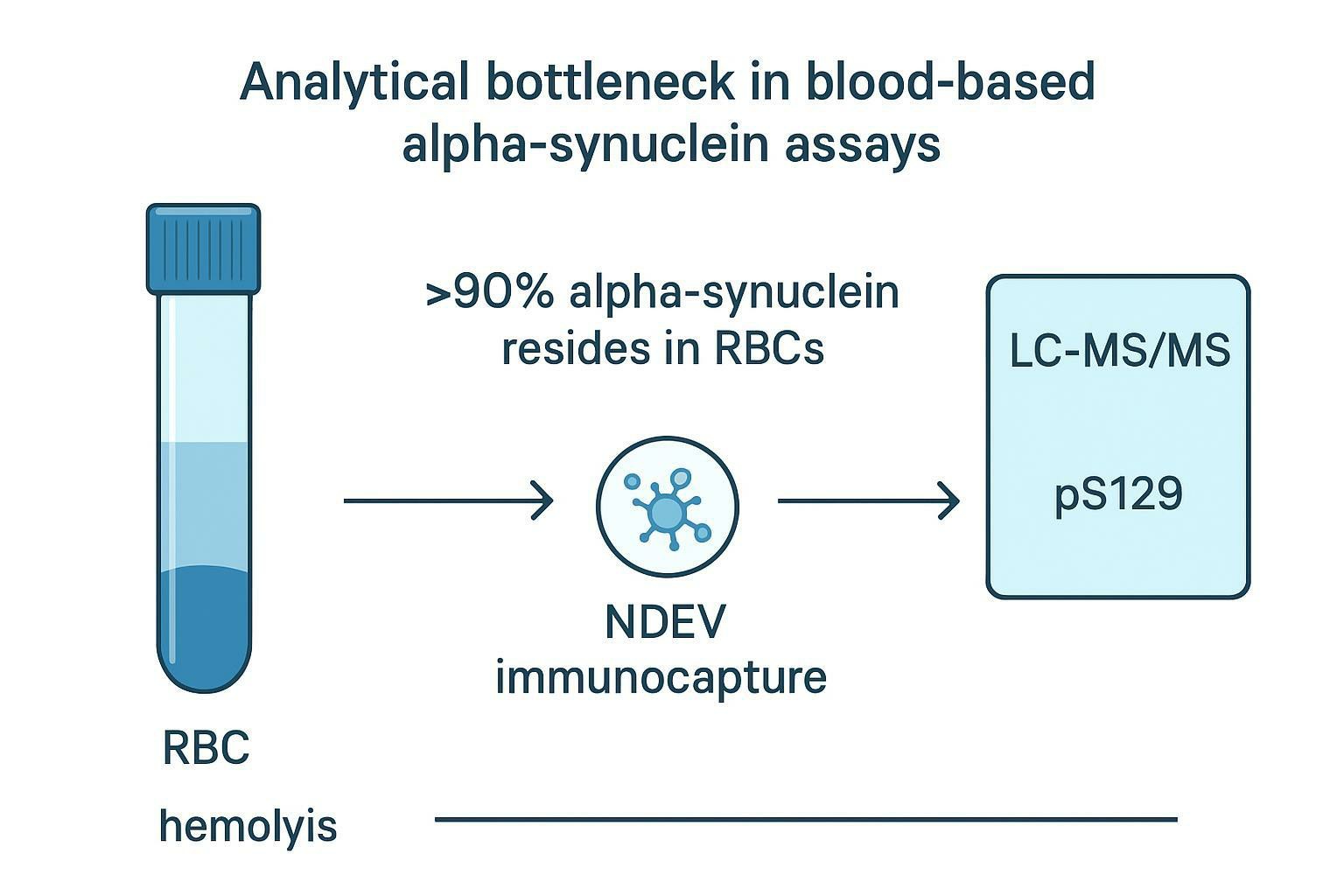
The Erythrocyte Matrix Problem
Red blood cells carry the vast majority of alpha-synuclein in whole blood. That means even minimal hemolysis can release large amounts of monomeric α-syn into plasma or serum and swamp the signal you hoped reflected CNS biology.
Mollenhauer and colleagues highlighted this confounding problem in the context of peripheral α-syn measurements and pre-analytical variability (Significance and confounders of peripheral DJ-1 and alpha-synuclein in Parkinson's disease (2010)). The translational point is straightforward: if hemolysis isn't controlled, you can "discover" biomarkers that are really just sample handling artifacts.
A workflow that aims to recover a CNS-relevant signal typically combines:
- Strict hemolysis QC (exclude visibly hemolyzed samples; quantify hemoglobin/hemolysis index where feasible)
- Standardized plasma processing (consistent anticoagulant choice, centrifugation, time-to-freeze, and freeze–thaw handling)
- Specific enrichment of the vesicle fraction of interest (for example, immunocapture toward NDEVs)
⚠️ Practical caution: In blood-based α-syn studies, "small" hemolysis differences can be bigger than the biological effect size you're trying to detect.
The Proteoform Problem: Moving Beyond Total Alpha-Synuclein
Quantifying total α-syn via generic ELISA is attractive because it's accessible. It's also frequently unreliable for translational interpretation. Total α-syn collapses multiple biologically distinct entities into one number:
- soluble monomer
- oligomeric species
- fibrillar/aggregated forms
- truncated variants
- post-translationally modified (PTM) forms
In PD biology, the pathogenic "culprits" are widely considered to be aggregated and misfolded species, often in specific PTM states. One reason different studies disagree is that different antibody pairs recognize different epitopes and conformations, and plasma is a difficult matrix.
Why pS129 matters
Phosphorylation at serine 129 (pS129) is one of the most widely used pathology-associated signatures of α-synucleinopathy. Importantly, pS129 is best treated as a disease-associated readout, not a guarantee of causality. For example, Walker and colleagues reported that S129 phosphorylation occurs after initial aggregation and is not required for Lewy body formation (2022; PMID: 35227419). That nuance matters for interpretation: pS129 can be a strong marker of pathological processing even if it is not the initial trigger.
Why LC–MS/MS improves proteoform-resolved measurement
Where immunoassays struggle, high-resolution LC–MS/MS can be decisive because it can explicitly quantify the peptide carrying the phosphorylation site. Instead of asking an antibody to discriminate between similar proteoforms, you design the assay to detect the exact modified peptide.
This matters most when you're trying to measure low-level CNS signals in a background-rich peripheral matrix. If your question is "is pathological α-syn processing increasing in this cohort?", the answer is more likely to come from proteoform-resolved MS than from total α-syn abundance.
For readers building EV proteomics workflows that include PTM-aware detection (including phosphorylation-focused assays), Extracellular Vesicles Proteomics Services is a relevant internal resource for EV proteomics and modification-focused workflows.
Beyond Alpha-Synuclein: The Multi-Pathway Biomarker Landscape
Alpha-synuclein is central to PD biology, but PD progression cannot be fully explained by a single protein. It is also a disorder of organelle failure and stress response, especially in endolysosomal and mitochondrial systems. EVs provide a multiplexed readout of these stress-response pathways.
A practical advantage of EV proteomics is that it can move beyond a single-marker mindset. Instead of asking "does α-syn go up?", you can profile pathway-linked signatures that are closer to mechanism and drug action.
Lysosomal Dysfunction: LRRK2 and GCase
Mutations in LRRK2 and GBA1 are major genetic drivers of PD risk and progression, and they converge on autophagy–lysosomal pathway (ALP) biology.
- LRRK2 is tied to vesicle trafficking and Rab GTPase phosphorylation, with broad downstream effects on endolysosomal dynamics.
- GBA1 encodes glucocerebrosidase (GCase), a lysosomal enzyme; reduced GCase activity is associated with impaired lysosomal function and α-syn accumulation.
A key translational angle is target engagement. If you're testing a therapy aimed at lysosomal function or LRRK2 signaling, you want a peripheral readout that reports on those pathways without requiring CNS tissue.
EVs can serve as that readout because they can carry:
- LRRK2 and pathway-relevant phospho-signatures
- lysosomal proteins and enzymes (including GCase-related signals)
However, interpretation requires discipline. EV-associated "LRRK2" might reflect changes in secretion biology, immune cell activity, or vesicle composition, not just neuronal signaling. That's exactly why multiplex design and normalization strategy matter.
Glial EVs, Complement Systems, and Atypical Synucleinopathies
Cell-type-specific EV isolation expands what's measurable. In PD, glial-derived EVs are especially important because neuroinflammation is not a side effect; it is a modulatory force that shapes progression and synaptic integrity.
Complement biology offers a concrete example. Complement proteins such as C1q and C3 are central to synaptic pruning and microglial phagocytic targeting. Schafer and Stevens reviewed complement's role in synaptic pruning and disease (2012; PMID: 22715882). More recently, D'Arrigo and colleagues reported a mechanism where microglial EVs mediate C1q deposition at presynapses and promote pruning (2025; PMID: 41397032).
For PD researchers, the practical implication is not "measure complement because it's interesting." It's this:
- If you see complement-related shifts in glial EV fractions, they can be interpretable pathway signals for neuroinflammatory synaptic remodeling.
- Those signals may also help stratify cohorts where inflammatory biology is a stronger driver of progression.
Differentiating PD from MSA using cell-type EVs
Multiple system atrophy (MSA) is an atypical synucleinopathy with oligodendroglial-predominant pathology. That means cell-type EV resolution can become a differentiator: neuronal vs oligodendroglial EV cargo may provide a molecular window into which cell type is "carrying" pathology.
A key reason this idea is plausible is that oligodendrocyte EV biology is perturbed in MSA. Dutta and colleagues reported reduced oligodendrocyte exosome secretion in MSA (Reduced oligodendrocyte exosome secretion in multiple system atrophy (2020)).
A translational strategy that follows from this literature is to:
- isolate neuron-enriched and oligodendroglia-enriched EV fractions
- quantify α-syn species and pathway proteins in each fraction
- evaluate ratios or differential patterns as cohort-level discriminators
This approach is still evolving, but it offers a mechanistic alternative to purely clinical differentiation in early or prodromal cohorts.
Preclinical Workflows: From iRBD Cohorts to Data Normalization
Mechanistic plausibility is not enough. Translational EV proteomics in PD lives or dies on workflow rigor: cohort design, pre-analytics, enrichment, targeted quantification, and normalization.
Prodromal Biomarker Discovery in iRBD
Idiopathic REM sleep behavior disorder (iRBD) is widely studied as a prodromal stage enriched for future synucleinopathy conversion risk. The biomarker logic is simple: if EVs carry pathology-linked α-syn species, you want to detect those shifts before motor symptoms appear.
Recent work supports the feasibility of neuron-derived EV α-syn as a prodromal signal. For example, a JAMA Neurology study evaluated neuronally derived EV α-synuclein as a serum biomarker for prodromal PD and dementia with Lewy bodies (JAMA Neurol, 2023; PMID: 38199054). A 2024 study analyzed α-syn species in plasma neuron-derived EVs in iRBD (2024; PMC11572749).
Two practical takeaways for iRBD cohort design:
- iRBD is ideal for longitudinal EV biomarker tracking because baseline between-subject variability is high.
- The closer your readout is to a specific proteoform (e.g., pS129) rather than total α-syn, the more interpretable the directionality can be.
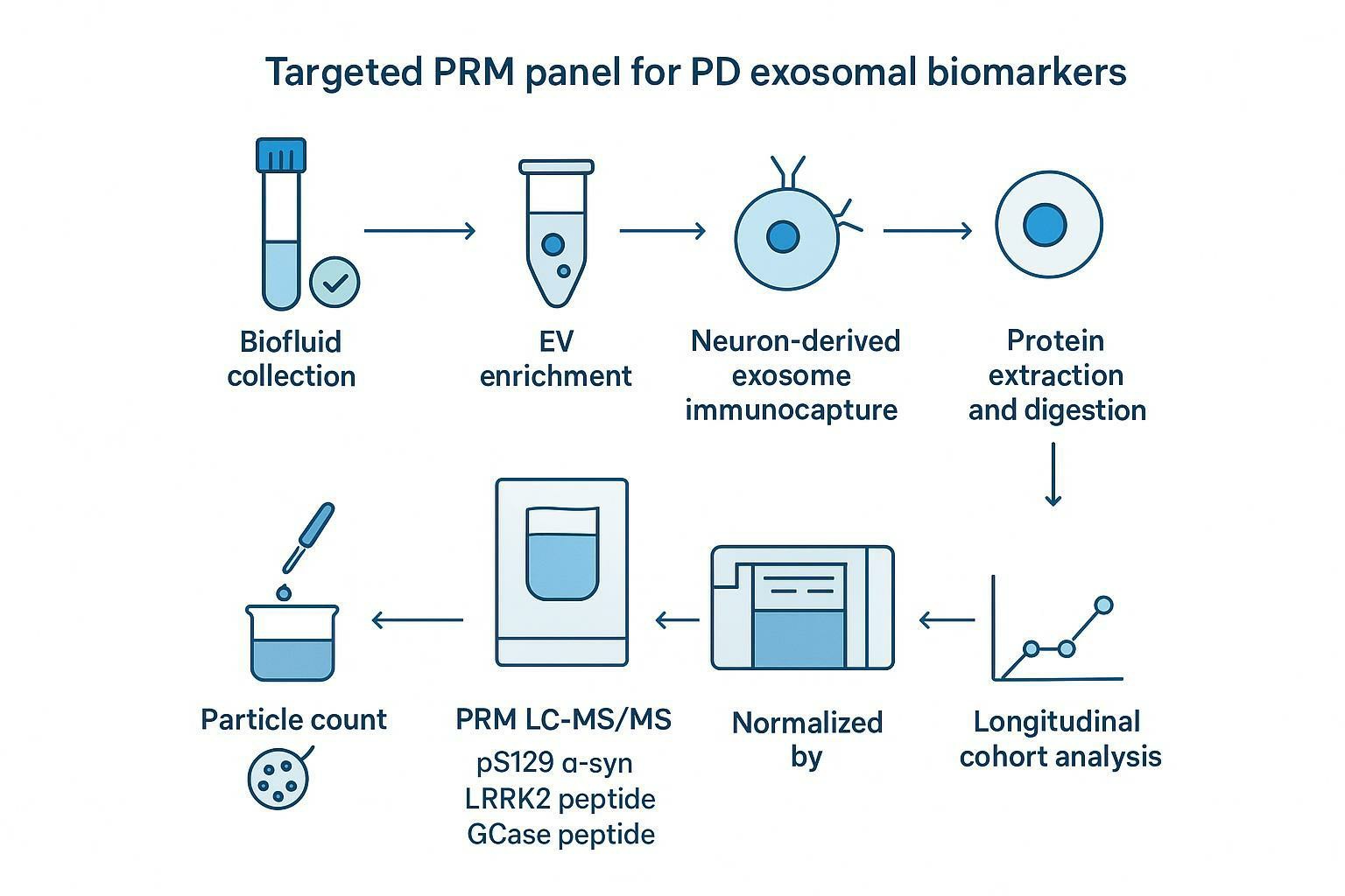
Targeted PRM Panels and Proteomic Normalization Strategies
Discovery proteomics can show you what's present. But if your goal is cohort-scale robustness, targeted panels are often the more translational choice.
Why PRM fits EV biomarker panels
Parallel Reaction Monitoring (PRM) is a targeted MS approach that can multiplex a small set of predefined peptides with high specificity and quantitative stability. In a PD EV context, a PRM panel can be designed to cover:
- pS129 α-syn peptide (pathology-associated proteoform)
- LRRK2 pathway peptide(s) (and/or phospho-Rab substrates when appropriate)
- GCase-linked peptide(s) (for lysosomal function context)
The key is that PRM lets you keep the biology narrow but the measurement deep: you can optimize transitions, quantify modified peptides, and run large cohorts with controlled acquisition.
A practical decision table: normalization strategies
Normalization is not an afterthought in EV proteomics. It is one of the main reasons two labs can run "the same" study and get different answers.
Below is a workflow-facing comparison of common normalization strategies used in EV proteomics datasets.
| Normalization approach | What it corrects for | Where it can fail | When it's most defensible |
|---|---|---|---|
| Normalize to starting biofluid volume | Collection-volume differences | Doesn't correct isolation efficiency or EV yield | Highly standardized collection sites and SOPs |
| Normalize to particle count (NTA/NFCM) | EV number proxies | Counts can include non-EV particles; purity affects counts | When EV purity/QC is strong and methods are consistent |
| Normalize to total protein | Bulk material in the prep | Contaminants inflate protein; purity differences dominate | When prep is highly clean and contamination is controlled |
| Normalize to EV marker(s) (e.g., TSG101) | EV-enrichment surrogates | Marker varies by EV subtype, cell type, or biology | When marker stability is verified in your cohort |
| Hybrid approach (2+ measures) | Multiple axes of variability | More complexity in analysis | Longitudinal cohorts and multi-site studies |
A disciplined EV program often treats normalization as an empirical choice: test which approach stabilizes technical variance without erasing plausible biology.
EV enrichment, characterization, and QC
If your workflow relies on NDEV immunocapture or cell-type EV fractionation, you also need to prove what you enriched and what you excluded. That's where characterization methods (particle sizing, morphology, canonical markers, and negative controls) become part of the "assay," not just a checkbox.
For method design and characterization support across isolation and multi-omics workflows, Extracellular Vesicles Analysis Services is directly relevant. For EV composition confirmation and component-level identification workflows, Exosomes Identification Service can be a complementary internal reference.
Robust measurement of PD-relevant EV proteoforms requires more than mass spectrometer sensitivity alone. It also takes enrichment discipline, reproducible sample handling, and normalization that holds up across sites and timepoints. If your team needs a targeted EV proteomics workflow designed around PD biology, Creative Proteomics can support custom EV enrichment and LC–MS/MS panel development to keep longitudinal datasets interpretable.
Frequently Asked Questions
Why is measuring exosomal pS129-alpha-synuclein better than measuring total alpha-synuclein?
Measuring total alpha-synuclein in plasma is easily confounded because most blood alpha-synuclein is cell-associated, especially in red blood cells, and small hemolysis differences can dominate the signal. pS129 is a pathology-associated modification enriched in Lewy pathology, so quantifying that specific proteoform after neuron-enriched EV isolation is more likely to reflect CNS-relevant protein processing than a bulk "total alpha-syn" number.
How do you normalize proteomics data when analyzing PD exosomes?
Use a normalization strategy that matches your study's biggest source of technical variance, then validate it on your dataset. Common approaches include normalization to starting biofluid volume, EV particle counts (NTA/NFCM), total protein in the EV prep, and EV markers such as TSG101. In large cohorts, a hybrid approach (for example, particle count plus an EV marker) is often more robust than relying on a single proxy.
How does red blood cell hemolysis affect PD exosome biomarker research?
Hemolysis releases large amounts of alpha-synuclein from red blood cells into plasma, inflating apparent alpha-syn levels and masking EV-derived signals. That can create false group differences or wash out real ones. Strict hemolysis QC and standardized processing are mandatory before EV enrichment and proteomics.
People also ask: What is a neuron-derived exosome, and how do you isolate it from blood?
A neuron-derived exosome is an extracellular vesicle subpopulation enriched using immunocapture against surface proteins associated with neuronal origin. In practice, researchers isolate total small EVs first (or work directly from plasma), then immunocapture the neuron-enriched fraction, and verify enrichment using EV markers plus neuron-associated markers. Because "neuronal" markers are not perfectly specific, include negative controls and report the full enrichment and QC workflow.
People also ask: Can pS129 alpha-synuclein be measured reliably with immunoassays?
It can be measured with immunoassays, but reliability depends on antibody specificity for the exact modified epitope and on matrix control. Cross-reactivity and different recognition of oligomeric vs monomeric species can distort results, especially in plasma. LC–MS/MS has an advantage because it can quantify the exact phosphorylated peptide, which reduces ambiguity when you need proteoform-level confidence.
People also ask: How can EVs help differentiate PD from MSA in early cohorts?
Cell-type-specific EV isolation can align the biomarker readout with the cell type most affected by each disease. PD pathology is neuron-centered, while MSA is oligodendroglial-centered. Comparing neuronal-enriched versus oligodendroglial-enriched EV signatures (including alpha-syn species and pathway proteins) can provide a mechanistic differentiation signal, especially when paired with rigorous enrichment validation and consistent normalization.
People also ask: What controls are needed to claim a CNS-relevant EV signal in plasma?
At minimum: document hemolysis and platelet contamination risk, use consistent pre-analytics, include EV characterization (size, morphology, canonical markers), validate enrichment specificity (positive and negative markers), and run technical replicates. For targeted MS, include stable isotope-labeled internal standards when possible and monitor batch effects across runs.
References
- Lysosomal dysfunction increases exosome-mediated alpha-synuclein release and transmission.
- Significance and confounders of peripheral DJ-1 and alpha-synuclein in Parkinson's disease.
- Exosomes in Parkinson disease.
- α-Synuclein phosphorylation at serine 129 occurs after initial protein aggregation and is not required for Lewy body formation.
- Neuronally Derived Extracellular Vesicle α-Synuclein as a Serum Biomarker for Prodromal Parkinson Disease and Dementia With Lewy Bodies.
- α-Synuclein species in plasma neuron-derived extracellular vesicles as biomarkers for idiopathic REM sleep behavior disorder.
- Microglial Extracellular Vesicles Mediate C1q Deposition at the Pre-Synapse and Promote Synaptic Pruning.
- Reduced oligodendrocyte exosome secretion in multiple system atrophy.
* For Research Use Only. Not for use in diagnostic procedures.
