

Planning EV surface protein experiments is rarely limited by instrument time—it's limited by method selection, control design, and annotation discipline. This resource is a practical roadmap for EV surfaceome profiling in the consideration stage: how to choose an approach, build an end-to-end workflow, and interpret results without inflating false positives.
Key takeaways
- EV surfaceome profiling answers a different question than global EV proteomics: "Which proteins are surface-accessible on intact EVs under my conditions?"
- Biotinylation typically maximizes enrichment; protease shaving can support exposure/topology hypotheses but is more sensitive to EV integrity and protease bias.
- The strongest surface-accessible lists are built from matched negative controls + topology-aware annotation (not from enrichment alone).
- Reproducibility depends on reporting the isolation, labeling/shaving, and filtering rules clearly enough that another lab can repeat them.
Why the EV Surfaceome Matters in Extracellular Vesicle Research
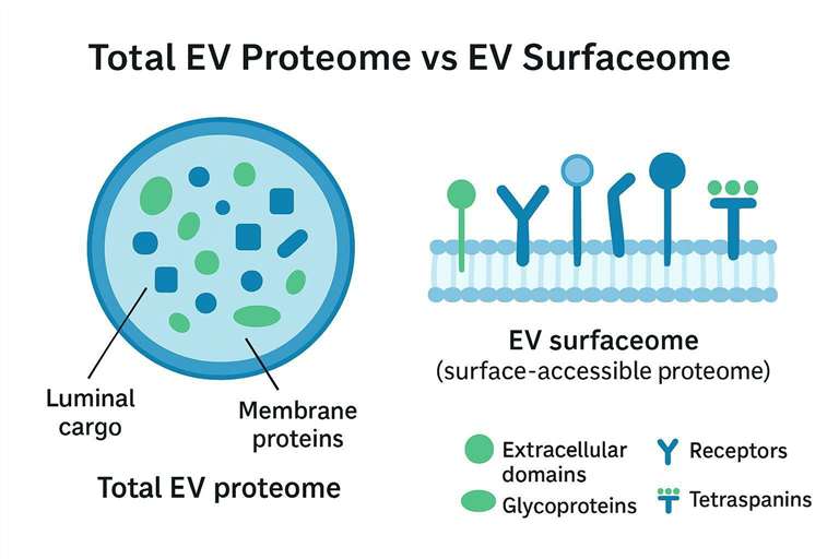
The EV surface proteome (the EV "surfaceome") is the set of proteins whose extracellular-facing domains are accessible on intact vesicles—i.e., the extracellular vesicle surface proteome you can actually probe without lysing the vesicle. This matters because an extracellular vesicle surface proteome readout is what enables actionable, follow-upable assays (antibody detection, ligand binding, uptake studies) rather than a general inventory.
Studying the surfaceome is useful because surface-accessible proteins are the most direct candidates for receptor–ligand interactions, immune recognition, cell uptake routes, and affinity-based EV subpopulation enrichment.
The importance of studying EV surface proteins in cell communication and disease mechanisms
EV surface proteins help encode where an EV can go and what it can do upon arrival. In many systems, uptake and tropism are more tightly coupled to surface accessibility than to bulk abundance in a lysis-based EV proteome.
Surface-focused profiling is also practical for biomarker workflows: surface candidates can often be targeted with antibodies or affinity reagents in follow-up assays, creating a path from discovery proteomics to scalable validation.
Understanding how surface proteins differ from the overall EV proteome and why they are crucial for targeted functional studies
A standard EV proteomics workflow (lysis → digestion → LC–MS/MS) answers: "What proteins are present in the EV preparation?" A surfaceome workflow aims to answer: "Which proteins are exposed on intact EVs under the conditions of my experiment?"
Those are different questions. The second requires you to preserve vesicle integrity during labeling/shaving, quantify background capture, and apply topology-aware annotation rules.
If you're planning to enrich EV subpopulations by immunocapture, validate candidates by bead-based flow or microscopy, or map receptor–ligand hypotheses, surface-accessible lists are the most practical starting point. That's why people often look for an EV surface marker discovery workflow (and why exosome surface proteins mass spectrometry results need topology-aware interpretation to be useful).
Key research questions that surfaceome analysis helps answer (biomarkers, receptor-ligand interactions, cell uptake)
A well-controlled surface-accessible dataset can help you answer:
- Which candidate surface markers are consistently accessible across replicates and preparations?
- Which receptors/adhesion molecules are positioned to mediate EV–cell interactions?
- Which surface-accessible proteins change between conditions in ways that plausibly affect uptake?
Experimental Strategies for EV Surfaceome Profiling
EV surfaceome profiling typically combines an intact-surface access step (chemical labeling or protease exposure) with enrichment and MS readout. The strategy you choose determines your dominant bias: biotinylation can pull along adsorbed proteins, while shaving can blur surface vs lumen if vesicles are disrupted.
Biotinylation vs. Protease Shaving: Which Method to Choose?
A useful way to decide is to be explicit about what you need:
- Do you need maximal enrichment for LC–MS/MS depth?
- Do you need exposure/topology evidence (which domains are accessible)?
- Is your sample likely to have high background (biofluids, protein corona, lipoproteins)?
Biotinylation
Biotinylation labels surface-exposed primary amines (often with membrane-impermeant sulfo-NHS reagents) or uses proximity/tethered biotinylation strategies. After labeling, streptavidin-based capture enriches labeled proteins (or peptides) prior to MS.
Advantages
Strong enrichment handles can improve detectability of low-abundance surface-accessible proteins and streamline downstream validation planning. EV surface biotinylation proteomics has been used in EV surfaceome studies including large EV surfaceome profiling (PMID: 34817906) and senescent-cell EV surfaceome mapping (PMID: 37862381).
Disadvantages
Biotinylation can capture proteins that are not truly EV-integrated (e.g., adsorbed proteins, co-isolated complexes) unless EV purity and wash stringency are high. In biofluids, the "protein corona" problem becomes a first-order design constraint.
Protease Shaving
Protease shaving exposes intact vesicles to a protease under conditions intended to cleave only outward-facing domains; the released peptides are analyzed by LC–MS/MS.
Advantages
When vesicle integrity is preserved, shaving can support exposure and topology hypotheses because detected peptides should come from externally accessible regions.
Disadvantages
Shaving is sensitive to EV integrity and protease/accessibility bias: partial permeabilization can make lumenal proteins look "surface," while highly exposed loops dominate identifications. If you plan to use protease shaving EVs, it's worth pre-defining an integrity assay and a background-cleavage subtraction plan before committing to scale.
Comparison Table
| Method | Advantages | Disadvantages | Best Use Case |
|---|---|---|---|
| Biotinylation | Strong enrichment; good sensitivity for low-abundance surface-accessible proteins | Risk of labeling/retaining non-EV-associated proteins without strict purification and controls | Candidate discovery for surface marker validation panels |
| Protease Shaving | Potentially higher surface specificity; exposure/topology hints | Vesicle disruption risk; protease/accessibility bias; needs strong controls | Exposure mapping and hypothesis-driven surface accessibility checks |
Mass Spectrometry Workflow for EV Surfaceome Profiling
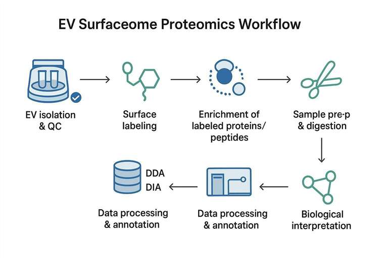
Most EV surfaceome proteomics pipelines have the same backbone: (1) preserve intact EVs, (2) create a selective handle for surface-accessible material, (3) enrich, (4) acquire by LC–MS/MS, and (5) interpret with topology-aware annotation.
Enrichment of Surface-Labeled Peptides
Enrichment design answers: "How do we make sure MS time is spent on the surface-accessible fraction?" Typical approaches include:
- Streptavidin bead enrichment after surface biotinylation (protein-level or peptide-level capture)
- Functionalized surfaces/chips for capturing labeled material in micro-scale workflows
Decision points that drive specificity:
| Decision point | Why it matters | What to document |
|---|---|---|
| EV purification stringency before labeling | Reduces co-isolated proteins that can be labeled/captured | Isolation method, wash steps, fraction logic (if SEC) |
| Capture at protein vs peptide level | Changes background and ID characteristics | Capture chemistry, digestion order |
| Negative controls | Quantifies non-specific binding and carryover | No-label, bead-only, mock digestion |
If you need a global baseline to contextualize enrichment, pair surfaceome runs with a conventional EV proteome run from the same prep batch. For MS-based profiling support pathways, Extracellular vesicles proteomics services can be a useful complement for establishing a global EV proteome baseline alongside surface-focused enrichment.
MS Analysis (DDA/DIA)
For experimental planning, the acquisition choice is mostly about data completeness and comparability:
- DDA often supports exploratory identification and spectral library generation.
- DIA often supports more consistent quantification across cohorts and batches—useful when you want to compare conditions or sample groups.
In surface-focused workflows where input complexity is already constrained, prioritizing reproducibility over "one-run depth" typically makes downstream interpretation and validation easier.
Data Interpretation
Most interpretation failures fall into three buckets:
- Overcalling surface hits (equating "enriched" with "surface-accessible")
- Ignoring topology (treating any membrane-associated protein as externally exposed)
- Underestimating matrix effects (letting biofluid background dictate the protein list)
A practical safeguard is to predefine evidence tiers:
- Tier 1: transmembrane or GPI-anchored annotation + enrichment over controls + consistent replicate behavior
- Tier 2: membrane-associated candidates with orthogonal support (e.g., intact-EV antibody detection)
- Tier 3: enriched proteins without membrane topology support (flag as likely adsorbed/co-isolated)
When your outcome is an antibody-based confirmation panel, build the validation plan into the workflow. For example, Exosome marker analysis aligns naturally with the validation stage after discovery.
Data Processing and Annotation for EV Surfaceome Proteomics
The core annotation challenge is defining what "surface-accessible" means in a way that is consistent across studies and robust to contaminants.
How to Define Truly Surface-Accessible Proteins: Data Denosing Strategies
Surfaceome denoising is a combination of topology annotation, matched controls, and conservative filtering.
Annotation Criteria
A pragmatic rule set:
- Annotate topology using prediction/annotation tools (e.g., TMHMM, Phobius) and curated resources.
- Explicitly categorize proteins (transmembrane, GPI-anchored, secreted, cytosolic, nuclear/mitochondrial, ribosomal).
- For transmembrane proteins, check whether identified peptides plausibly map to extracellular regions when peptide-level mapping is available.
This is also where you should intentionally include long-tail concepts like surface-accessible membrane proteins in EVs: surface accessibility is the biological property you are testing, and it must be defined operationally.
Negative Control Definition
Negative controls should represent what you can capture even when proteins are not surface-accessible:
- Proteins present in bead-only or no-label controls
- High-abundance matrix proteins expected to co-isolate (especially for plasma/serum)
- Canonical intracellular proteins used as integrity/contamination indicators
Data Filtering Strategy
Filtering should remove likely non-surface proteins without stripping true surface biology.
| Data Filtering Criteria | Validating True Surface Proteins | Common Pitfalls |
|---|---|---|
| Membrane Prediction | Use TMHMM/Phobius + curated annotations | Prediction ≠ extracellular exposure |
| Negative Control | Subtract proteins in no-label/bead controls; track enrichment factors | Missing controls inflate false positives |
| Peptide Hydrophobicity | Supports membrane association when paired with topology calls | Hydrophobic soluble proteins can be misclassified |
Functional Enrichment and Biological Interpretation of the EV Surfaceome
Once you have a conservative (or tiered) surface-accessible list, interpretation should be framed around functions plausible for exposed proteins.
Enrichment Analysis
Using GO, KEGG, and Reactome is common, but for surfaceome datasets it's often most informative to focus on:
- receptor activity and ligand binding
- cell adhesion
- immune interaction pathways
- membrane trafficking and endocytosis-related processes
Interpreting Biological Roles
To avoid overinterpretation:
- Check whether enrichment is driven by a few abundant proteins (possible bias).
- Ask whether top drivers have topology consistent with extracellular exposure.
- Stress-test whether you are seeing "matrix biology" (e.g., plasma proteins) instead of EV interface biology.
Integrating Surfaceome Data
Surfaceome signals are strongest when triangulated:
- RNA-seq for plausibility in producing cells
- lipidomics to interpret membrane-context shifts that affect accessibility
- global EV proteomics to separate "abundance change" from "accessibility change"
If you need a deeper baseline EV proteome to interpret surface candidates, Exosome proteomics services can complement surfaceome enrichment with broader EV proteome coverage.
Identifying and Mitigating Biases in EV Surfaceome Profiling
Bias is inevitable. The practical goal is to recognize where it enters and design controls and filters that bound it.
Contaminant Capture
Common background sources include lipoproteins and abundant soluble proteins that co-isolate with EVs.
Mitigation strategies:
- Use isolation workflows that reduce non-EV carryover and document fraction logic for SEC.
- Use matched control pull-downs to quantify non-specific binding.
- Track canonical non-EV proteins as a contamination signature rather than treating them as "invisible."
Surface vs. Internal Protein Ambiguity
Surfaceome workflows can convert internal proteins into apparent surface hits if EVs are damaged.
Mitigation moves:
- Monitor EV integrity before and after the surface-access step.
- Use negative markers and lysis-susceptible proteins as integrity flags.
- Prefer tiered reporting ("high-confidence surface-accessible" vs "enriched but ambiguous").
Matrix Effects and Pre-Analytical Bias
Biofluids can dominate capture and suppress low-abundance EV surface proteins.
Practical steps:
- Tighten pre-analytical variables (collection, storage, freeze–thaw counts).
- Optimize for the sample matrix you actually have, not for a generic EV.
- Process negative controls alongside real samples to avoid control mismatch.
Best Practices for EV Surfaceome Data Annotation and Reproducibility
If your goal is a surface marker list you can defend in peer review (or across labs), reporting and reproducibility are first-class outputs.
Quality Control for Surfaceome Data
Metrics that are often more meaningful than raw protein counts:
- proportion of IDs annotated as transmembrane/GPI vs cytosolic
- enrichment consistency across replicates
- control subtraction behavior (what survives no-label controls)
- peptide-to-protein evidence thresholds and FDR behavior
A compact QC checklist:
| QC question | "Good" looks like | Common failure mode |
|---|---|---|
| Are replicates consistent? | Similar enrichment patterns and membrane fraction | One replicate dominated by matrix proteins |
| Do controls explain background? | Many background proteins appear in controls and subtract cleanly | Controls missing or not processed identically |
| Does topology support exposure? | Many candidates have membrane topology support | "Surface list" is mostly cytosolic proteins |
Data Sharing and Reporting
Follow community reporting expectations and align surface-marker claims with EV characterization transparency. A practical approach is to include (1) isolation parameters, (2) characterization evidence, (3) contaminant assessment, and (4) the exact filtering rules used to define surface-accessible proteins.
Ensuring Cross-Study Comparability
Surfaceome comparability is often broken by inconsistent:
- EV isolation and purity controls
- surface-access step conditions (labeling time, buffer, enzyme exposure)
- negative control definitions
- annotation thresholds and topology rules
Cross-study comparability improves when you standardize:
- a tiered "surface-accessible evidence" definition
- a stable control panel
- a minimal reporting table of key parameters
Case Studies and Examples of Surfaceome Profiling in EV Research
These examples illustrate what surfaceome profiling can reveal when paired with controls and topology-aware interpretation.
Example 1: Surfaceome profile of EVs from cancer cell lines: identifying key surface markers involved in tumor progression
Cancer-focused EV surface proteome studies frequently use labeling and enrichment to propose candidate surface marker panels for follow-up assays. One example is a biotinylation-based study proposing breast cancer-associated surface candidates (PMID: 38339272).
Example 2: EV surface proteins in neuroinflammatory diseases: linking surface protein signatures to immune modulation
In immune-driven conditions, the most defensible approach is to treat surface signatures as candidate panels and confirm with orthogonal assays (e.g., intact-EV antibody detection, single-EV surface profiling platforms) before making mechanistic claims.
Example 3: Profiling extracellular vesicle surface proteins in exosome-based drug delivery systems
For engineered EV delivery systems, surfaceome profiling is useful for a simple question: "Which surface features are actually present and accessible after engineering and formulation?" Benchmark surface accessibility before and after engineering steps using identical controls.
FAQs
1) What is the EV surfaceome?
The EV surfaceome is the set of proteins with domains accessible on the outside of intact extracellular vesicles under defined experimental conditions. It's a functional subset of the total EV proteome because it focuses on what can directly contact recipient cells.
2) How do you profile surface proteins on extracellular vesicles?
You typically label or cleave surface-accessible proteins on intact EVs (e.g., surface biotinylation or protease exposure), enrich the labeled proteins/peptides, and identify/quantify them by LC–MS/MS. The most reliable workflows include matched negative controls (no-label, bead-only) and topology-aware annotation.
3) Surface biotinylation vs protease shaving: which is better for EV surface proteins?
Neither is universally better; they bias in different directions. Biotinylation offers strong enrichment but can pull along adsorbed or co-isolated proteins, while shaving can be more exposure-focused but is sensitive to EV integrity and protease/accessibility bias.
4) What are common exosome surface markers?
Commonly used markers for small EVs/exosomes include tetraspanins such as CD9, CD63, and CD81, but marker suitability depends on EV source, isolation method, and the question you're asking. For discovery work, treat these as characterization aids—not proof of a specific EV subtype.
5) How can I tell if a "surface marker" is truly surface-accessible and not a contaminant?
A strong indicator is when the protein is (1) annotated as transmembrane or GPI-anchored, (2) enriched over matched negative controls, and (3) supported by orthogonal validation on intact EVs. Proteins that lack membrane topology support but appear enriched should be reported as ambiguous rather than promoted as definitive markers.
6) What are the most common reasons EV surfaceome studies fail to reproduce?
The most common causes are inconsistent EV isolation/purity, missing or non-matched negative controls, and inconsistent annotation rules for what counts as surface-accessible. Reproducibility improves when you standardize the surface-access step conditions, report parameters explicitly, and apply the same filtering logic across batches.
References
- Proteomic dissection of large extracellular vesicle surfaceome reveals optimised proteins and molecular leads
- Surfaceome analysis of extracellular vesicles from senescent cells uncovers uptake repressor DPP4
- Surface Proteome of Extracellular Vesicles and Correlation Analysis with Parent Cell Proteome Reveal Potential Breast Cancer Biomarkers
- Minimal information for studies of extracellular vesicles (MISEV2023)
* For Research Use Only. Not for use in diagnostic procedures.
