

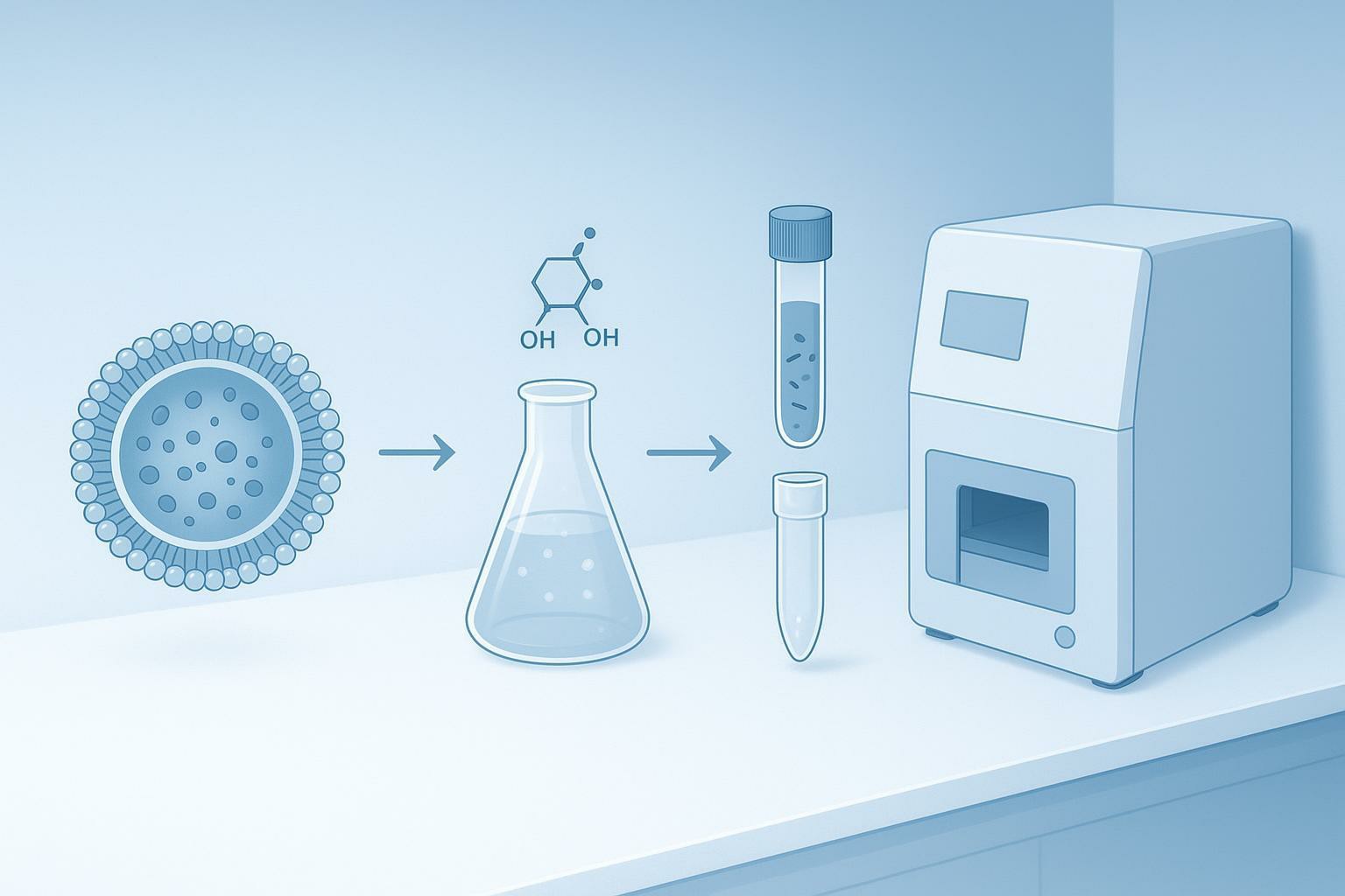
Extracellular vesicles (EVs) are an attractive proteomics specimen because they can carry transmembrane receptors, tetraspanins, and disease-linked signaling proteins that don't appear in the same form in bulk cell lysate. The catch is that EVs make sample prep feel "unfair" in a way standard proteomics does not: your starting material is tiny, your matrix is lipid-heavy, and the proteins you most care about are often the hardest to solubilize and digest.
This resource walks through a practical, low-loss approach to EV protein digestion for LC-MS/MS. It's written as a hybrid: a decision guide to match lysis chemistry with MS-compatible cleanup, plus compact checklists you can lift into an SOP.
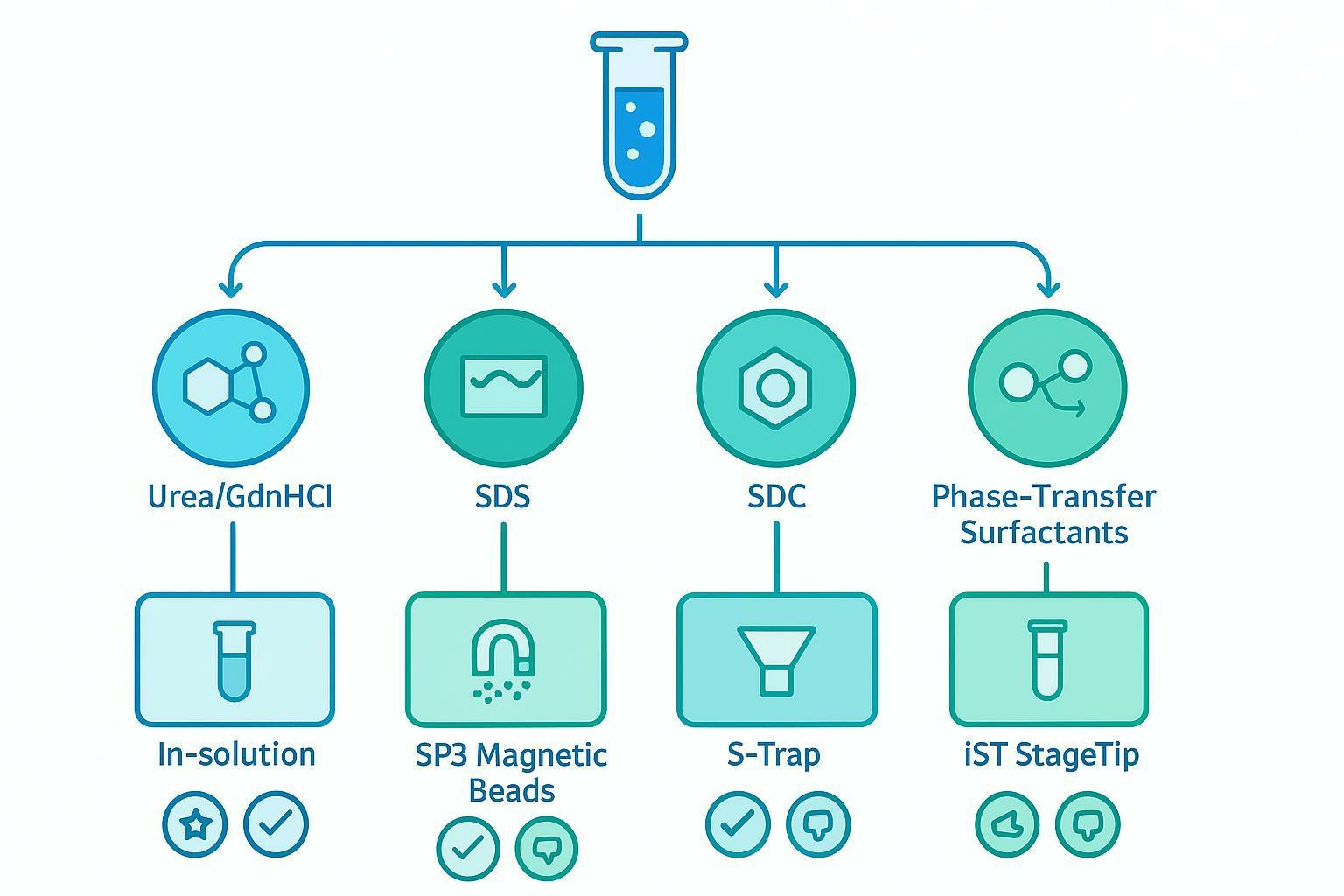
Key Takeaway: In EV proteomics, the best workflow is the one that keeps lipids and detergents under control while minimizing transfers. Choose lysis strength first, then choose the cleanup method that makes that chemistry safe for nanoLC-MS/MS.
The "High-Lipid, Low-Protein" Dilemma of EV Proteomics
EVs are lipid-bilayer particles designed by biology to protect cargo. That design is great for intercellular communication, and terrible for bottom-up proteomics.
The barrier is physical and chemical. The EV membrane is enriched in lipids and membrane proteins, and it can shield protease-accessible cleavage sites. Meanwhile, the total protein mass per prep is often small enough that normal tube-to-tube handling becomes a dominant source of loss. Peer-reviewed reviews of MS-based small EV proteomics repeatedly point to low input, contaminants, and preparation variability as major constraints on identification depth and reproducibility (for example, discussions in Challenges of MS-based small extracellular vesicles proteomics).
That sets up the core conflict:
- You need aggressive disruption to extract hydrophobic, transmembrane EV markers.
- Aggressive detergents are among the most common causes of ion suppression, poor chromatography, and instrument contamination.
If you apply a standard cell-lysate workflow directly to EVs, three failure modes show up fast: (1) poor recovery of membrane proteins, (2) high missed cleavage rates and low sequence coverage, and (3) persistent matrix contaminants that compress dynamic range.
Lysis Buffer Selection: Matching Chemistry to MS Compatibility in EV Protein Digestion for LC-MS/MS
A good EV lysis buffer is not "the strongest one you can find." It's the strongest chemistry you can fully clean up without losing your sample.
The practical way to think about this is to decide which of the following is your main constraint:
- Extraction-limited: you suspect transmembrane proteins are not coming into solution.
- Compatibility-limited: you suspect detergents/chaotropes are harming digestion or MS performance.
- Loss-limited: you have 1–10 µg total protein and can't afford dead volume or transfers.
Chaotropes vs. Detergent-Assisted Disruption
Chaotropes (8 M urea, GdnHCl) are often the starting point because they denature proteins in a way that is broadly compatible with proteases and LC-MS workflows after dilution or cleanup. Their limitation in EV work is that "denature" is not the same as "solubilize." Highly hydrophobic EV proteins can remain poorly recovered without detergent-like assistance.
- 8 M urea is a classic choice for in-solution workflows. It supports sequential digestion (Lys-C first, then trypsin after dilution). The main practical risk is artifact formation when urea is old, heated, or left at conditions that promote carbamylation. If you must heat, do it briefly and keep pH controlled.
- Guanidine hydrochloride (GdnHCl) is a stronger denaturant than urea but typically demands a cleanup step that can tolerate high chaotrope and still deliver good peptide recovery. In EV workflows, GdnHCl is most defensible when you know extraction is the limiting factor.
Strong detergents (SDS) can solve the extraction problem immediately. SDS is exceptionally effective at disrupting lipid membranes and keeping hydrophobic proteins in solution. But SDS is also one of the most MS-toxic reagents you can carry into digestion and LC-MS/MS, because it suppresses electrospray and can contaminate chromatographic systems.
General guidance on detergent interference and removal strategies in proteomics is well established and worth revisiting when you troubleshoot suppression and spray instability.
For EV proteomics, SDS is reasonable only when paired with a workflow designed to eliminate it (and ideally to keep sample transfers minimal).
Acid-Cleavable and Phase-Transfer Surfactants (PTS)
For many labs, sodium deoxycholate (SDC) and related phase-transfer surfactant (PTS) strategies are the most practical "middle ground" for EVs.
SDC and PTS reagents behave like detergents during lysis: they help solubilize membranes and hydrophobic proteins. The difference is how they leave the workflow. After digestion, acidification can drive precipitation and phase behavior that removes the surfactant from the peptide mixture. That lets you keep strong extraction chemistry early, but still protect LC-MS/MS performance at the end.
This approach is particularly useful when you want SDS-like membrane disruption without committing to an SDS-only cleanup ecosystem.
A practical compatibility table
Use this as a quick pre-flight check before you commit precious EV material.
| Lysis chemistry | Extraction strength for EV membranes | Main risk to LC-MS/MS | Cleanup that most naturally fits | When it's the right choice |
|---|---|---|---|---|
| 8 M urea | Medium | Carbamylation risk if mishandled; trypsin inhibition if not diluted | In-solution (after dilution), SP3, iST | You need a conventional workflow with good enzyme compatibility |
| GdnHCl | Medium-high | Protease inhibition and LC issues unless cleaned | SP3, S-Trap | Extraction is difficult and you can commit to robust cleanup |
| SDS | High | Severe ion suppression; instrument contamination | S-Trap, SP3 (optimized) | You are extraction-limited and must solubilize stubborn membrane proteins |
| SDC / PTS | High (often) | Surfactant carryover if acid removal is incomplete | Phase-transfer removal + C18, SP3, iST | You want strong solubilization with a realistic path to MS compatibility |
Micro-Proteomics Cleanup: Rescuing the 5 µg Sample
Once EV proteins are in solution, the next failure point is not digestion. It's losing the sample while trying to make it digestible and MS-safe.
A useful mental model is this: in low-input EV work, every additional surface is a "silent competitor" for your proteins and peptides. Filters, tubes, pipette tips, and membranes can bind enough material to change your results.
The FASP Bottleneck in Low-Input Workflows
Filter-Aided Sample Preparation (FASP) is a well-known approach for detergent removal and buffer exchange. It works by retaining proteins on an ultrafiltration membrane while detergents and small molecules are washed away, followed by on-filter digestion.
For EV samples, especially when you are in the single-digit microgram range, FASP can become a bottleneck because:
- ultrafiltration devices have dead volume,
- membranes can bind proteins nonspecifically,
- repeated centrifugation/washing steps increase handling time and loss.
The result is predictable: in ultra-low-input EV workflows, FASP can produce near-total loss or inconsistent recovery, even if the chemistry is "correct."
SP3, S-Trap, and iST Methodologies
If your constraint is low input, prioritize methods that combine three properties: (1) minimal transfer, (2) robust detergent/lipid tolerance, and (3) digestion in a contained environment.
SP3 (Single-Pot Solid-Phase-enhanced Sample Prep) uses magnetic beads to capture proteins in a way that supports aggressive washing and low transfer loss. SP3-style workflows have also been adapted explicitly for extracellular vesicles (see Automated Proteomics Sample Preparation of Phosphatidylserine-Positive Extracellular Vesicles Using Magnetic Beads and SP3).
S-Trap (Suspension Trapping) traps proteins in a matrix that enables rapid detergent removal and on-matrix digestion. It is widely used when SDS is involved because it provides a strong, predictable path to SDS removal.
iST (in-StageTip) approaches integrate lysis, reduction/alkylation, digestion, and desalting inside a single tip-like format. For EV micro-samples, the main advantage is fewer exposures and fewer transfers.
Method comparison for low-input EV samples
| Method | Strengths in EV proteomics | Common failure mode | Best paired with |
|---|---|---|---|
| FASP | Effective buffer exchange and detergent removal in many standard inputs | High loss at very low input; dead volume and membrane binding | Higher-input samples; well-optimized workflows |
| SP3 | Low transfer loss; scalable; strong for micro-proteomics | Binding/wash conditions must be tuned; can be sensitive to lipid carryover | Urea, GdnHCl, SDC/PTS; can handle detergent with optimization |
| S-Trap | Very strong detergent removal; efficient on-column digestion | Loss if overloaded or washed improperly; consumable variability | SDS lysis; high-lipid extracts |
| iST | Minimal handling; integrated workflow suited for trace inputs | Performance depends on lysis chemistry and matrix load | SDC/PTS or urea-based micro workflows |
Pro Tip: If you only have one chance with a cohort, choose the workflow that minimizes transfers. In EV proteomics, handling is often a bigger enemy than enzyme kinetics.
Protease Strategies for EV Transmembrane Proteins
Even after successful lysis and cleanup, EV proteins present a digestion challenge. EV surface proteins are frequently glycosylated, and glycan density can create steric barriers around cleavage sites. The result is a familiar pattern in the MS data: missed cleavages, weak sequence coverage in extracellular loops, and underrepresentation of key transmembrane markers.
Reduction, Alkylation, and Lys-C/Trypsin Sequential Digestion
Reduction and alkylation aren't optional "nice-to-haves" in EV digestion. They are the step that turns folded, disulfide-stabilized membrane proteins into substrates enzymes can actually access.
A conservative, widely transferable strategy is sequential digestion:
- Lyse/denature the EV proteins.
- Reduce disulfides (commonly DTT).
- Alkylate cysteines (commonly IAA) in the dark.
- Digest with Lys-C under denaturing conditions.
- Dilute chaotrope/detergent as needed.
- Add trypsin for completion.
Lys-C is useful here because it remains active in conditions that inhibit trypsin. It "opens up" structured proteins so trypsin can finish the job with fewer missed cleavages.
Quick checklist: sequential digestion for low-input EVs
- Done when lysis is complete: the solution is uniform with no visible flocculation; pellets are minimal after clarification.
- Done when reduction/alkylation is complete: no visible precipitation after alkylation; sample remains soluble.
- Done when digestion is acceptable: pilot MS shows low missed-cleavage rate for representative EV markers and stable chromatography.
Accelerated Digestion Options
For low-input EV samples, long incubations can increase the risk of nonspecific degradation and chemical artifacts. They also increase exposure to surfaces and repeated handling.
Accelerated approaches can help when you need rapid completion with controlled artifacts:
- microwave-assisted digestion
- ultrasound-enhanced digestion
- heat-stable or high-temperature trypsin formulations
The tradeoff is that acceleration can amplify protocol sensitivity. If you adopt an accelerated digestion, validate it with a small pilot and review missed cleavages before running a cohort.
Final C18 Desalting and Lipid Depletion
Even if your cleanup method removed detergents, the last gate before LC-MS/MS is usually a peptide-level solid-phase extraction step.
The purpose is not just desalting. In EV workflows it is also about protecting the analytical column from residual lipids and other hydrophobic contaminants that can foul the stationary phase and destabilize retention times.
C18 StageTip best practices
A C18 StageTip works when three things are right: wetting, equilibration, and load conditions.
- Wetting: ensure the C18 bed is fully wetted with a high-organic solvent before equilibration.
- Equilibration: equilibrate with an aqueous acidic solution to create peptide-binding conditions.
- Loading: load peptides under acidic, low-organic conditions so they bind efficiently.
- Washing: wash to remove salts and acidification byproducts without eluting peptides.
- Elution: elute with a high-organic solvent into low-bind vessels; avoid overdrying.
Residual lipid removal
Residual membrane lipids are not just an "ion suppression" problem. They can permanently foul LC columns and generate retention time shifts that look like instrument drift.
If your EV prep is lipid-heavy (common in plasma-derived EVs), consider building lipid depletion into the workflow at one of two points:
- during protein-level cleanup (e.g., bead/trap-based methods that shed lipids during washing),
- during peptide-level cleanup (StageTip wash strategy that reduces lipid carryover).
⚠️ Warning: If your chromatogram shows progressive retention time shifts across injections, treat residual lipid carryover as a primary suspect, not a minor contaminant.
Pre-MS Peptide Quantification and Quality Control
QC is where EV proteomics becomes reproducible.
The limitation of standard assays
Traditional protein assays such as BCA or Bradford are designed for intact proteins and are often incompatible after digestion. At that stage you have peptides in buffers that can interfere with colorimetric readouts, and the assay no longer measures what you intend.
Fluorometric peptide quantification
For trace EV digests, fluorometric peptide quantification is usually the most practical approach to avoid under- or over-loading nanoLC columns. Fluorometric assays designed for low concentrations (for example, Qubit-style peptide quantification) or UV-based A205 measurements can help you standardize injection load across samples.
The goal is not to "maximize load." It is to load consistently at an amount your nanoLC system can handle without peak distortion.
Monitoring missed cleavages
Before you commit a full cohort, run a pilot injection and check:
- missed cleavage rate for representative EV markers,
- chromatography cleanliness (baseline, peak shape),
- evidence of detergent carryover (signal suppression, unstable spray),
- signs of lipid carryover (retention shifts).
For broader discussion of EV proteomics challenges and the importance of standardized workflows, it can help to read widely across EV-proteomics methodology reviews and isolation-focused best-practice papers.
Improving low-input EV proteomics workflow reliability
Low-input EV proteomics leaves little margin for sample loss or preparation variability. For clinical samples, membrane-rich EV populations, or cohort studies where reproducibility is critical, workflows that minimize sample transfers, such as SP3 or iST-based preparation, can help improve consistency. Matching lysis and cleanup chemistry, including PTS-compatible approaches, can further reduce preparation-related bias.
To reduce workflow risk and protect limited samples, Creative Proteomics supports EV proteomics studies through Extracellular Vesicles Proteomics Services, with optional EV identity confirmation via Exosomes Identification Service and upstream workflow support informed by Exosomal Biogenesis and Identification.
Frequently Asked Questions
Can I use SDS or RIPA buffer to extract exosome proteins for LC-MS/MS?
Yes, but only if SDS (or SDS-containing RIPA variants) is removed with a detergent-compatible cleanup method before digestion and LC-MS/MS. In practice, pair SDS lysis with an approach built for harsh detergents (commonly S-Trap, or an optimized bead-based workflow), then confirm suppression-free MS performance with a small pilot injection.
What is the best lysis buffer for extracellular vesicle proteomics?
There isn't a single best buffer for every EV prep. Urea is often a safe starting point for enzyme compatibility, SDS is strongest for membrane solubilization but demands robust detergent removal, and SDC/PTS approaches can offer strong extraction with a simpler removal step after digestion. The best buffer is the one you can remove completely while keeping peptide recovery high.
Why should I use Lys-C alongside trypsin for EV digestion?
Because EV surface and transmembrane proteins can be structurally constrained and glycan-shielded, trypsin alone often leaves missed cleavages. Lys-C remains active under stronger denaturing conditions and can pre-digest proteins into more accessible fragments, which typically improves completeness and sequence coverage once trypsin is added.
How do I reduce missed cleavages in exosome proteomics?
Improve denaturation and confirm reduction/alkylation, then use sequential Lys-C followed by trypsin. Also treat residual detergents and lipids as first-line suspects: incomplete surfactant removal and lipid carryover can inhibit proteases and suppress ionization.
My EV total protein yield is only 2 µg. Should I use FASP?
Usually no. At very low input, FASP can lose material to membrane binding and dead volume. A single-pot workflow that minimizes transfers (SP3, iST, or a carefully executed S-Trap workflow) is generally a safer choice when you have only a few micrograms total.
Do I still need C18 desalting after SP3, S-Trap, or iST?
Often yes. These methods remove many contaminants, but C18 desalting is still a reliable final step to remove residual salts, surfactants, and lipid-like carryover that can suppress ionization and foul nanoLC columns.
References
- Challenges of MS‐based small extracellular vesicles proteomics
- A Perspective on Extracellular Vesicles Proteomics
- An Update on Isolation Methods for Proteomic Studies of Extracellular Vesicles
- Automated Proteomics Sample Preparation of Phosphatidylserine-Positive Extracellular Vesicles Using Magnetic Beads and SP3
- Size-exclusion chromatography combined with DIA-MS enables reproducible EV proteomics
* For Research Use Only. Not for use in diagnostic procedures.
