

Extracellular vesicle (EV) lipidomics is deceptively hard. The analytical stack (LC–MS/MS, annotation, statistics) is mature, but EV samples push every weak point in the workflow: tiny lipid mass, high risk of co-isolated contaminants, and pre-analytical variability that can quietly remodel the lipidome before the first injection.
This guide focuses on the parts that most often decide whether EV lipidomics is interpretable: extraction method choice, purity-aware QC, and downstream interpretation that separates biology from artifacts. It is written for researchers working with plasma/serum EVs, urine EVs, and cell-culture EVs who need reproducible, defensible lipid readouts.
Key takeaways: EV lipidomics succeeds when you (1) treat low biomass as a design constraint, (2) proactively manage lipoprotein co-isolation, (3) choose an extraction system that minimizes interphase carryover, (4) spike internal standards before extraction, and (5) interpret lipid shifts with class- and contamination-aware frameworks.
Core challenges in extracellular vesicle lipidomics
Low biomass is not a minor inconvenience
EV lipidomics is often limited by total lipid mass rather than instrument sensitivity. That changes the risk profile: losses during transfers matter more, adsorption to plastics is easier to miss, and a small amount of carryover or background can dominate true signal.
Practically, "low biomass" should shape decisions upstream of mass spectrometry. You want fewer handling steps, fewer phase collections, and extraction conditions that are tolerant of low input while still providing broad lipid coverage.
Lipoprotein interference can overwhelm true EV signal
For clinical EV lipidomics (especially plasma/serum), the dominant failure mode is interpreting lipoprotein lipids as EV membrane lipids. HDL, LDL, and VLDL can co-isolate with EVs depending on the isolation method, and their lipid signatures can mask or mimic EV biology.
A useful mindset is to treat purity assessment as part of lipidomics QC rather than an upstream checkbox. If you cannot quantify or at least strongly bound contamination, downstream statistics can be technically correct and biologically wrong.
Pre-analytical variables reshape labile lipids
Lipid oxidation and hydrolysis can occur during storage and handling. Freeze–thaw cycles, prolonged time at room temperature, and oxygen exposure disproportionately affect oxidation-prone lipids, introducing oxylipins and other artifacts that look like biology.
Even at −80 °C, storage duration and container headspace can matter for sensitive species. If you are comparing cohorts or time points, standardize the sample history as much as possible, and document what cannot be standardized.
Sample stability: manage oxidation rather than hoping it doesn't happen
Oxidation control is easiest when it is built into the protocol: work cold, reduce exposure to air/light where feasible, and consider antioxidants (commonly butylated hydroxytoluene, BHT) when appropriate for your downstream workflow.
The goal is not "zero oxidation" (rarely realistic) but reproducibility: the same sample handled twice should yield the same lipidome within expected technical variance.
EV lipid extraction methods: MTBE vs. Bligh–Dyer (and where BUME fits)
Most EV lipidomics workflows trace back to biphasic liquid–liquid extraction. The method you choose affects not only recovery, but also how often you accidentally pull proteins/salts into the extract, which matters a lot for low-input EVs.
Quick comparison table (decision support)
| Decision factor | MTBE / Matyash | Bligh–Dyer / Folch (chloroform-based) | What it means for EVs |
|---|---|---|---|
| Organic phase position | Upper | Lower | Upper-phase recovery is typically easier and reduces interphase disturbance in low biomass |
| Common failure mode | Incomplete phase separation if ratios are off | Interphase carryover during lower-phase collection | Interphase carryover can dominate EV lipid signal when starting material is limited |
| Safety/handling | Avoids chloroform | Uses chloroform | Lab safety and waste handling may influence method choice |
| Automation | Generally easier | More manual caution required | Automation helps reproducibility when cohorts are large |
| Polar lipid behavior | Often strong for many classes | Strong broad recovery; matrix dependent | Either can work, but low-input EVs are less forgiving to collection errors |
Bligh–Dyer and Folch methods (chloroform-based)
Bligh–Dyer and Folch methods are widely used biphasic systems built around chloroform, methanol, and water. They can deliver broad lipid recovery across polar and nonpolar species and have decades of precedent in lipid research.
For EV lipidomics, the operational drawback is practical: the lipid-rich organic phase settles at the bottom. Collecting it requires passing a pipette tip near the interphase, where proteins and other matrix components accumulate. With high-lipid tissues, a little contamination can be tolerable. With EVs, it can be fatal to interpretability.
If you use a chloroform-based method for EVs, focus on risk management: stable phase boundaries, conservative pipetting, and explicit controls to detect carryover.
MTBE (Matyash) extraction method
The MTBE/Matyash method inverts the biphasic system so the lipid-rich organic phase is on top. That single physical change is why many labs prefer MTBE for low-biomass lipidomics. Upper-phase collection is typically cleaner, easier to automate, and less likely to scrape the protein-rich interphase.
For EV samples where each aliquot is precious, MTBE often wins because it reduces the number of "high-risk moments" in the workflow. The method is also compatible with high-throughput LC–MS/MS, where reproducibility depends on minimizing operator-dependent steps.
When you need broad coverage and clean extracts from small EV inputs, MTBE is a practical default.
BUME and single-phase systems (when they help)
BUME (butanol/methanol) and other solvent systems can improve recovery for certain highly polar sphingolipids or signaling mediators, depending on matrix and analytical targets. Single-phase approaches (for example, isopropanol/methanol mixes) reduce handling steps and can help technical reproducibility for direct-injection or streamlined workflows.
The tradeoff is that "simpler" is not always "cleaner." For EV lipidomics, any method that increases non-lipid carryover can increase ion suppression and complicate annotation. If you adopt a single-phase strategy, confirm performance with blanks, pooled QCs, and class-level recovery checks.
Quality control (QC) standards in EV lipidomics
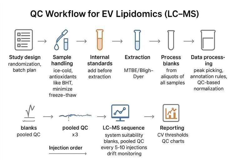
QC in EV lipidomics is not only about instrument stability. It is also about proving that your signal is (a) reproducible, (b) not dominated by background, and (c) meaningfully tied to EV biology rather than lipoproteins or handling artifacts.
Internal standard (IS) strategy: add before extraction
If you take only one QC step seriously, make it this: spike internal standards before extraction. Pre-extraction spiking allows you to monitor extraction recovery, matrix effects, and ionization behavior in a way that post-extraction spikes cannot fully recover.
A practical approach is to use a mixture of isotope-labeled standards that cover major lipid classes (for example, mixed internal standard cocktails used in many lipidomics labs) and ensure the standards span your expected dynamic range. The goal is not perfect correction for every species; it is stable normalization that makes technical variance visible.
Pooled QC and batch design: engineer reproducibility
Pooled QC samples (aliquots combined from across study samples) let you measure system drift, retention time stability, and intensity reproducibility across the run. They also give you a stable anchor for QC-based normalization.
Batch design matters because EV lipidomics is often run as multi-day sequences. Without a plan, you will "discover" batch effects late, when re-running is expensive.
A typical sequence design includes:
- system suitability and blanks at the start,
- several pooled QCs to condition the column,
- randomized study injections,
- pooled QCs injected regularly (for example, every 5–10 injections), and
- blanks at the end (and between high-load samples if carryover is suspected).
Lipoprotein interference: monitor with orthogonal markers
For plasma/serum EVs, QC should include at least one orthogonal readout of lipoprotein contamination. Protein markers (for example, apolipoproteins) are often used in EV characterization workflows, but lipidomics can also carry its own warning flags.
One pragmatic lipidomics-aware check is to monitor lipid class patterns that look "too plasma-like" relative to the EV subtype and isolation method, and to compare lipid signatures against EV protein markers when available. The objective is not to banish every contaminant; it is to quantify purity enough that you can interpret differences between groups.
If isolation and lipidomics are being planned together, choosing a purification approach that reduces lipoprotein co-isolation can improve interpretability downstream. When you need a dedicated workflow for EV isolation and purification, Exosome Isolation and Purification Service is one practical reference point for common method options (density-based, SEC, immunoaffinity) and purity testing.
Blank controls: treat background as a quantitative signal
With low-input EVs, blanks are not formalities. Solvent blanks, process blanks, and extraction blanks help you quantify background features that should be removed or down-weighted.
Two common pitfalls:
- ignoring low-intensity background that becomes significant after normalization, and
- removing features solely based on presence in blanks without considering relative abundance.
A better approach is to set explicit rules (for example, feature intensity must exceed blank by a defined fold-change in a defined fraction of samples) and to apply those rules consistently.
A practical framework for EV lipidomics data interpretation

Interpretation is where many EV lipidomics projects quietly drift away from biology. Species-level lists can look impressive while hiding the real questions: Are changes driven by membrane remodeling, lipoprotein carryover, oxidation artifacts, or biogenesis shifts?
A defensible interpretation strategy moves from global structure to targeted hypotheses.
Step 1: Confirm the dataset is QC-stable before you interpret biology
Start with QC metrics that tell you whether your downstream interpretation is worth doing:
- pooled QC clustering (should be tight),
- retention time drift and mass accuracy trends,
- internal standard stability,
- blank feature burden.
If pooled QC variance is high, any "significant" lipid differences can easily be analytical noise.
Step 2: Annotation quality is the foundation (and should be explicit)
EV lipidomics conclusions are only as strong as lipid identification. Annotation should be anchored to:
- high mass accuracy,
- consistent retention time behavior,
- MS/MS fragmentation matching,
- and naming standards consistent with community conventions (for example, LIPID MAPS classification).
When annotation confidence differs by class (common), report that explicitly. It will guide which biological interpretations are safe.
Step 3: Use multivariate analysis for structure, not storytelling
PCA (and related unsupervised methods) is useful to detect batch effects, outliers, and broad group separations. It is less useful as a biological explanation by itself.
If PCA separates groups, ask the unglamorous questions first:
- Does the separation track injection order?
- Does it track extraction day?
- Does it track a known contamination marker?
Only after those checks should you interpret it as biology.
Step 4: Interpret at the lipid-class level before you interpret individual species
Species-level volcano plots are attractive, but class-level shifts are often more robust and more interpretable.
A practical pattern is:
- summarize by class (PC, PE, PS, PI, SM, Cer, CE, TAG, etc.),
- examine ratios that map to membrane properties (for example, PC/PE as a rough curvature proxy),
- then drill down to species that plausibly drive the class shift.
This is also where contamination detection becomes easier. Lipoprotein carryover tends to distort class distributions in ways that can be spotted before you over-interpret a handful of "biomarker" species.
Step 5: Fatty acyl chain analysis links lipids to biophysics
Fatty acyl chain length and unsaturation influence membrane packing, curvature, and fusion propensity. For EVs, this can connect lipidomics to plausible functional hypotheses (biogenesis pathways, membrane rigidity, or uptake behavior).
Rather than listing dozens of species, summarize:
- chain length distribution by class,
- double bond index,
- and whether shifts are concentrated in a few classes (often the most interpretable case).
Step 6: Map lipid shifts to EV biology cautiously (but meaningfully)
Some interpretations are more defensible than others:
- Enrichment in cholesterol and sphingolipids can be consistent with raft-like membrane organization.
- Ceramides can be associated with membrane remodeling and signaling pathways.
- Lysophospholipids can indicate remodeling, enzymatic activity, or handling artifacts, depending on context.
The key is to bind interpretation to evidence: EV subtype, isolation method, QC stability, and orthogonal markers.
If you plan to connect lipid signatures to broader EV characterization and multi-omics profiling, Extracellular Vesicles Analysis Services is a relevant overview page for how lipidomics fits alongside EV isolation, characterization, proteomics, and metabolomics in an integrated workflow.
MTBE vs. Bligh–Dyer in EV lipidomics: a more nuanced view
The "best" extraction method depends on matrix, lipid classes of interest, and operational constraints. But for EVs, operational failure modes are unusually important.
When MTBE is often the safer default
MTBE tends to be a safer default when:
- EV input is low (small plasma volume, limited conditioned medium),
- you need to minimize interphase disturbance,
- you plan high-throughput sequences where operator variability matters, and
- you care about reproducible recovery across many lipid classes.
When chloroform-based methods can still be justified
Chloroform-based extraction can still be justified when:
- your lab has deep experience and strong SOP control,
- you are benchmarking against legacy datasets,
- or you have specific class-level targets with validated recovery under your matrix.
If you choose a chloroform-based method, consider explicitly documenting:
- phase volumes and ratios,
- collection technique,
- whether a re-extraction step is used,
- and what steps prevent interphase carryover.
Practical workflow tips (without turning this into a protocol dump)
A resource article should not pretend every lab has identical instruments and sample types. But a few operational choices consistently improve EV lipidomics outcomes:
- Add internal standards before extraction and keep the spiking method constant.
- Keep handling cold and minimize time in partially dried states (oxidation risk).
- Use low-bind plastics where possible, and avoid unnecessary transfers.
- Build blanks and pooled QCs into the run plan, not as optional "if time allows."
- Treat purity assessment as part of lipidomics QC, especially for plasma/serum EVs.
If your project requires dedicated exosome lipid profiling with standardized workflows and reporting, Exosomes Lipidomics Service is the most directly relevant internal service page to reference.
FAQs (AI Answer Engine format)
What is EV lipidomics?
EV lipidomics is the qualitative and quantitative measurement of lipid species carried by extracellular vesicles, typically using LC–MS/MS, to study membrane composition, biogenesis, and disease-associated changes.
What is the difference between exosome lipidomics and EV lipidomics?
Exosome lipidomics focuses on lipids from exosomes (endosome-derived small EVs), while EV lipidomics can include multiple vesicle subtypes. The distinction matters because lipid composition and contamination risk can differ by subtype and isolation method.
Why is lipoprotein contamination such a big problem in plasma EV lipidomics?
Because lipoproteins are abundant and lipid-rich, even small co-isolation can dominate the measured lipid signal and create false "EV biomarkers." Purity-aware controls and orthogonal markers are essential for interpretable results.
MTBE vs Bligh–Dyer: which extraction is better for low-input EV samples?
MTBE is often preferred for low-input EVs because the organic phase is on top, making it easier to collect without disturbing the protein-rich interphase. That reduces carryover risk and improves reproducibility in low-biomass workflows.
When should internal standards be added in EV lipidomics?
Internal standards should be added before extraction so they capture extraction losses and matrix effects, not only instrument variation.
How often should pooled QC samples be injected in an LC–MS lipidomics run?
A common approach is to inject pooled QCs regularly throughout the sequence (often every 5–10 injections) to monitor drift, retention time stability, and intensity variability, and to support QC-based normalization.
How can you tell if an EV lipid signature is driven by contaminants rather than biology?
Look for convergence of evidence: blank burden, pooled QC stability, class-level patterns that look inconsistent with the EV subtype, and agreement with orthogonal purity markers (for example, apolipoproteins for lipoproteins). If group separation tracks batch or injection order, treat it as an analytical artifact until proven otherwise.
What is the most defensible way to interpret EV lipidomics results?
Start with QC stability checks, then interpret at the lipid-class level, then connect species-level shifts to fatty acyl patterns and plausible EV biology. Bind any mechanistic claims to the isolation method, EV subtype, and purity evidence.
Integrating EV lipidomics with proteomics for holistic insights
EV lipidomics answers questions about membrane composition and biophysical properties, but it becomes more powerful when interpreted alongside proteins that report on EV subtype, biogenesis pathways, and contamination.
A practical integration strategy is:
- use proteomics to confirm EV markers and assess contaminants,
- use lipidomics to quantify membrane remodeling and signaling lipids,
- then interpret convergence: do lipid shifts align with changes in biogenesis markers or uptake-related proteins?
This integrated framing can be especially valuable for translational studies, where a single-omics signature is rarely robust enough to generalize.
If you want a single end-to-end workflow that includes isolation, characterization, and multi-omics on EVs, Exosome Analysis Services is an internal page that aligns with that combined approach.
References
- Lipid extraction by methyl-tert-butyl ether for high-throughput lipidomics
- Optimization of Folch, Bligh-Dyer, and Matyash sample-to-extraction solvent ratios for human plasma-based lipidomics studies
- Recommendations for good practice in MS-based lipidomics
- Quantitative Lipid Analysis of Extracellular Vesicle Preparations: A Perspective from the Lipidomics Working Group of ISEV
- The lipid composition of extracellular vesicles
* For Research Use Only. Not for use in diagnostic procedures.
