Get Your Custom Quote

Online Inquiry

Immunopeptidomics Service — MHC Peptide Discovery, HLA Ligandome Profiling & Neoepitope Identification for T-Cell Immunotherapy Research

Comprehensive HLA class I and class II ligandome analysis using immunoaffinity purification, high-resolution LC-MS/MS on timsTOF Pro and Orbitrap platforms, and deep immunoinformatics — from sample to prioritised neoepitope candidates.

Research Use Only (RUO) Notice: All services and data provided are strictly for non-clinical research purposes. Our analytical results are not intended for clinical diagnosis, patient management, or therapeutic decision-making.

Services Technologies Demo Case Study FAQ Related Services

CORE SERVICE

Comprehensive Immunopeptidomics for T-Cell Immunotherapy Research

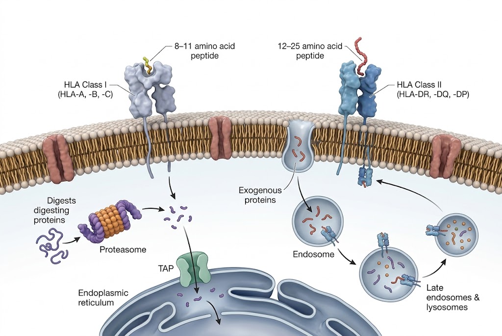
The peptides presented by major histocompatibility complex (MHC) molecules on the cell surface constitute the immune system's window into the cellular proteome. These HLA-bound peptide repertoires — the immunopeptidome — are the molecular basis for T-cell recognition in infection, autoimmunity, and cancer. Identifying which peptides are presented in a given biological context is the essential first step in understanding T-cell responses, discovering vaccine targets, and advancing T-cell-based immunotherapy programmes.

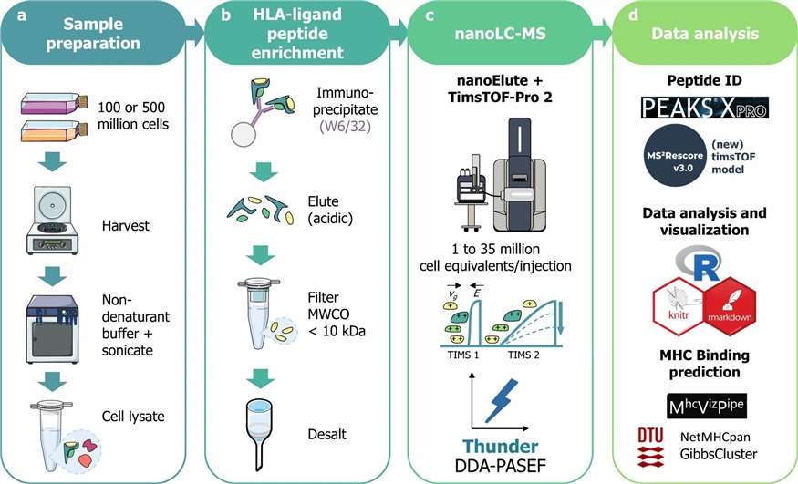
At Creative Proteomics, our Immunopeptidomics service provides end-to-end HLA ligandome profiling — from immunoaffinity-based MHC peptide enrichment through high-resolution LC-MS/MS identification to comprehensive bioinformatics analysis. We support both HLA class I (HLA-I) and HLA class II (HLA-II) workflows, processing a wide range of sample types including cell lines, primary tissues, tumour biopsies, FFPE specimens, and PBMCs.

  • HLA-I and HLA-II dual capability: Dedicated workflows for class I (W6/32 antibody-based) and class II immunopeptidomics, both optimised for non-tryptic peptide identification
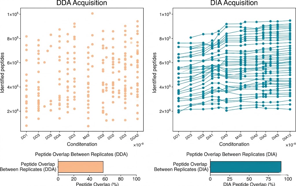
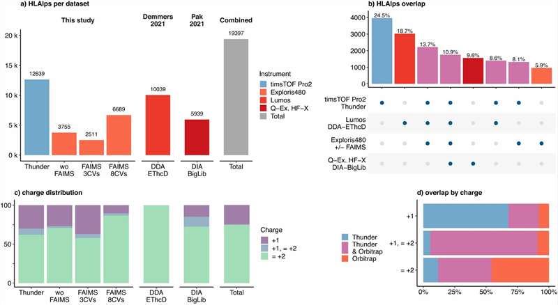
  • Deep coverage with DIA/4D-DIA: Data-independent acquisition on Bruker timsTOF Pro and Orbitrap platforms extends detectable ligand depth by 30–50% over standard DDA
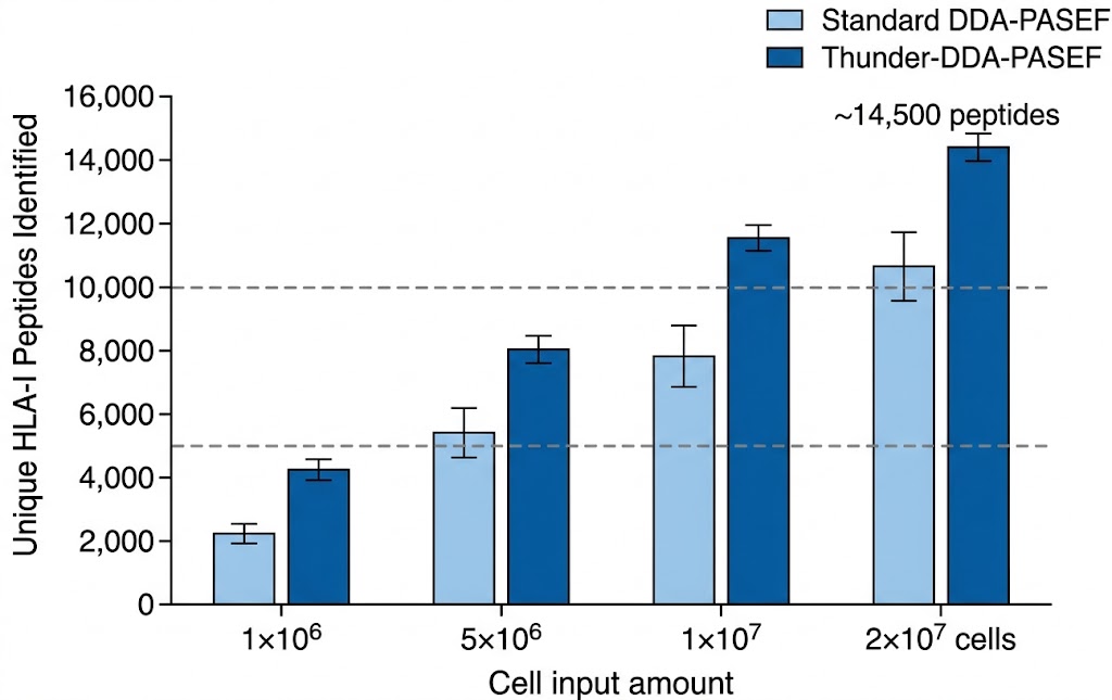
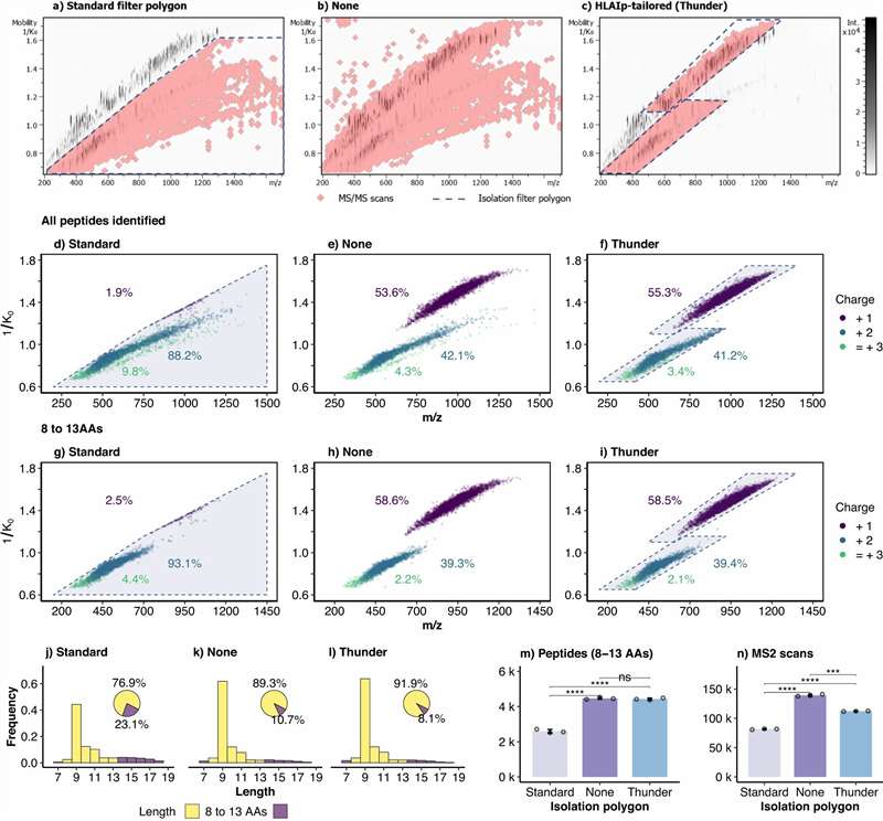
  • Low-input compatibility: Validated workflows from as few as 5×10⁶ cells; Thunder-DDA-PASEF achieves >5,000 HLA-I peptides from 1×10⁶ cells
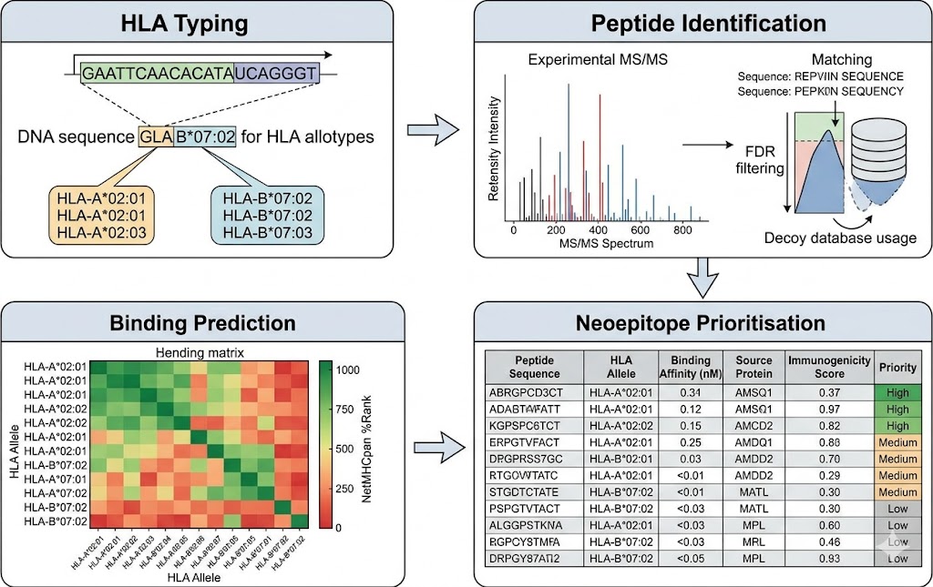
  • Comprehensive bioinformatics: HLA typing, allele-specific binding prediction, multi-allelic deconvolution, and neoepitope prioritisation
Immunopeptidomics workflow showing MHC peptide elution, LC-MS/MS analysis, and bioinformatics pipeline

Figure 1. Immunopeptidomics workflow: MHC immunoaffinity purification → LC-MS/MS acquisition → HLA peptide identification → neoepitope prioritisation.

What Is Immunopeptidomics and Why It Matters for Immunotherapy Development

Immunopeptidomics is the systematic characterisation of the complete repertoire of peptides bound to MHC molecules — both HLA-I and HLA-II — on the surface of cells. Unlike whole-proteome or transcriptome analysis, which measures what a cell can express, immunopeptidomics reveals what a cell actually presents to the immune system. This distinction is critical: the thousands of proteins expressed in a cell are processed into millions of potential peptide fragments, but only a small fraction — roughly 0.1–1% — are loaded onto MHC molecules and displayed on the cell surface. The immunopeptidome is therefore a highly filtered, functionally relevant subset of the proteome that directly governs T-cell recognition. For studies requiring analysis at the resolution of individual cell types within heterogeneous tissues, our single-cell proteomics platform provides complementary capabilities for deep protein profiling of defined cell populations.

For drug discovery and immunotherapy research teams, immunopeptidomics addresses three fundamental questions that no other technology can answer at scale. First, which tumour-specific peptides (neoantigens) are presented on cancer cells — the targets for personalised cancer vaccines, TCR-T cell therapies, and bispecific T-cell engagers. Second, which self-peptides are presented in autoimmune target tissues, enabling the rational design of antigen-specific therapies. Third, how does immunotherapy — checkpoint blockade, T-cell engagers, or other modalities — change the presented peptide repertoire over the course of treatment, providing a mechanistic window into therapy response and resistance.

Integration with Broader Proteomics Capabilities

The full power of immunopeptidomics is realised when it is integrated with orthogonal proteomics data. Combining immunopeptidomics with cell surface proteomics provides a complete picture of the tumour-immune interface — both the peptide antigens presented by MHC and the co-stimulatory/inhibitory receptors that modulate T-cell responses. Integration with deep proteome profiling and targeted protein quantification by PRM provides quantitative data on source protein expression and peptide-level validation, enabling prioritisation of neoantigens whose source proteins are confirmed at the protein level. This multi-modal approach — a unique capability of Creative Proteomics' proteomics platform — bridges the gap between peptide discovery and functional prioritisation in a way that standalone immunopeptidomics services cannot.

HLA class I and class II peptide presentation on cell surface

Immunopeptidomics Approaches and Analytical Workflow

HLA Class I Ligandome Profiling

HLA class I molecules (HLA-A, -B, -C) present peptides of 8–11 amino acids derived primarily from endogenously expressed proteins. Our HLA-I workflow uses the W6/32 monoclonal antibody for immunoaffinity purification of HLA-I complexes, followed by mild acid elution of bound peptides. The analytical challenge lies in the unique characteristics of HLA-I peptides: they are predominantly non-tryptic, short, and present at extremely low quantities. We deploy acquisition methods specifically tuned for immunopeptidomics — including Thunder-DDA-PASEF on the timsTOF Pro platform, which uses semi-selective precursor ion selection based on ion mobility and m/z windows tailored to HLA-I peptide properties, typically doubling identification rates compared to standard DDA-PASEF acquisition.

HLA Class II Ligandome Profiling

HLA class II molecules (HLA-DR, -DQ, -DP) present longer peptides (12–25 amino acids) derived predominantly from exogenous proteins internalised through endocytosis. HLA-II peptidomics is methodologically more challenging due to longer peptide lengths producing more complex MS/MS spectra, lower surface expression reducing total peptide yield, and greater structural heterogeneity across allotypes. Our HLA-II workflow uses HLA-DR-specific (L243) or pan-HLA-II antibody cocktails for enrichment, with LC-MS/MS acquisition parameters optimised for longer, more highly charged precursor ions, combined with hybrid search strategies that maximise identification rates from the complex spectra typical of HLA-II ligands.

Comparative Immunopeptidomics & Bioinformatics

One of the most powerful applications of immunopeptidomics in drug development is comparative analysis across conditions — treated versus untreated, responder versus non-responder, tumour versus matched normal tissue. Our comparative workflow uses label-free DIA quantification to measure relative changes in HLA peptide presentation, providing quantitative data on how therapeutic intervention modulates the antigen landscape. Downstream bioinformatics integrates HLA allotype assignment, NetMHCpan-based binding affinity prediction, multi-allelic deconvolution, source protein annotation, and prioritised candidate ranking based on binding strength and predicted immunogenicity.

Standardized Immunopeptidomics Workflow

1. Sample preparation: Cells, tissues, or PBMCs are lysed in detergent-based buffer with protease inhibitors. Tissue samples undergo mechanical homogenisation. MHC complexes are extracted from clarified lysate.

2. Immunoaffinity purification: HLA-I or HLA-II complexes are captured using conformation-specific antibodies (W6/32 for class I; L243 or pan-HLA-II cocktail for class II) immobilised on protein A/G agarose beads. Non-specifically bound material is removed by extensive washing.

3. Peptide elution and cleanup: HLA-bound peptides are eluted under mild acid conditions, separated from higher-molecular-weight HLA components by ultrafiltration (10 kDa MWCO), desalted by C18 StageTips or SPE, and concentrated.

4. LC-MS/MS acquisition: Purified HLA peptides are analysed by nanoLC-MS/MS on the selected platform: Thunder-DDA-PASEF (timsTOF Pro) for maximum sensitivity from low-input samples; DDA-PASEF or DIA-PASEF for routine depth; Orbitrap-based DDA or DIA (Fusion Lumos, Q Exactive HF-X) for established platform consistency.

5. Peptide identification and HLA binding prediction: Raw MS data are searched against the species-appropriate proteome database using pic-immunopeptidomics-optimised settings. Identified peptides are filtered by length (8–11 aa for HLA-I, 12–25 aa for HLA-II) and allele-specific binding affinity is calculated using NetMHCpan or NetMHCIIpan.

6. Neoepitope prioritisation and reporting: Candidate peptides are prioritised based on binding affinity, source protein expression, mutant versus wild-type discrimination (for tumour neoantigens), and predicted immunogenicity. A comprehensive report is delivered with full peptide lists, binding predictions, annotated MS/MS spectra, and prioritised candidate rankings.

Sample Requirements for Immunopeptidomics Analysis

Sample Type Recommended Input Minimum Input Key Notes
Cell lines (suspension / adherent) 2×10⁷ – 1×10⁸ cells 5×10⁶ cells Thunder workflow validated to 1×10⁶ cells; lower input yields proportionally fewer HLA peptides
Primary cells / PBMCs 2×10⁷ – 1×10⁸ cells 1×10⁷ cells Activation status affects HLA expression; IFN-γ-treated cells yield higher peptide counts
Fresh frozen tissue 50–200 mg 20 mg HLA expression varies by tissue; lymphoid tissues typically yield highest peptide counts
FFPE tissue 5–10 sections (10 µm) 3 sections HLA peptide recovery from FFPE is reduced; feasibility assessment recommended
Tumour biopsies (core needle) 2–3 cores (12G) 1 core Low-input optimisation required; contact for feasibility assessment

Immunopeptidomics Performance Benchmarks

The following representative data illustrate the performance of optimised immunopeptidomics workflows across different sample inputs and platforms. These benchmarks are derived from published method development studies and internal validation runs using our standard operating procedures.

HLA class I peptide identification counts across sample input amounts using Thunder-DDA-PASEF

Figure 2. HLA-I peptide identification counts across cell input amounts using Thunder-DDA-PASEF workflow on timsTOF Pro. From 1×10⁶ cells, >5,700 unique HLA-I peptides are identified; from 2×10⁷ cells, >14,500 peptides are routinely detected.

Comparison of DDA vs DIA acquisition for immunopeptidomics coverage

Figure 3. Comparison of DIA versus DDA acquisition for immunopeptidomics. DIA-based workflows provide more complete data with reduced missing values across samples — critical for comparative and cohort-scale studies.

Bioinformatics pipeline for HLA binding prediction and neoepitope prioritisation

Figure 4. Immunopeptidomics bioinformatics pipeline: identified HLA peptides are mapped to HLA allotypes, binding affinity is predicted using NetMHCpan, and candidates are prioritised based on binding strength, source protein expression, mutation status, and immunogenicity score.

CASE STUDY

Thunder-DDA-PASEF Enables High-Coverage HLA-I Immunopeptidomics from Clinically Relevant Sample Inputs

Optimised Acquisition Strategy for Low-Input Immunopeptidomics

Background & Purpose

A fundamental limitation of current immunopeptidomics workflows is the large sample input required — typically tens of millions of cells — which restricts the analysis of clinical specimens such as core needle biopsies, FACS-sorted cell populations, and rare primary cell types. While DDA-PASEF on timsTOF instruments had improved immunopeptidomics sensitivity, standard acquisition methods are optimised for tryptic peptides rather than for the shorter, differently charged HLA class I peptides. The researchers aimed to develop a dedicated acquisition strategy that maximises HLA-I peptide identification from minimal cell input.

Methods

The team developed Thunder-DDA-PASEF, a tailored LC-IMS-MS/MS method using extended TIMS ramp duration (300 ms) with fewer MS/MS frames (3 frames per ramp), combined with a custom isolation polygon designed to semi-selectively fragment singly and multiply charged HLA-I peptides based on their characteristic ion mobility and m/z properties. A timsTOF-specific peak intensity prediction model (MS2PIP) was trained for both tryptic and non-tryptic peptides and implemented in MS2Rescore (v3) alongside a CCS predictor. The method was validated on JY lymphoblastoid cells and Raji B cells, including cells transfected to express the SARS-CoV-2 spike protein.

Results Overview

Thunder-DDA-PASEF doubled the number of identified HLA-I peptides compared to standard DDA-PASEF from the same sample. The method identified 5,738 HLA-I peptides from 1×10⁶ JY cell equivalents and 14,516 HLA-I peptides from 2×10⁷ cells — substantially exceeding prior benchmarks for timsTOF-based immunopeptidomics. MS2Rescore with the custom MS2PIP model further boosted identification rates by 33–41.7%. In the SARS-CoV-2 spike protein case study, 16 spike-derived HLA-I peptides were identified, of which 13 had been previously reported to elicit T-cell responses in human patients.

Thunder-DDA-PASEF method workflow diagram for immunopeptidomics analysis

Figure 5. Thunder-DDA-PASEF acquisition strategy: extended TIMS ramp with tailored isolation polygon for semi-selective HLA-I peptide fragmentation.

Comparison of HLA-I peptide identifications between Thunder-DDA-PASEF and standard DDA-PASEF

Figure 6. Thunder-DDA-PASEF achieves approximately 2× the HLA-I peptide identifications of standard DDA-PASEF across different cell input amounts.

SARS-CoV-2 spike protein HLA-I peptide identification and validation

Figure 7. SARS-CoV-2 spike protein case study: 16 HLA-I peptides identified, 13 known to elicit T-cell responses — confirming biological relevance of detected ligands.

Why This Matters for Your Immunopeptidomics Project

This case study demonstrates two advances directly relevant to our immunopeptidomics service: optimised acquisition strategies specifically designed for HLA peptide properties can dramatically improve identification rates from limited sample inputs; and the method performs reliably at input levels compatible with clinical samples — 1×10⁶ cells, within the range of core needle biopsies. The Thunder-DDA-PASEF workflow, deployed on the Bruker timsTOF Pro platform available at Creative Proteomics, represents the current state of the art in low-input immunopeptidomics and is available as a core component of our service offering.

Frequently Asked Questions

Q1: What is the typical number of HLA-I peptides identified per sample?

From cell lines with standard input (2×10⁷ cells), we typically identify 8,000–14,000 unique HLA-I peptides using our optimised Thunder-DDA-PASEF workflow. From tissue samples, the yield depends on MHC expression level — tumour tissues and lymphoid tissues generally produce higher peptide counts than non-lymphoid tissues. From lower-input samples (5×10⁶ cells), we typically identify 3,000–6,000 HLA-I peptides. We provide a study-specific estimate during project planning.

Q2: Can you analyse HLA class II peptides in addition to class I?

Yes. Our service includes both HLA-I and HLA-II ligandome profiling, each with dedicated immunoaffinity reagents and acquisition methods. HLA-II peptide yields are typically lower and the peptides are longer (12–25 amino acids), which requires different database search parameters and binding prediction tools. We recommend discussing your specific HLA-II requirements during project design.

Q3: What bioinformatics analysis is included with the service?

Our standard bioinformatics package includes: HLA typing from RNA-seq or genomic data; peptide identification with FDR control; allele-specific binding affinity prediction using NetMHCpan/NetMHCIIpan; multi-allelic deconvolution for samples with multiple allotypes; source protein annotation and pathway analysis; and a prioritised candidate list based on binding strength, source protein expression, and predicted immunogenicity. For tumour neoantigen projects, we also provide mutant versus wild-type peptide discrimination and integration with genomic variant data.

Q4: What is the minimum sample input for immunopeptidomics analysis?

Our validated minimum input is 5×10⁶ cells for cell lines and 1×10⁷ cells for primary samples and PBMCs. Using the Thunder-DDA-PASEF workflow, we have demonstrated >5,700 HLA-I peptide identifications from 1×10⁶ cell equivalents — suitable for core needle biopsies and FACS-sorted populations. For tissue samples, we recommend 20–50 mg as the minimum. Inputs below these thresholds are possible but require a feasibility assessment.

Q5: Can you detect post-translationally modified HLA peptides?

Yes. A substantial fraction of HLA-presented peptides carry post-translational modifications — including phosphorylation, citrullination, and deamidation — that can generate neoepitopes recognised by T cells. Our search pipelines include variable modification searching for common PTMs, and we can perform dedicated PTM-enriched immunopeptidomics analyses for specific modifications of interest.

References

  1. Gomez-Zepeda D, Arnold-Schild D, Beyrle J, Declercq A, Gabriels R, Kumm E, et al. Thunder-DDA-PASEF enables high-coverage immunopeptidomics and is boosted by MS²Rescore with MS²PIP timsTOF fragmentation prediction model. Nat Commun. 2024;15:2288.
  2. Marcu A, Bichmann L, Kuchenbecker L, Kowalewski DJ, Freudenmann LK, Backert L, et al. HLA Ligand Atlas: a benign reference of HLA-presented peptides to improve T-cell-based cancer immunotherapy. J Immunother Cancer. 2021;9:e002453.

GET STARTED

Advance Your Immunotherapy Research with Comprehensive Immunopeptidomics

From HLA ligand discovery to prioritised neoepitope candidates — partner with Creative Proteomics for end-to-end immunopeptidomics analysis tailored to your research question and sample type.

Ready to discover what your cells are presenting?

Request a Customized Quote