Limited Proteolysis–Mass Spectrometry (LiP-MS) Service
What is Limited Proteolysis (LiP)?
Limited proteolysis (LiP) involves using specific proteases to partially digest proteins without fully denaturing them. Proteases selectively cleave accessible regions of proteins, typically flexible loops or surface-exposed domains, generating characteristic peptide fragments. These fragments serve as "structural fingerprints" that reflect protein conformation and changes induced by binding events, post-translational modifications, or environmental conditions.
What are the Principles of LiP-MS in Structural Proteomics?
LiP-MS relies on selective proteolysis to probe protein conformations. Specific proteases, such as trypsin, protease K, or chymotrypsin, cleave proteins at accessible or flexible regions while leaving the overall structure intact. The resulting peptides reflect the protein's conformational state, allowing researchers to identify structural changes induced by ligands, post-translational modifications, or disease conditions.
Enzymatic digestion strategies: Proteases are selected based on their cleavage specificity and the experimental goal. Broad-spectrum proteases probe unfolded or flexible regions, whereas targeted proteases focus on specific domains.
Protein structure and conformation mapping: By comparing peptide signatures across conditions, LiP-MS can detect subtle and large conformational changes. This capability enables mapping of protein domains, identification of allosteric effects, and detection of ligand-induced structural perturbations.
Figure 1. Illustration of LiP-MS (Elsasser F, et al., 2026).
Advantages of LiP-MS Compared with Traditional Proteomics
- High Sensitivity and Structural Resolution: The technique detects both subtle and large-scale structural alterations, including changes in low-abundance proteins, offering unmatched insights into proteome dynamics.
- Label-Free and Non-Invasive Analysis: LiP-MS requires no chemical modification or protein purification, preserving native protein structures and enabling studies under near-physiological conditions.
- Compatibility with Multiple Quantitative Proteomics Approaches: LiP-MS integrates seamlessly with DDA, DIA, PRM, and SRM workflows. It complements services such as DIA Quantitative Proteomics Analysis and PRM Targeted Proteomics Analysis, enabling both global discovery and targeted validation studies.
Integrating LiP-MS With Other Proteomics Solutions
Integrating with Thermal Proteome Profiling (TPP)
TPP measures how the overall stability of a protein responds to environmental factors or small molecules. Together, these methods reveal not only that a protein is affected, but also how it responds at a structural and stability level, giving a more complete picture of its functional state.
Integrating with Targeted Proteomics Services
After LiP-MS identifies structural changes across the proteome, targeted methods can validate and quantify these changes in specific proteins with high precision. This is particularly valuable when studying low-abundance proteins, membrane proteins, or proteins with complex modifications.
Integrated into Drug Discovery and Translational Proteomics
LiP-MS can also be integrated into Drug Discovery and Translational Proteomics workflow. Structural insights from LiP-MS reveal where compounds bind and how they affect protein conformation. When used alongside quantitative proteomics tools like DIA, researchers can track not only structural changes but also overall protein abundance, giving a multi-dimensional view of how molecules interact with cellular networks.
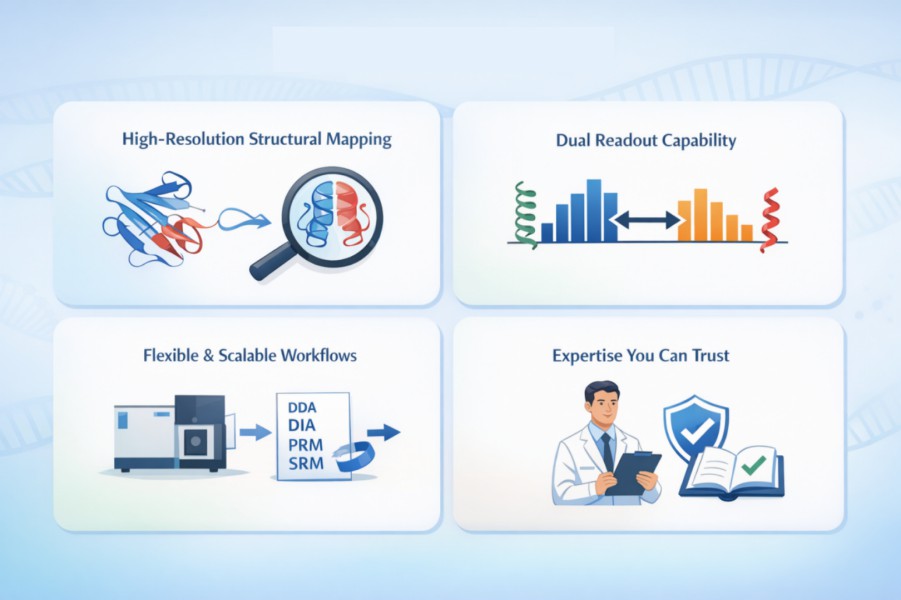
Applications of LiP-MS in Proteomics and Drug Discovery
Protein Structural Dynamics and Conformation Studies
LiP-MS enables high-resolution detection of conformational changes, providing insights into protein folding, post-translational modifications, and environment-induced structural alterations.
Mapping Protein-Protein and Protein-Small Molecule Interactions
LiP-MS identifies accessible regions affected by protein-protein and protein-small molecule interactions, allowing the study of protein networks and ligand binding. It is essential for understanding complex regulatory mechanisms in cellular pathways.
Drug Target Identification and Mechanistic Studies
By revealing drug-induced structural changes, LiP-MS assists in mapping compound binding sites and validating potential targets. Researchers can prioritize targets based on conformational impact, aiding mechanistic studies and structure-based research.
Biomarker Discovery in Disease Research
LiP-MS can uncover novel structural biomarkers, such as amyloid-to-soluble protein ratios in neurodegenerative disorders, providing insights into pathological mechanisms and protein dysfunction.
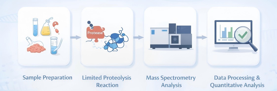
Creative Proteomics' LiP-MS Service Workflow
- Sample Preparation: Samples can include cells, tissues, protein solutions, serum, and cerebrospinal fluid. Proteins are extracted under non-denaturing conditions to preserve native conformations.
- Limited Proteolysis Reaction: A broad-spectrum or specific protease is applied to partially digest proteins. TProteolysis is stopped through denaturation, and proteins are subsequently digested with trypsin.
- Mass Spectrometry Analysis: Peptide fragments are separated by LC and analyzed by MS. LiP-MS supports multiple acquisition strategies. Quantitative analysis is typically performed in label-free mode or using stable isotope-labeled standards for absolute quantification.
- Data Processing and Quantitative Analysis: Advanced bioinformatics tools are employed to map peptide fragments to protein sequences, quantify changes, and identify statistically significant structural alterations.

Sample Requirements LiP-MS Service
| Sample Type | Minimum Amount per Sample | Notes | Shipping Conditions |
| Cultured cells | 2 × 10⁶ cells | Cell pellets preferred; avoid harsh lysis buffers | Dry ice |
| Tissues | 10 mg | Fresh or properly snap-frozen tissue recommended | Dry ice |
| Protein solution | ≥200 μL at 1 μg/μL | Buffer should be compatible with protease activity | Ice packs or dry ice |
| Serum | 100 μL | Avoid repeated freeze–thaw cycles | Dry ice |
| Cerebrospinal fluid (CSF) | 200 μL | Low-abundance proteins supported | Dry ice |
Why Choose Creative Proteomics for LiP-MS Service
- Deep Proteomics Expertise: Rich experience in MS-based proteomics ensures scientifically rigorous LiP-MS experimental design and execution.
- Flexible and Sample-Adaptive Workflows: LiP-MS pipelines are optimized for diverse sample types, including cells, tissues, protein solutions, serum, and cerebrospinal fluid, under non-denaturing conditions.
- Advanced Mass Spectrometry Platforms: High-resolution LC-MS systems support discovery-scale and targeted analyses using DIA, PRM, and SRM acquisition strategies.
- Robust Bioinformatics and Data Interpretation: Delivered results include structure-sensitive peptide identification, quantitative analysis, functional annotation, and interaction network interpretation.
- Support for Discovery and Mechanistic Studies: Services are designed to support drug target mapping, protein-protein and protein-small molecule interaction analysis, and structural biomarker discovery.
FAQ
-
Q1: Why are two protease digestion steps used?
A1: First, limited proteolysis probes protein structure. Second, complete tryptic digestion under denaturing conditions generates peptides amenable to high-resolution MS and facilitates peptide identification and quantification.
-
Q2: What proteases are commonly used in LiP-MS?
A2: Protease K, thermolysin, chymotrypsin, elastase, and others are used for structural probing. Tryptic digestion is typically used in a second step to generate peptides for MS analysis.
-
Q3: Can LiP-MS be applied to living cells?
A3: In-cell LiP-MS methods introduce proteases into intact cells to probe structural states in a closer-to-native context, capturing conformational changes with minimal perturbation.
-
Q4: How many proteins can LiP-MS analyze simultaneously?
A4: Proteome-wide LiP-MS can interrogate thousands of proteins and structural sites, depending on MS sensitivity and sequence coverage.
Demo
Demo: Systematic identification of structure-specific protein-protein interactions
Figure 2. LiP-MS detects protein-protein interactions in purified systems (Holfeld A, et al., 2024).
Figure 3. LiP-MS detects interactors of integral membrane proteins in crude membranes (Holfeld A, et al., 2024).
-
Case Study
Case: Limited proteolysis-coupled mass spectrometry captures proteome-wide protein structural alterations and biomolecular condensation in living cells
Author: Elsässer F, et al.
Journal: Molecular Systems Biology
Publication: 2026
DOI: 10.1038/s44320-025-00182-6
Abstract
The study aimed to develop and optimize an in-cell LiP-MS approach to detect structural changes across the proteome within intact cells, preserving intracellular organization and molecular concentrations. This method seeks to provide peptide-level resolution of dynamic protein structural changes under physiological or stress conditions.
Methods
- Cell culture and treatment: HEK293 and Flp-In T-Rex 293 cells were cultured under standard conditions. Perturbations included rapamycin treatment and arsenite-induced stress.
- In-cell LiP-MS workflow: Proteinase K (PK) was electroporated into live cells, followed by controlled proteolysis, protease inactivation, tryptic digestion, and peptide cleanup. Classical LiP-MS in lysates was used for comparison.
- Quantitative proteomics: Peptides were analyzed using high-resolution mass spectrometry (DIA and PRM) and quantified with label-free methods. Peptide mapping was performed on AlphaFold2-predicted structures.
- Validation: Fluorescence-based PK activity assays, Seahorse assays, polysome profiling, immunoblotting, and imaging of nuclear speckles and stress granules were used to validate cellular effects and structural changes.
- Bioinformatics: Differential analysis, gene ontology enrichment, and mapping of structurally altered peptides onto protein structures were performed to interpret results.
Results
- The method revealed early structural changes in proteins such as CKB, PPP1R12A, and SERBP1 before visible stress granule formation.
- Arsenite stress induced structural rearrangements in stress granule proteins and nuclear speckles, highlighting changes in RNA-binding and intrinsically disordered regions.
- Comparisons with classical LiP-MS demonstrated that in-cell LiP-MS is particularly suited for studying complex biological processes dependent on intact cellular organization.
- Functional insights included potential early regulators of stress granules and pathways affected by small molecules like rapamycin.
Figure 4. Organelle and domain coverage of LiP-MS in cells and in lysate.
Figure 5. In-cell LiP-MS analysis of structural changes upon arsenite treatment of mammalian cells.
Conclusion
In-cell LiP-MS is a robust, proteome-scale method for detecting structural changes in proteins within intact cells. It provides peptide-level resolution, preserves native cellular architecture, and enables the study of dynamic assemblies, cellular stress responses, and small-molecule effects. The approach offers a versatile platform for functional proteomics, with potential applications in studying molecular condensates, post-translational modifications, and complex biological processes under physiological or stress conditions.
Related Services
References
- Lu H, et al. Mass Spectrometry Structural Proteomics Enabled by Limited Proteolysis and Cross‐Linking. Mass Spectrometry Reviews, 2024.
- Elsässer F, et al. Limited proteolysis-coupled mass spectrometry captures proteome-wide protein structural alterations and biomolecular condensation in living cells. Molecular Systems Biology, 2026: 1-32.
- Holfeld A, et al. Systematic identification of structure-specific protein–protein interactions. Molecular systems biology, 2024, 20(6): 651-675.
