What is cell migration?
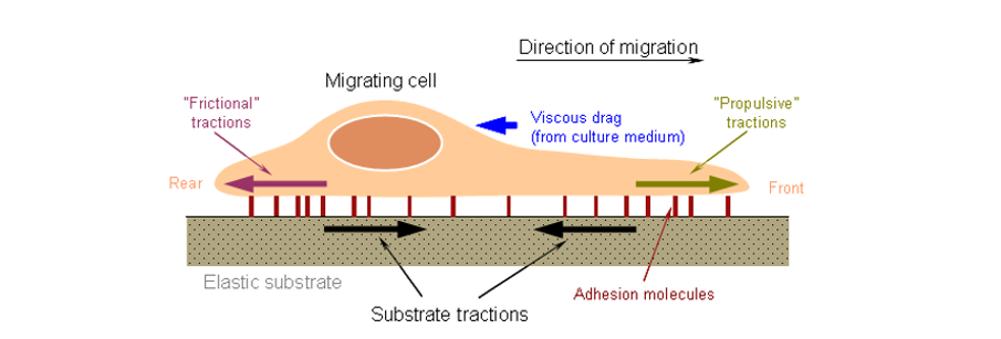
Cell migration refers to the movement of cells after they receive the migration signal or feel the gradient of certain substances. Cell migration is an alternate process of the extension of the pseudopod, the establishment of new adhesions, and the contraction of the cell body and tail in space and time. Cell migration is one of the basic functions of normal cells, a physiological process of normal growth and development, and a form of exercise that is common in living cells. It involves multiple physiological processes including embryonic development, angiogenesis, wound healing, immune response, inflammatory response, atherosclerosis, and cancer metastasis.
How do cells migrate?
The process of cell migration follows four steps: (1) the front end of the cell protrudes from the sheet-like pseudopod; (2) pseudopod in front of cell and extracellular matrix form new cell adhesion; (3) cell shrinkage; (4) adhesion and dissociation of the cell tail and surrounding matrix. The cell then moves forward. Cell migration requires coordination between the driving forces provided by extracellular and intracellular signaling molecules regulating cytoskeletal powerplants and the anchoring forces provided by actin-mediated adhesion. Many studies have shown that adhesive plaques, adhesive kinase, integrins and Rho family proteins play important roles in regulating cell migration.
How to measure cell migration?
Wound Healing Assay
1. Draw horizontal lines evenly on the back of the 6-well plate using a marker pen.
2. Seed 5X10⁵ cells to each well. Cell number can be adjusted accordingly depending on the cell type.
3. The next day, scratch the wells with a pipette tip against a ruler. The ruler should be as perpendicular to the horizontal line as possible. The tip of the pipette must be vertical and cannot be tilted.
4. Wash 3 times with PBS. Remove the excised cells before adding serum-free medium.
5. Culture the cells in a 37 °C 5% CO2 incubator. Harvest the samples at 0, 6, 12, 24 hours and take images.
Transwell Migration Assay
Place transwell in a culture plate. Culture medium in the upper and lower chambers are different, separated by a polycarbonate membrane. Seed cells in the transwell. Due to membrane permeability, cells are exposed to the medium from the lower chamber. This assay can be used to study the effect of the components in the lower chamber medium on cell growth and movement.
Track Cell Trajectories in Real Time
Single-cell migration real-time tracking is particularly suitable for the study of some physiological and pathological activities involving single-cell migration, such as inflammatory response, immune response, and tumor migration. A single cell is located at (t, X, Y) coordinates. The trajectory of the cell is established by tracking the entire movement of the cell, thereby qualitatively and quantitatively characterizing the dynamic characteristics of the cell, such as the distance, speed, direction, and duration of migration of the cell.
References:
1. Yamaguchi H, Wyckoff J, Condeelis J. Cell migration in tumors. Current opinion in cell biology, 2005, 17(5): 559-564.
2. Van Helvert S, Storm C, Friedl P. Mechanoreciprocity in cell migration. Nature cell biology, 2018, 20(1): 8.
