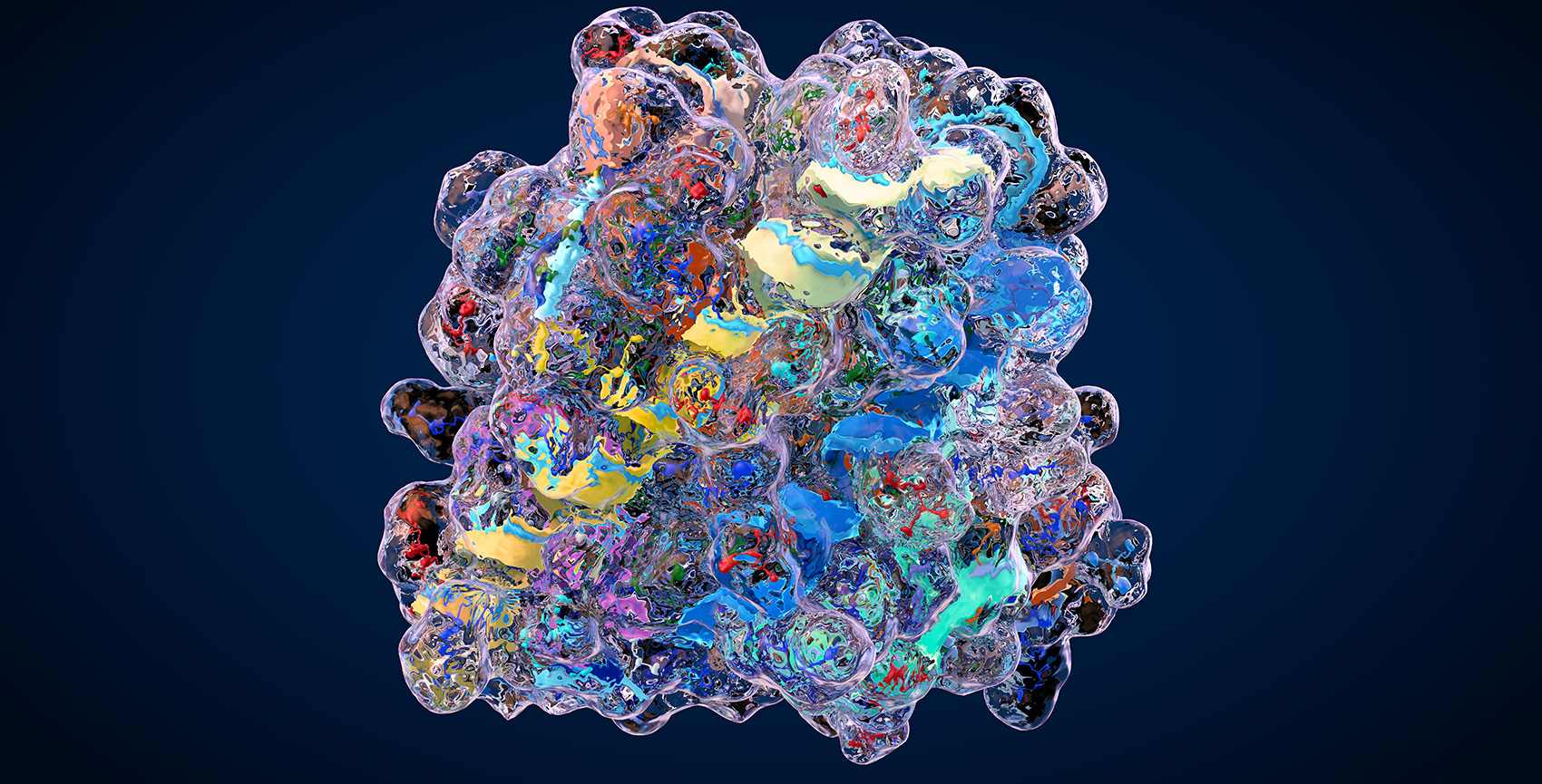
Protein crotonylation (Kcr) is a newly recognized type of protein modification, where a crotonyl group is added to lysine amino acids. First found on histone proteins—the core components of chromatin—it quickly gained attention as a key regulator of gene activity. By changing how tightly DNA is packaged, crotonylation makes certain genes more or less accessible for transcription.
Although it resembles acetylation, crotonylation has unique chemical features that affect protein behavior in distinct ways. These differences give it a specialized role in controlling biological processes. Recent studies have linked crotonylation to critical functions such as cell development, immune system signaling, energy metabolism, and disease mechanisms like cancer progression.
As its importance grows, scientists rely on advanced tools like mass spectrometry and disease model systems to accurately detect and study crotonylation. These methods help uncover how this modification shapes health and disease at the molecular level..
Biological Significance of Crotonylation in Health and Disease
Lysine crotonylation acts as a flexible regulator of gene activity and cell metabolism, with wide-ranging roles in both healthy and diseased states. On chromatin, crotonyl groups are added by the enzyme p300/CBP and removed by specific deacetylases (class I HDACs and sirtuins). These crotonyl marks loosen the DNA–protein complex, boosting the expression of genes that control the cell cycle, help cells respond to stress, and guide cell specialization.
Beyond histones, crotonylation also modifies key non-histone proteins—such as the tumor suppressor p53, the metabolic enzyme enolase 1, and various signal-relay molecules. Adding a crotonyl group can change a protein's shape, stability, or binding partners, directly tying crotonylation to how cells process energy and pass along signals.
In disease models, abnormal crotonylation patterns appear across several areas:
Cancer: Tumors in the colon and liver often show higher overall crotonylation. When p300 is overactive, it fuels cancer-promoting genes and can make cells less responsive to chemotherapy.
Brain Disorders: In models of Alzheimer's and Parkinson's, shifts in crotonylation change gene programs that control nerve connections and inflammation, contributing to memory loss and cell damage.
Immune Dysregulation: In infection or autoimmunity, crotonylation in macrophages and T cells fine-tunes the on/off switches for cytokine production, balancing defense against microbes with the risk of chronic inflammation.
Metabolic Disease: Levels of crotonyl-CoA (the building block for crotonylation) rise and fall with fat breakdown and compounds made by gut bacteria. These shifts alter crotonylation of liver and muscle enzymes, affecting how the body responds to insulin and linked to fatty liver disease.
Disease Models Where Crotonylation Matters
Protein crotonylation has been functionally characterized in several disease contexts, with direct evidence highlighting its regulatory roles in cancer, inflammation, metabolism, and neurobiology:
Cancer Models
Colorectal cancer & ENO1 crotonylation: In colorectal cancer (CRC), crotonylation at ENO1 K420 is significantly upregulated and promotes tumor cell growth, migration, and invasion through enhanced ENO1 activity and tumor associated gene expression pathways (Hou J Y, et al., 2024).
H2BK12cr in blood cells as CRC biomarker: A study measured H2BK12 crotonylation in peripheral blood mononuclear cells (PBMCs) from CRC patients, finding elevated levels correlated with distant metastasis and late TNM stage, with strong diagnostic performance (Hou J Y, et al., 2023).
H3K27cr and DNA damage in colon cancer: In colon cancer cell lines treated with DNA damaging agents (etoposide, camptothecin), H3K27cr decreased and was negatively regulated by SIRT6; promoters occupied by H3K27cr were enriched in genes associated with reduced DNA damage activity (Liao M J, wt al., 2022).
 Figure 1. Working model illustrates that PTBP1 crotonylation promotes colorectal cancer progression through alternative splicing-mediated upregulation of PKM2 (Hou J Y, et al., 2024).
Metabolic Reprogramming & Tumor Immunity
GCDH and H4 crotonylation in glioblastoma stem cells (GSCs): Dysregulated lysine catabolism in GSCs elevates crotonyl-CoA levels via upregulated GCDH and downregulated ECHS1, increasing histone H4 Kcr and promoting tumor growth. Disruption of this metabolic crotonylation axis triggers interferon signaling and suppresses GSC proliferation (Wei F C, et al., 2023).
Inflammatory Models (Macrophage Activation & Neuroinflammation)
Crotonyl-CoA induced macrophage activation: In a murine trigeminal ganglia (TG) inflammation model, administration of crotonyl-CoA lithium significantly upregulated IBA 1, and mRNA/protein levels of TNFα, IL 1β, and IL 6 by ~2 to 12 fold; effects were reversed by the p300 inhibitor C646, demonstrating Kcr’s sufficiency to induce macrophage activation and pro inflammatory signaling Zou Y, et al., 2022).
 Figure 2. Schematic diagram illustrating the possible mechanism of KCR in peripheral nerve injury-induced neuropathic pain (Zou Y, et al., 2022).
Acute Kidney Injury & Organ-Specific Stress
Histone crotonylation in kidney injury: Folic acid or cisplatin-induced acute kidney injury (AKI) in mice leads to increased histone crotonylation enriched at genes like PGC-1α and Sirt3 involved in mitochondrial biogenesis. Crotonate administration in vivo protected renal function and enhanced expression of protective genes, suggesting a potentially beneficial role of Kcr in organ stress response (Jun H W, wt al., 2019).
Workflow: From Disease Model to Crotonylome Map
- Sample Collection: Obtain tissues or cells from relevant disease models (e.g., xenografts, knockout mice, treated cell lines).
- Protein Extraction: Lyse cells/tissues using buffers compatible with downstream mass spectrometry and PTM preservation.
- Proteolytic Digestion: Digest extracted proteins into peptides using enzymes like trypsin.
- Crotonylated Peptide Enrichment: Enrich crotonylated peptides using pan-anti-Kcr antibodies via immunoaffinity purification.
- LC-MS/MS Analysis: Analyze enriched peptides using high-resolution liquid chromatography-tandem mass spectrometry.
- Data Processing & Identification: Identify crotonylation sites using proteomics software and annotated databases.
- Quantitative Analysis: Perform label-free or label-based quantification to compare crotonylation levels across samples.
- Bioinformatics & Interpretation: Map crotonylated proteins to pathways, networks, and disease-relevant functions.
Select Service
Related Article
Applications in Drug Discovery and Therapeutic Targeting
Target Identification and Validation
Crotonylation can modify key proteins involved in cancer progression, inflammatory signaling, and neurodegeneration. For instance:
- In colorectal and liver cancers, crotonylation of histone H3K18 is enriched at oncogene promoters and driven by increased crotonyl-CoA levels and p300 activity.
- In immune cells, crotonylation regulates proinflammatory gene expression, making immune signaling proteins promising targets for autoimmune and inflammatory disease therapies.
By profiling the crotonylome in diseased versus healthy tissues or in drug-treated versus untreated models, researchers can pinpoint crotonylated proteins that act as functional disease drivers—ideal candidates for small-molecule or biologic targeting.
Mechanistic Evaluation of Epigenetic Drugs
Crotonylation is closely interconnected with acetylation. Many HDAC inhibitors—such as vorinostat and romidepsin—not only increase acetylation but also indirectly elevate global crotonylation levels. Profiling crotonylation allows researchers to:
- Track on-target effects of HDAC and sirtuin inhibitors in preclinical models.
- Identify off-target pathways impacted through global epigenetic remodeling.
- Determine compound-specific PTM profiles to guide lead optimization.
3. Biomarker Discovery for Therapy Response
Disease- or treatment-specific crotonylation patterns can serve as predictive or prognostic biomarkers. For example:
- Elevated histone crotonylation has been correlated with poor prognosis in some cancers.
- Dynamic changes in crotonylation during drug treatment may indicate early resistance mechanisms, informing adaptive therapy strategies.
Rational Design of Combination Therapies
Crotonylation profiling helps uncover functional crosstalk with other PTMs such as acetylation, methylation, and phosphorylation. This enables:
- Discovery of co-regulated epigenetic pathways for synergistic targeting.
- Development of multi-target epigenetic therapies with increased efficacy.
- Optimization of drug scheduling and dosing to minimize resistance.
Future Directions and Emerging Research
Non-Histone Crotonylation
Although crotonylation was initially discovered on histones, recent research has uncovered many non-histone proteins that also carry this modification. These proteins play key roles in metabolism, cell cycle regulation, and immune responses. Crotonylation on non-histone proteins can influence their stability, where they are located within the cell, and how they interact with other proteins.
Single-Cell Crotonylomics
Emerging technologies aim to measure crotonylation at the single-cell level. Integrating MS with microfluidics or proximity labeling could uncover cellular heterogeneity in tumor microenvironments or developmental processes, offering insights not visible in bulk analysis.
Dynamic Crotonylation Mapping
Time-course studies and real-time monitoring of crotonylation in live cells or in response to treatment are gaining interest. These approaches can reveal transient regulatory events and help characterize PTM kinetics in signaling pathways.
Multi-Omics Integration
Combining crotonylome data with transcriptomics, metabolomics, and other PTM datasets (e.g., acetylation, phosphorylation) allows researchers to build comprehensive regulatory networks. This systems biology approach enhances our understanding of how crotonylation coordinates with other layers of cellular control.
Crotonylation in Clinical Contexts
Ongoing studies are exploring the diagnostic and prognostic potential of crotonylation signatures in patient samples. Differentially crotonylated proteins are being evaluated as biomarkers for cancer progression, drug resistance, and treatment response.
Relevant FAQ
Why profile crotonylation in disease models specifically?
Disease models such as cancer xenografts or neurodegenerative cell lines allow researchers to observe how crotonylation patterns change in pathophysiological states. This profiling helps identify disease-related regulatory mechanisms, potential drug targets, and biomarkers.
Which experimental techniques are best for detecting crotonylation?
The most effective method combines antibody-based enrichment with high-resolution LC-MS/MS. Label-free or TMT-labeled quantification can then be used for comparative analysis of crotonylation levels across samples or conditions.
How should I prepare biological samples for crotonylation analysis?
Samples should be rapidly collected and preserved under cold conditions to prevent PTM degradation. Proteins are then extracted, digested with trypsin, and crotonylated peptides are enriched using specific anti-Kcr antibodies before LC-MS analysis.
Can crotonylation serve as a reliable biomarker in disease research?
Yes. Altered crotonylation patterns have been linked to various diseases, including cancer, inflammatory disorders, and metabolic syndromes. Identifying disease-specific crotonylation signatures can support diagnostic and therapeutic development.
Are there any published databases of known crotonylation sites?
Yes. Databases like PhosphoSitePlus and CPLM (Compendium of Protein Lysine Modifications) list experimentally validated crotonylation sites. These resources can guide target selection and validation.
References
- Ji Y, Liu S, Zhang Y, et al. Lysine crotonylation in disease: mechanisms, biological functions and therapeutic targets. Epigenetics & Chromatin, 2025, 18(1): 13. DOI: 10.1186/s13072-025-00577-7
- Guo Y, Li J, Zhang K. Crotonylation modification and its role in diseases. Frontiers in Molecular Biosciences, 2024, 11: 1492212. DOI: 10.3389/fmolb.2024.1492212
- Jiang G, Li C, Lu M, et al. Protein lysine crotonylation: past, present, perspective. Cell death & disease, 2021, 12(7): 703. DOI: 10.1038/s41419-021-03987-z

Related Articals

Our products and services are for research use only.
