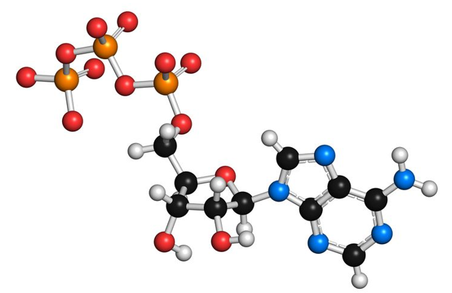
Protein phosphorylation refers to a process catalyzed by protein kinase to transfer the γ-position phosphate group of ATP or GTP to amino acid residues (serine, threonine, tyrosine, etc.) of the substrate protein. Protein phosphorylation is a common regulation method in the body and plays an important role in the process of cell signal transduction. Almost all proteins undergo some form of modification during or after synthesis. Abnormal post-translational modifications are associated with the occurrence of diseases. Certain specific post-translational modifications were developed as molecular markers or therapeutic targets for diseases. The most common and important protein post-translational modification, protein phosphorylation is a major mechanism that converts external stimuli into intracellular signals.
In the phosphorylation reaction, a strong negatively charged phosphate group is added to the amino acid side chain of the protein to cause esterification, which changes the configuration, activity, and performance of its interaction with other molecules. Esterification regulates many biological processes, such as signal transduction, gene expression, and cell division. The encoded protein will only become phosphorylated by specific amino acids. In eukaryotes, it is mainly phosphorylated by amino acid residues such as serine, threonine and tyrosine. In bacteria, residues such as aspartic acid, glutamic acid and histidine phosphorylate proteins. Some proteins are phosphorylated in both prokaryotes and eukaryotes, and their phosphorylation sites are usually arginine, lysine, and cysteine residues. Reversible protein phosphorylation regulates most functions of cells, such as energy storage, morphological changes, protein synthesis, gene expression, signal factor release, muscle contraction, and biochemical metabolism. Therefore, protein phosphorylation analysis and site identification have become one of the focuses in proteomics research.
The Main Types and Functions of Protein Phosphorylation
According to the different phosphate amino acid residues, phosphorylated proteins can be divided into 4 categories: O-phosphate protein, N-phosphate protein, acyl phosphate protein and S-phosphate protein.
- O-phosphate proteins are formed by phosphorylation of hydroxy amino acids (such as serine, threonine or tyrosine), but the phosphorylation of hydroxyproline or hydroxyIlysine is still unclear.
- N-phosphate proteins are formed by phosphorylation of arginine, lysine or histidine.
- Acyl phosphate proteins are formed by phosphorylation of aspartic acid or glutamic acid.
- S-phosphate proteins are formed by cysteine phosphorylation.
The functions of protein phosphorylation are diverse.
- articipate in other important enzymatic reactions (phosphorylation reaction produces intermediate products, mostly S or N phosphate, etc.)
- Mediate protein activities (protein molecules change performance through phosphorylation of protein kinases), such as protein kinase A (phosphorylated serine and threonine residues) or various receptor tyrosine kinases (phosphorylated tyrosine residues)
- Exert a variety of unique physiological effects. For example, aspartic acid, glutamic acid, and histidine phosphorylated proteins dissociate in the sensory transmission of bacterial chemotactic responses. Certain hormones also exist in the form of specific phosphorylated proteins in target tissues.
Research Methods for Protein Phosphorylation
In recent years, more researches focus on protein phosphorylation. Some topics include determining the site-specific phosphorylation of a protein, plant protein phosphorylation, and animal nerve cell phosphorylation. New research methods continue to emerge. The most widely used techniques are mass spectrometry and isotope labeling combined with immunoblotting-chemiluminescence analysis.
Mass spectrometry is a method of detecting the physical and chemical properties of charged moving particles (including atoms, molecules, and even molecular structure fragments) according to their mass-to-charge ratio by using electric and magnetic fields. The exact mass of a nuclide is a number of decimal places. The masses of any two nuclides are different, and one kind of nuclide is not exactly an integer multiple of the mass of another. Therefore, by measuring the accurate mass of the charged particles (ions), the corresponding compound composition, structure, and cracking rules can be determined.
Compared with unmodified peptides, the relative molecular mass of peptides modified with a phosphate group increases by 79.983. Generation of phosphopeptide means that a fragment ionic structure can be used for diagnosis, which is different from the unmodified peptide. Mass spectrometry can be used to identify protein phosphorylation. Different peptides are obtained by hydrolyzing the gel-separated protein by using a specific sequence hydrolase (such as trypsin). High-performance liquid chromatography is used to isolate the peptide of interest, followed by mass spectrometric analysis.
- Isotope labeling combined with immunoblotting-chemiluminescence analysis
This method uses radioisotope labeling, two-dimensional electrophoresis, and autoradiography to establish a proteome map. The target protein can be selectively identified and analyzed by immunoblotting persistent chemiluminescence, which can also be used to compare the expression of the same protein in multiple samples at the same time.
When the protein is phosphorylated, the incorporation of radioactive 32P and changes in molecular mass will cause gel shift, which makes immunoblotting occupy an important position in analytical detection.
When the energy produced by a chemical reaction is released in the form of light, it is called chemiluminescence. For example, one of the most commonly used chemiluminescence reagents, luminol, is oxidized by peroxide to produce an aminophthalate. When this product decays to a low energy state, a photon is released. Using patented additives to increase the luminous intensity and duration (stability) of the luminox reaction, an enhanced chemiluminescence (ECL) reagent was produced. In immunoblotting using ECL, chemiluminescent agents produce detectable light when enzymes and reaction substrates interact. The stable light signal output produced by the specific antibody at the appropriate dilution concentration helps maintain the consistency and sensitivity of protein detection.
References
1. Marchbanks R M. Protein phosphorylation. 1998.
2. Bou M, Guedouari H, et al. Role of Protein Phosphorylation among Oxidative Phosphorylation Components// eLS. 2017.