Direct answer
For protein hydrolysates, measure the native peptide mixture to define sequence coverage and cleavage products. For culture supernatants, design collection and controls to distinguish released or processed peptides from media background, cell leakage, and collection-time effects before comparing abundance across conditions.
Study Overview
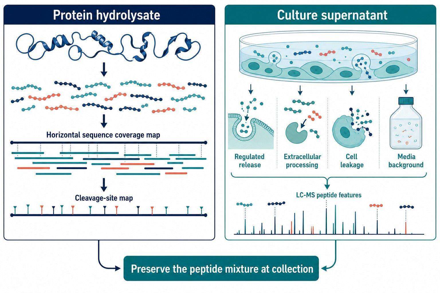
Peptidomics of protein hydrolysates and culture supernatants should measure the native peptide mixture that exists at collection—not create a new tryptic digest before analysis. For hydrolysates, the main question is which peptide sequences and cleavage products were generated from the starting substrate. For culture supernatants, the question expands to which peptides are released, processed, or accumulated in the extracellular environment and how confidently those changes can be quantified across conditions. Both projects use LC-MS/MS, but they require different experimental logic. A hydrolysate is often a controlled proteolysis system where sequence coverage and cleavage distribution are core outputs. A culture supernatant is a dynamic matrix in which peptide signals can reflect secretion, extracellular cleavage, intracellular leakage, media background, and collection timing. Treating both as generic “peptide identification” risks an answer that is technically correct yet scientifically unhelpful.
Key Takeaways for Hydrolysate and Culture Supernatant Peptidomics
- Preserve the peptide mixture present at sampling. Do not apply a routine proteomics digestion when the original cleavage products are the analytes of interest.
- For protein hydrolysates, report peptide sequences together with substrate coverage, cleavage-site patterns, and abundance distribution—not just the number of identified peptides.
- For culture supernatants, define media background, cell integrity, collection interval, and normalization before comparing peptide intensities across conditions.
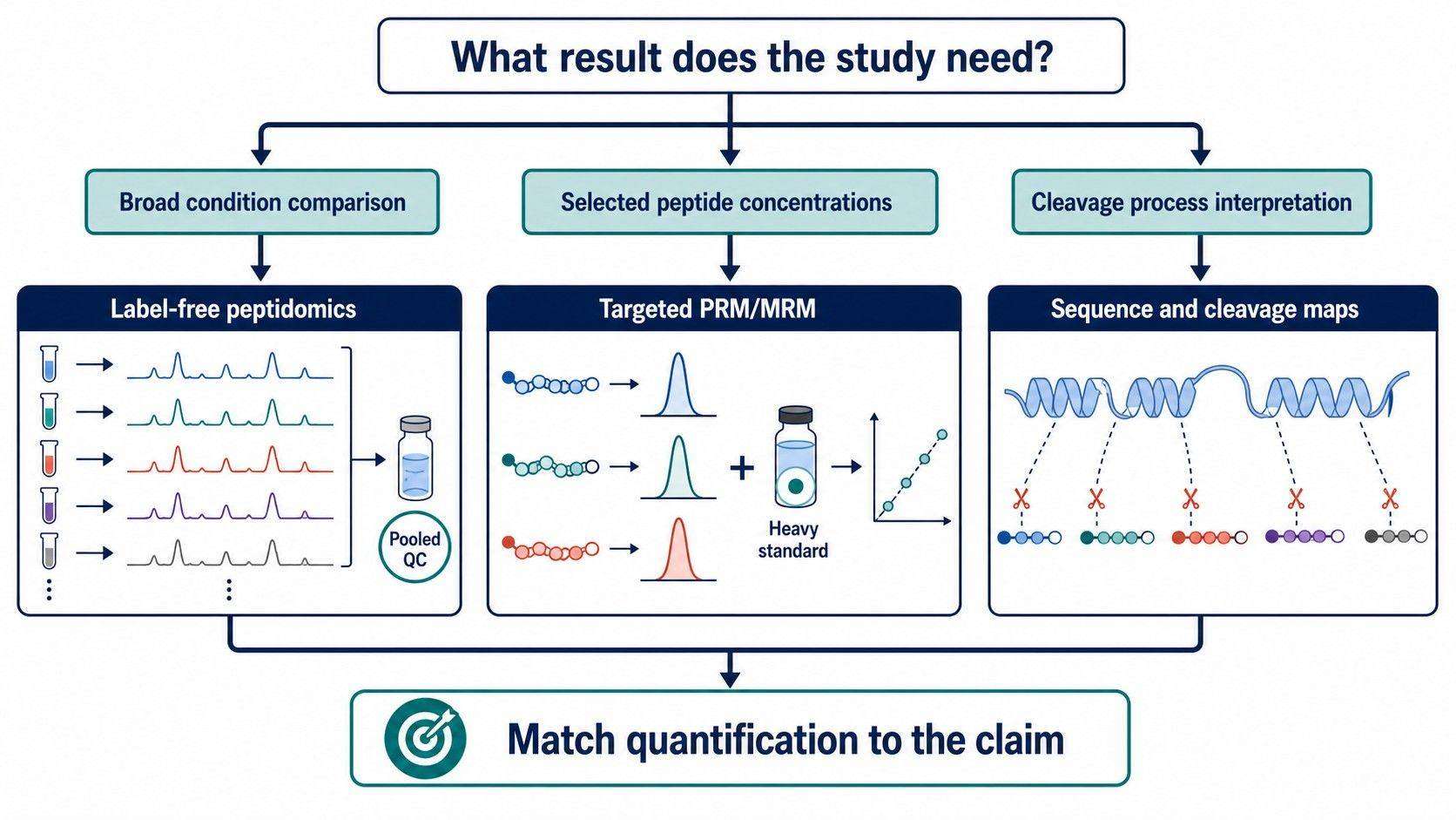
- Relative label-free quantification is appropriate for broad condition comparisons; stable-isotope-supported targeted quantification is appropriate when a limited peptide panel requires defensible concentration estimates.
- A peptide sequence observed in a supernatant is not automatically a secreted signaling peptide. Its origin must be interpreted using controls, source-protein context, and cleavage evidence.
Why Hydrolysate Peptidomics and Supernatant Peptidomics Need Different Study Designs
Protein hydrolysates and culture supernatants both contain complex mixtures of peptides, but the biological or process interpretation is different. In a hydrolysate, the sample usually begins with a known or partly known protein substrate and a defined processing event such as enzymatic hydrolysis, fermentation, digestion, or stability testing. The goal is to map products back to the substrate and understand which peptide bonds were cleaved or retained.
In culture supernatants, the observed peptide mixture is shaped by the cells, the culture medium, the collection interval, and extracellular processing. Some peptides may be actively released, some may arise from precursor processing or protease activity, and others may reflect cell damage or media-derived background. The study must be designed to distinguish these origins as far as the sample system allows.
Neither design should default to a bottom-up proteomics workflow. In conventional bottom-up proteomics, proteins are intentionally digested to create analyte peptides. In peptidomics, an in-workflow digestion can destroy the sequence boundaries and cleavage information that the study seeks to measure (Checco, 2023). This distinction should appear in the project brief, sample-handling plan, and final report.
For studies where native peptide composition and processing context matter, an endogenous peptidomics LC-MS/MS profiling service can be scoped around the starting material and intended biological or process claim rather than around a generic protein-identification workflow.
Protein Hydrolysate Peptidomics: What Sequence Coverage Should Mean
In a hydrolysate project, “sequence coverage” is often used too loosely. A high count of identified peptides may indicate analytical depth, but it does not by itself show whether the starting protein sequence is represented evenly, whether key regions are absent, or whether quantitative peptide recovery is balanced across the substrate.
Three complementary coverage measures
Peptide identity coverage asks which unique peptide sequences are identified. It is useful for cataloguing the hydrolysate but can be inflated by many overlapping forms from one substrate region.
Amino-acid or substrate-sequence coverage asks which positions in the parent sequence are represented by one or more observed peptides. It reveals regions that are repeatedly recovered, absent, or only represented by low-confidence signals.
Concentration-weighted sequence coverage asks whether the amount represented across the substrate sequence is balanced rather than merely detected. This is particularly relevant when the objective is to compare hydrolysis conditions, enzyme selectivity, or enrichment performance. A region represented by many trace-level peptides does not necessarily carry the same quantitative contribution as a region represented by one dominant peptide.
Vreeke and colleagues developed an untargeted identification and quantitative framework for protein hydrolysates that explicitly evaluated annotation completeness, protein recovery, and concentration-based sequence coverage. Their work showed why coverage metrics should be interpreted alongside peptide concentrations rather than treated as a single detection percentage (Vreeke et al., 2022).
Build the peptide search around the substrate knowledge
When the starting substrate is known, include the relevant protein sequence(s), species information, processing enzymes where applicable, and expected modifications in the data-analysis plan. This enables mapping of observed peptides to specific parent proteins and reconstruction of cleavage patterns.
When the substrate is heterogeneous, partially characterized, fermented, or contains unknown processing products, the search may need a broader sequence database and complementary de novo sequencing. The trade-off is a larger search space and a greater need for peptide-level confidence control. A clear report should distinguish sequence assignments supported by database evidence from de novo-only candidates.
If the aim is to compare products of hydrolysis rather than to identify every theoretical fragment, focus the analysis on reproducible, confidently identified peptides and the substrate regions they represent. The most useful output is a map of what changed, where it changed, and how large the change was.
How to Interpret Cleavage Patterns in Protein Hydrolysates
Cleavage-pattern analysis turns a peptide list into a process or mechanism result. Each observed peptide has an N- and C-terminus that can be mapped to the parent sequence. Across the dataset, those termini reveal which bonds are repeatedly cut, which regions remain protected, and whether different conditions shift the apparent specificity of the processing system.
Questions a cleavage map can answer
- Which parent-protein regions yield the largest diversity of peptide products?
- Do different enzymes, fermentation states, or treatment conditions enrich distinct cleavage sites?
- Are long intermediates accumulating because a processing step is incomplete?
- Are shorter peptides appearing in a way that suggests additional trimming or degradation?
- Do observed peptides cluster around known functional, structural, or modification-rich regions of the substrate?
The answer should not be reduced to a list of enzyme names inferred from a motif. Cleavage patterns are consistent with an active processing environment, but a motif alone does not prove that one protease caused every event. A defensible analysis combines the observed termini, condition-dependent abundance changes, substrate context, and, when available, orthogonal evidence about the protease system.
For example, a hydrolysate may show extensive overlapping peptides across a protein region. That pattern can indicate multiple cleavage events, incomplete processivity, or differential stability of related products. The interpretation becomes stronger when the same region and terminal pattern recur in independent processing replicates.
When a project specifically asks which proteolytic processes shape the peptide mixture, degradomics and protease profiling can be incorporated to keep cleavage-site evidence distinct from broad peptide abundance reporting.
Culture Supernatant Peptidomics: What Is Actually Measured?
A culture supernatant is not a blank extracellular compartment. It contains the media formulation, supplements, cell-derived material, extracellular enzymes, vesicles, and molecules released during the chosen collection interval. Peptidomics can reveal native peptides in this environment, but the project must define which biological interpretation is realistic.
Possible origins of a supernatant peptide
An observed supernatant peptide can arise from several, sometimes overlapping, sources:
- Regulated release or secretion of a mature peptide.
- Extracellular processing of a precursor protein or a released protein fragment.
- Shedding or cleavage of a membrane-associated protein.
- Release from extracellular vesicles or other particulate material, depending on sample preparation.
- Cell damage, lysis, or apoptosis-associated leakage.
- Background from serum, supplements, carrier proteins, or culture reagents.
The peptide sequence alone cannot always assign one origin. A convincing study includes matched medium-only controls, defined collection windows, cell-state or viability metadata where relevant, and a sample-preparation strategy that is consistent across conditions. The purpose is not to eliminate every background signal; it is to ensure that a reported condition difference is interpretable.
Define the collection window before the experiment
Time is part of the analyte definition. A short collection interval can emphasize acute release events but may yield less material. A longer interval can increase cumulative peptide recovery while also increasing the opportunity for extracellular degradation, nutrient depletion, and cell-stress-related release.
If the study compares treatments, all conditions should use matched collection duration, media composition, cell density or culture scale, and handling. A pooled composite across several time windows can be useful for discovery, but it cannot answer a time-resolved question. Preserve separate intervals if kinetic behavior is part of the research objective.
For neuronal cultures and organoid models, functional secretome profiling can be aligned with the relevant stimulation, culture, and collection context rather than assuming that every extracellular peptide reflects regulated release.
Peptide Identification Strategy: Database Search, De Novo Sequencing, or Both
Hydrolysate and supernatant peptidomics both benefit from a layered identification strategy. The appropriate balance between database search and de novo sequencing depends on how much is known about the possible sequences.
Database-informed identification
Database-informed searches are efficient when the substrate proteins, organism, construct, or culture components are known. They support parent-protein mapping, cleavage-position analysis, and a transparent link between observed peptide and possible source. Search parameters must allow non-tryptic termini and relevant modifications; otherwise, the workflow can systematically miss the very peptides of interest.
De novo sequencing for unanticipated peptide forms
De novo sequencing is valuable when hydrolysis creates unanticipated products, the substrate has incomplete sequence coverage, or a supernatant may contain peptides not adequately represented in the chosen reference database. It can extend discovery, but sequence proposals require careful confidence assessment and, where needed, confirmation with synthetic standards or targeted follow-up.
A practical hybrid approach
For a known hydrolysate, begin with a substrate-aware non-specific database search and use de novo analysis to investigate high-quality spectra that remain unassigned. For a complex supernatant, begin with a database that includes the cell system and defined media components, then use de novo analysis selectively for important unassigned features.
If the study must identify a short list of novel or ambiguous peptides with strong sequence evidence, peptide de novo sequencing should be planned as a confirmatory layer, not represented as a substitute for peptide-level QC.
How to Quantify Peptides in Hydrolysates and Culture Supernatants
Quantification should be fit for purpose. “Quantitative peptidomics” can mean relative comparison of many peptides or concentration measurement of a limited set. The correct choice depends on the scientific claim and the matrix.
Label-free relative quantification for broad comparisons
Label-free quantification is useful when the study needs to compare many peptide features across hydrolysis conditions, batches, cell states, or collection times. Each sample is measured independently, and intensities are aligned by m/z, retention time, and MS/MS evidence. The design should include biological replicates, pooled QC where a batch is large enough to need it, procedural blanks, and an appropriate normalization strategy.
For hydrolysates, normalization should account for starting substrate amount and process sampling. For supernatants, it should also be tied to the chosen biological normalization basis, such as collection volume together with a pre-specified culture-scale metric. Do not choose the denominator after inspecting the apparent peptide changes.
Targeted quantification for a defined peptide panel
Use targeted PRM/MRM with suitable reference materials when the study needs robust measurement of a small candidate set, comparison to a calibration relationship, or a concentration estimate for selected peptides. Heavy synthetic peptides can help control recovery and signal variability, but they need to be chosen for the actual non-tryptic peptide sequence and matrix behavior.
A 2025 study of Vicia faba hydrolysate peptides illustrates this point: non-tryptic peptides in a complex hydrolysate required an in-sample calibration strategy with heavy synthetic peptides rather than a simple transfer of tryptic-proteomics assumptions (Vanhoutte et al., 2025). This is an example of a general principle, not a mandatory design for every hydrolysate.
If the project needs broad discovery first and a small quantitative panel later, use untargeted peptidomics to choose candidates, then transition to peptide absolute quantification or targeted PRM/MRM only for peptides that meet the evidence and biological-priority criteria.
Side-by-Side Planning Table: Hydrolysates vs Culture Supernatants
| Planning element | Protein hydrolysates | Culture supernatants | Implication |
|---|---|---|---|
| Main biological/process question | What peptide products were generated from the substrate? | What peptides accumulate or are released in the extracellular environment? | Define the desired claim before selecting extraction and controls. |
| Key reference information | Starting protein sequence, species, processing condition, enzyme or fermentation context | Cell system, medium and supplements, collection interval, perturbation, cell-state metadata | The database and interpretation framework differ. |
| High-value output | Sequence/substrate coverage, cleavage maps, condition-dependent peptide abundance | Peptide identities, time- or condition-dependent abundance, source-context evidence | Do not use one reporting template for both matrices. |
| Major confounder | Co-elution, incomplete substrate annotation, process variation | Media background, cell leakage, density differences, extracellular degradation | Include matrix-specific controls. |
| Broad quantification | Relative peptide profiles across processing conditions | Relative peptide profiles across matched culture conditions | Use biological replication and pre-specified normalization. |
| Focused quantification | Selected product peptides or marker sequences | Selected released/processed peptides | Use targeted methods only after candidates are defined. |
Common Design Pitfalls in Hydrolysate and Supernatant Peptidomics
Digesting away the native peptide information
Adding a standard protease digestion before analysis can erase the endogenous termini and processing signatures that distinguish a hydrolysate or extracellular peptide profile. Preserve the sampled peptide mixture unless the project is explicitly a protein-level proteomics study.
Reporting peptide counts without coverage or abundance context
A list of hundreds of sequences does not reveal whether important substrate regions were covered or whether a few dominant peptides account for most of the mixture. Include sequence mapping and abundance-aware coverage where the substrate is known.
Calling every supernatant peptide “secreted”
Supernatant detection alone cannot resolve secretion from processing, leakage, or medium background. Use matched media controls, collection metadata, and cell-state context before attaching a biological source label.
Comparing intensities across unmatched culture conditions
Different collection volumes, medium formulations, culture density, or collection durations can create apparent peptide changes unrelated to the perturbation of interest. Match these factors prospectively and document the normalization rationale.
Treating an in-silico cleavage motif as direct proof of one protease
Cleavage patterns are evidence of processing, not automatic proof of causality. Interpret them alongside the condition, substrate context, and any orthogonal information about the relevant enzyme system.
When Should You Choose Hydrolysate Peptidomics, Supernatant Peptidomics, or Targeted Follow-Up?
Choose protein hydrolysate peptidomics when the objective is to identify product peptides, compare hydrolysis or fermentation conditions, map substrate coverage, or characterize cleavage patterns.
Choose culture supernatant peptidomics when the objective is to compare extracellular peptide landscapes across matched cell states, perturbations, or collection times. Build controls that distinguish media background and cell-state effects before interpreting release biology.
Choose targeted PRM/MRM or absolute quantification when broad discovery has produced a short list of high-priority peptides and the project now needs focused, repeatable measurements for those sequences.
Choose a hybrid workflow when the research question begins with an unknown peptide composition and ends with a specific process or biological candidate. Use untargeted discovery to map the system, then use targeted quantification for selected peptides that can support a narrower claim.
Creative Proteomics can help define the appropriate extraction, identification, cleavage-pattern analysis, QC, and quantification layer for hydrolysate or culture-supernatant samples. A useful inquiry specifies the sample matrix, starting substrate or cell system, collection/processing conditions, intended comparison, and whether the priority is discovery, coverage, cleavage interpretation, or selected-peptide quantification.
FAQ: Protein Hydrolysate and Culture Supernatant Peptidomics
Can LC-MS/MS show which regions of a protein hydrolysate were processed?
Yes. When parent protein sequences are available, observed peptide termini can be mapped to the substrate to show covered regions and recurrent cleavage sites. The result is stronger when peptide abundance and replication are considered alongside sequence identity.
Should protein hydrolysates be digested with trypsin before peptidomics?
Usually no, if the aim is to characterize the hydrolysis products already present. Trypsin would create new fragments and obscure the original peptide sequences and cleavage boundaries. Use a bottom-up digestion only when the stated goal is protein-level proteomics rather than native-peptide analysis.
How can a supernatant peptide be distinguished from media background?
Include medium-only controls processed alongside samples, maintain a documented media formulation, and compare peptide features against the relevant cell and reagent sources. Cell-state or viability information and matched collection conditions help prevent background or leakage from being misinterpreted as regulated release.
What does sequence coverage mean for a protein hydrolysate?
It can refer to the fraction of unique peptides identified, the positions on the parent sequence represented by observed peptides, or the concentration-weighted representation of those positions. The most useful metric depends on whether the goal is a qualitative catalogue or a quantitative comparison of processing outcomes.
When is label-free quantification sufficient?
Label-free quantification is appropriate for broad, relative comparison of many peptides across matched conditions with adequate replication and QC. It is not the best standalone choice when the project needs defensible concentration estimates for a small, high-priority peptide panel.
When should synthetic peptide standards be used?
Use them when selected peptides require focused confirmation, robust relative control, or absolute quantification. Standards should match the native non-tryptic sequence and be evaluated in the relevant sample matrix rather than assumed to behave like tryptic proteomics standards.
References
- Checco JW. Identifying and Measuring Endogenous Peptides through Peptidomics. ACS Chemical Neuroscience. 2023;14(20):3728-3731. doi: 10.1021/acschemneuro.3c00546.
- Vreeke GJC, Lubbers W, Vincken J-P, Wierenga PA. A method to identify and quantify the complete peptide composition in protein hydrolysates. Analytica Chimica Acta. 2022;1201:339616. doi: 10.1016/j.aca.2022.339616.
- Poliseli CB, de Carvalho A, et al. Tri- and dipeptides identification in whey protein and porcine liver protein hydrolysates by fast LC-MS/MS neutral loss screening and de novo sequencing. Journal of Mass Spectrometry. 2021;56(2):e4701. doi: 10.1002/jms.4701.
- Vanhoutte I, et al. Quantification of Peptides in Food Hydrolysate from Vicia faba. Foods. 2025;14(7):1180. doi: 10.3390/foods14071180.
- Fricker LD, Lim J, Pan H, Che FY. Peptidomics: Identification and Quantification of Peptides in Biological Samples. Mass Spectrometry Reviews. 2006;25(2):327-344. doi: 10.1002/mas.20078.
- Schulz C, et al. A protocol for analyzing the protein terminome of human cancer cell line culture supernatants. STAR Protocols. 2021;2(3):100728. doi: 10.1016/j.xpro.2021.100728.
Build a Peptidomics Workflow Around the Native Peptide Mixture
Tell us the substrate or culture system, sample collection process, desired sequence or quantitative output, and available experimental groups. We can help plan native-peptide preservation, identification, controls, and fit-for-purpose quantification.
Request a Quote