Direct answer
For short peptides (10–30 residues), Far-UV Circular Dichroism (CD) is sufficient when evaluating global secondary structure content, tracking formulation stability, screening thermal unfolding transitions (Tm), comparing batch-to-batch consistency, or profiling folding propensity via trifluoroethanol (TFE) titrations. Conversely, 2D/3D solution NMR is strictly required when the project demands residue-level dihedral angle (φ, ψ) restraints, atomic confirmation of disulfide bond connectivity in multicyclic peptides, delineation of dynamic conformational ensembles, spatial verification of non-canonical/D-amino acids, or definitive 3D coordinates for IND regulatory filings and patent claims.
Key Takeaways: Analytical Selection for Peptide Structural Biology
- Global Average vs Atomic Resolution: Far-UV CD measures the collective optical activity of peptide backbones, providing fractional secondary structure percentages without residue-specific location. NMR provides atomic-level chemical shifts, coupling constants (3JHNα), and Nuclear Overhauser Effect (NOE) distance restraints.
- Dynamic Conformational Ensembles: Short linear peptides rarely adopt a single rigid ground-state conformation in aqueous solution. Instead, they sample dynamic conformational ensembles where CD reports an intensity-weighted average, while NMR resolves populated sub-states through chemical shift indexing and temperature coefficients.
- TFE Titrations Reveal Latent Propensity: Co-solvent titrations with 2,2,2-trifluoroethanol (0–50% TFE) in CD distinguish intrinsically disordered peptides from those possessing latent, inducible α-helices (such as amphipathic target-binding domains).
- Disulfide Regioisomer Verification: For peptides with two or more disulfide bridges (e.g., bicyclic peptides, conotoxins, defensins), CD cannot establish pairing topology. NMR is mandatory to confirm correct covalent connectivity through inter-cysteine NOE networks.
- Cost-Effective Staged Strategy: The most resource-efficient workflow utilizes Far-UV CD as an initial high-throughput triage filter (<50 μg peptide) to establish foldedness and stability, advancing only structurally validated lead candidates to multi-dimensional NMR characterization (milligram scale).
The Short Peptide Conformation Paradox: 10 to 30 Amino Acids
In biopharmaceutical discovery, therapeutic peptides occupying the 10- to 30-amino-acid range—such as glucagon-like peptide-1 (GLP-1) receptor agonists, bicyclic tumor-homing peptides, antimicrobial peptides (AMPs), and peptide-drug conjugate (PDC) linkers—represent a challenging structural category. Unlike globular proteins that possess extensive hydrophobic cores driving cooperative folding into rigid, unvarying tertiary structures, short linear peptides lack sufficient tertiary contacts to overcome the conformational entropy of the polypeptide chain.
Consequently, in aqueous solution at physiological temperature and ionic strength, short unmodified peptides rarely adopt a single, static ground-state structure. Instead, they exist in rapid, dynamic equilibrium across a broad conformational ensemble, sampling transient α-helical turns, nascent β-hairpins, polyproline II (PPII) conformations, and unstructured random coils on picosecond-to-nanosecond timescales. This intrinsic flexibility creates a pervasive analytical dilemma: when a drug development team requires structural characterization for lead optimization, target engagement modeling, or regulatory submission, which biophysical technique provides genuine, actionable insight without squandering budget and synthetic material?
Drug developers routinely debate whether Far-UV Circular Dichroism (CD) is legally and scientifically sufficient, or whether investing in two-dimensional Nuclear Magnetic Resonance (2D NMR) spectroscopy is non-negotiable. Answering this question requires moving past generic marketing claims and dissecting the exact physical phenomena captured by each instrument.
For research teams establishing formal structural pipelines, our validated peptide structure analysis services offer fully integrated biophysical characterization spanning chiroptical spectroscopy, high-field NMR, and mass spectrometry-based topological mapping.
Circular Dichroism: What Far-UV Measures and What It Misses
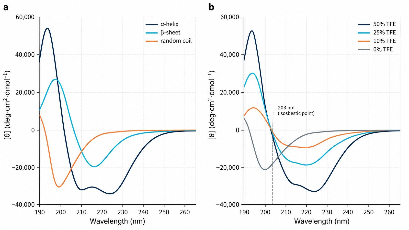
Far-UV Circular Dichroism (190–260 nm) measures the differential absorption of left- and right-circularly polarized light (ΔA = AL - AR) by chiral chromophores. In peptides, the primary absorbing chromophore in this spectral window is the repeating peptide bond (amide linkage), specifically the electronic transitions of the carbonyl π → π* (polarized along the C=O bond at ~190 nm) and n → π* (electron transfer from the non-bonding oxygen lone pair to the antibonding π orbital at ~222 nm).
Diagnostic Spectral Signatures
When peptide amides align into regular secondary structures, chiral exciton coupling between adjacent amide transitions produces highly distinct Far-UV spectral fingerprints:
- α-Helix: Characterized by pronounced negative minima at 222 nm (n → π*) and 208 nm (π → π* parallel), alongside a strong positive maximum at 193 nm (π → π* perpendicular). The mean residue ellipticity at 222 nm ([θ]222) serves as a quantitative benchmark for fractional helical content.
- β-Sheet: Displays a single broad negative minimum at 216–218 nm and a positive band between 195 and 200 nm. Due to geometric variability across parallel versus antiparallel strands and twist angles, β-sheet signatures exhibit wider spectral dispersion than α-helices.
- Random Coil / Unstructured State: Lacks long-range order, yielding a dominant negative absorption band near 198 nm, with near-zero or weakly positive ellipticity above 215 nm.
The Deconvolution Bottleneck in Short Peptides
To quantify secondary structure composition, researchers apply computational deconvolution algorithms (such as CONTINLL, CDSSTR, SELCON3, or BeStSel) that model the experimental spectrum as a linear combination of reference spectra derived from globular proteins of solved crystallographic structure. In short peptides, however, this mathematical approach suffers from severe systematic errors:
- End-Fraying and Non-Cooperative Terminal Effects: In a globular protein of 200 residues, terminal amides represent <2% of the signal. In a 15-amino-acid peptide, the unconstrained terminal residues constitute 25–30% of the entire chain. These fraying ends contribute disordered spectral components even within a fully folded functional core, causing standard algorithms to underestimate true helicity.
- Absence of Residue Localization: A CD deconvolution result stating "42% α-helix, 18% β-turn, 40% random coil" provides zero spatial information. It cannot reveal whether residues 1–6 form the helix while 7–15 are disordered, or whether the entire peptide samples a dynamic helical state 42% of the time.
- Aromatic and Disulfide Optical Interference: Side-chain chromophores from tyrosine, tryptophan, phenylalanine, and rigid cystine disulfide bridges contribute electronic transitions in the 190–240 nm range. In short peptides with high aromatic density, these side-chain transitions distort the amide signal, leading algorithms to report phantom β-sheet content where none exists.
The Power of TFE Titration in CD
Far-UV CD excels in evaluating structural plasticity via solvent-perturbation assays. By titrating 2,2,2-trifluoroethanol (TFE, 0% to 50% v/v) into the peptide solution, investigators alter the local dielectric constant and weaken peptide-solvent hydrogen bonding, thereby strengthening intramolecular hydrogen bonds. Peptides that exhibit an intrinsically disordered profile in water but adopt a classic double-minima α-helical spectrum upon TFE addition possess a high latent helical propensity. This transition frequently mimics the conformational induction that occurs when the peptide binds its physiological receptor membrane or protein target pocket.
For projects requiring high-precision chiroptical characterization, explore our dedicated CD-based peptide secondary structure analysis platform, optimized for micro-volume thermal melting and solvent titration profiling.
Solution NMR: Resolving Atomic Dihedrals, Side Chains, and Ensembles
Where Circular Dichroism provides a macroscopic optical average, high-resolution solution Nuclear Magnetic Resonance (NMR) spectroscopy (500–900 MHz) provides an atomic lens. In short peptides, NMR operates without the molecular weight limitations that complicate large multi-subunit proteins, providing sharp, high-intensity resonances that can be assigned to individual nuclei across the entire sequence.
Two-Dimensional Homonuclear and Heteronuclear Experiments
Structural characterization of unlabelled synthetic peptides relies primarily on homonuclear two-dimensional 1H-1H experiments, augmented by heteronuclear single quantum coherence (HSQC) at natural isotopic abundance:
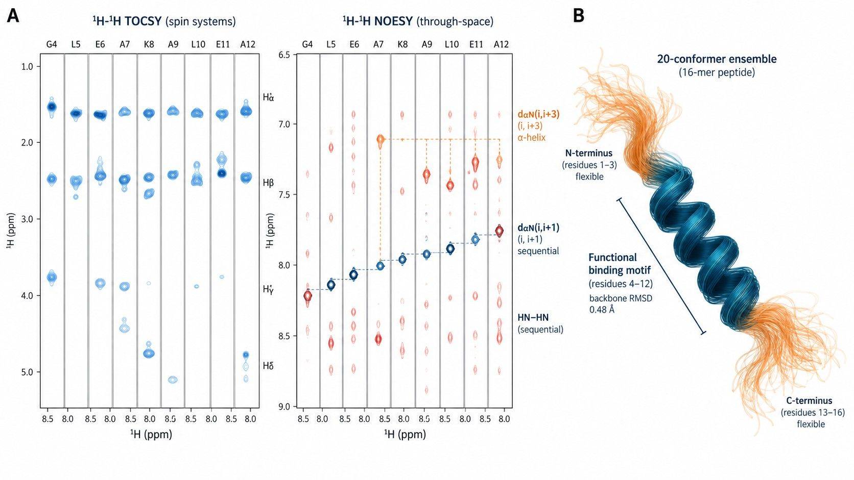
- 1H-1H TOCSY (Total Correlation Spectroscopy): Establishes scalar coupling across complete through-bond spin systems, allowing definitive identification of each amino acid residue's characteristic chemical shift pattern (NH → Hα → Hβ → Hγ → Hδ).
- 1H-1H NOESY / ROESY (Nuclear Overhauser Effect Spectroscopy): Measures dipolar cross-relaxation through space, yielding cross-peaks whose intensities are inversely proportional to the sixth power of the inter-proton distance (r-6). Characteristic sequential NOE patterns—such as dense dαN(i, i+3) and dαβ(i, i+3) contacts—provide indisputable proof of α-helical turns. Conversely, strong inter-strand dαα(i, j) contacts establish antiparallel β-sheet geometry.
- 1H-15N HSQC (at Natural Abundance): For peptides available at 1–5 mM concentrations, 1H-15N HSQC tracks backbone amide nitrogen chemical shifts without costly uniform 15N enrichment. Chemical shift dispersion in the 1H dimension (7.0–9.0 ppm) serves as a rapid diagnostic readout of peptide folding: collapsed, overlapping amide peaks indicate conformational disorder, while well-dispersed resonances confirm a single, stable chemical environment.
Quantitative Restraints and Ensemble Calculations
By compiling hundreds of experimentally derived upper-distance bounds from NOESY cross-peaks, three-bond scalar coupling constants (3JHNα, extracted from high-resolution 1D or DQF-COSY spectra to calculate backbone φ angles via the Karplus relation), and temperature coefficients of amide proton chemical shifts (ΔδNH/ΔT, where values > -4.5 ppb/K identify amides protected by intramolecular hydrogen bonds), computational molecular dynamics algorithms (such as CYANA or XPLOR-NIH) calculate a family of converged structures.
Rather than forcing a short peptide into a deceptive, single static PDB model, modern NMR reporting represents the peptide as an ensemble of 20 to 50 conformers that satisfy all experimental distance and dihedral boundaries. This structural ensemble directly visualizes which residues remain rigid (low root-mean-square deviation, RMSD < 0.5 Å) and which segments retain flexibility.
To inspect residue-resolved chemical shifts, spatial restraints, and conformer modeling, review our full-suite NMR-based peptide structure analysis capabilities.
Comparative Capability Matrix: Far-UV CD vs High-Resolution Solution NMR
Selecting the correct analytical technology requires balancing biophysical resolution against sample consumption, instrument time, and development cost. The following matrix contrasts the core analytical boundaries of both platforms:
| Analytical Metric / Capability | Far-UV Circular Dichroism (190–260 nm) | 2D/3D Solution NMR Spectroscopy (500–900 MHz) | Decision Implication for Short Peptides (10–30 aa) |
|---|---|---|---|
| Spatial Resolution | Global ensemble average across all amide chromophores; zero residue-specific resolution | Atomic, residue-by-residue resolution of backbone (φ, ψ) and side-chain (χ) dihedral angles | CD cannot pinpoint which residues participate in secondary structure; NMR maps the exact boundary of helical or folded segments. |
| Sample Quantity & Concentration | Low consumption: 0.1–0.5 mg/mL; ~20–50 μg total peptide per optical cuvette | Moderate to high consumption: 1.0–5.0 mM (1.5–10 mg total peptide in 500 μL NMR tube) | CD is ideal for scarce early-discovery screening; NMR requires milligram-scale synthetic investment. |
| Conformational Heterogeneity Handling | Linear spectral superposition of dynamic sub-states; deconvolution algorithms yield fractional secondary structure percentages | Measures ensemble-averaged chemical shifts, residual dipolar couplings (RDCs), and NOEs; computes conformational ensemble distributions | Linear short peptides exist as dynamic ensembles; CD yields ambiguous percentage estimates, while NMR characterizes the conformational landscape. |
| Disulfide Bond & Topology Mapping | Near-UV CD (250–320 nm) detects chiral disulfide presence, but cannot assign connectivity | Inter-cysteine NOEs (Hβ–Hβ, Hα–Hβ) and 13Cβ chemical shifts definitively establish regioisomeric disulfide pairings | For multi-cyclic or constrained peptides (e.g., bicyclic peptides, conotoxins), NMR is mandatory to confirm correct topological folding. |
| Thermodynamic & Formulation Profiling | Rapid, automated temperature ramping (thermal melting, Tm) and solvent titrations (TFE, pH, salt, detergents) | Variable-temperature NMR requires extensive acquisition time per temperature point (hours to days) | CD is vastly superior for high-throughput stability screening, excipient compatibility, and TFE-induced folding assays. |
| Regulatory & Patent Filings (IND / CQA) | Accepted as a qualitative batch-to-batch consistency and secondary structure identity test | Required for definitive 3D structural proof, atomic contact mapping, and stereochemical purity verification | CD suffices for routine lot release; NMR is indispensable for novel therapeutic IND filings and patent claims. |
Technical Decision Matrix: Selecting CD, NMR, or an Orthogonal Hybrid Workflow
Different synthetic peptide modalities present vastly distinct structural stability, folding mechanics, and regulatory hurdles. The matrix below defines the optimal primary and secondary analytical pathways across major peptide classes:
| Peptide Modality & Structural Architecture | Recommended Primary Analytical Tool | Secondary / Orthogonal Confirmation Tool | Scientific Rationale & Technical Risk Mitigation |
|---|---|---|---|
| Linear Hydrophilic Peptides (10–25 aa, No Cys) | Far-UV CD with TFE Titration (0–50% v/v) | 1D 1H NMR (backbone amide dispersion check) | Most linear short peptides are intrinsically disordered in water. CD rapidly evaluates latent α-helical propensity; expensive 2D NMR is unjustified unless a stable fold is detected. |
| Stapled / Macrocyclic α-Helical Peptides | Far-UV CD (Absolute Mean Residue Ellipticity [θ]222) | 2D 1H-1H NOESY / TOCSY NMR | CD provides quantitative, rapid calculation of percent helicity across chemical iterations. NMR is reserved for the lead candidate to verify the atomic stapling geometry. |
| Disulfide-Constrained Peptides (2–3 Disulfides) | 2D Solution NMR (NOESY / TOCSY) | LC-MS/MS Differential Alkylation Mapping | Multiple disulfide combinations generate topological isomers with indistinguishable mass and identical amino acid composition. Only NMR confirms exact spatial connectivity. |
| Peptide-Drug Conjugates (PDCs) & Lipid Lipopeptides | Far-UV CD (in vehicle-matched buffer) | 2D 1H-15N HSQC NMR (at natural abundance) | Bulky payloads or palmitoyl chains induce self-assembly and micelle scattering. CD screens monomeric vs aggregated states; NMR verifies whether conjugation perturbs target-binding secondary structure. |
| D-Amino Acid / Non-Canonical Backbone Peptides | 2D/3D Solution NMR | Chiral Amino Acid Analysis (AAA) | Non-canonical residues distort standard CD reference spectra (causing deconvolution algorithms to fail). NMR directly tracks chiral chemical shift perturbations and atomic packing. |
Staged Analytical Workflow: Mitigating Cost and De-risking Peptide Development
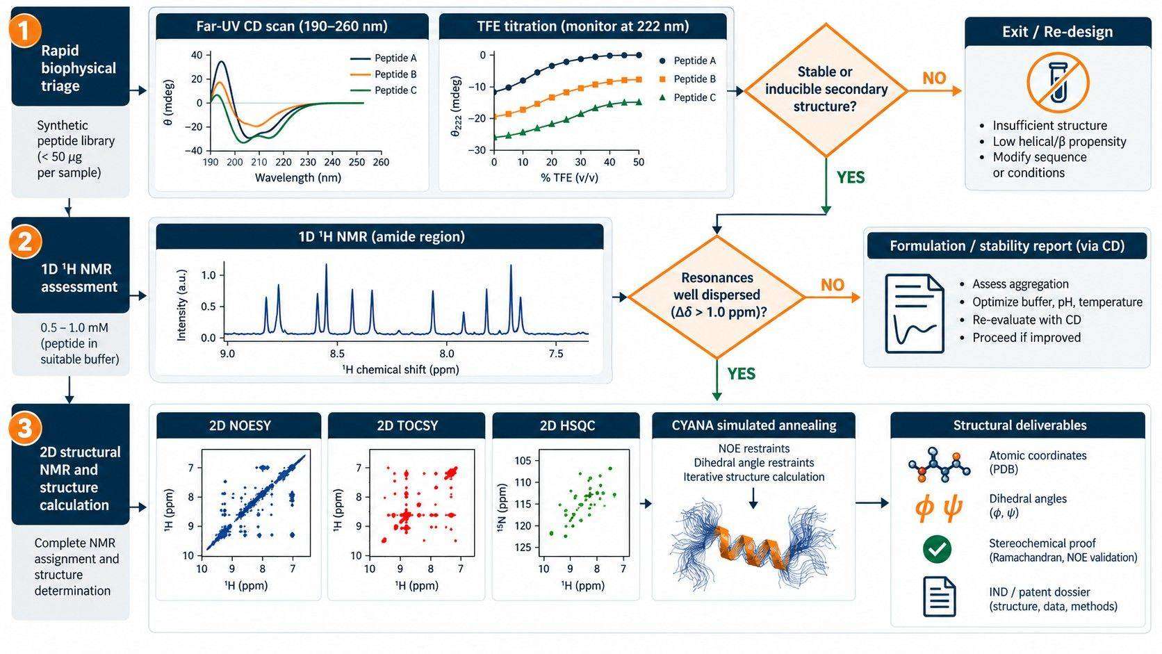
In commercial biotechnology and academic pharmacology, deploying full-scale 2D NMR on dozens of early chemical variants is neither cost-effective nor methodologically sound. A staged structural characterization funnel ensures that resources are allocated strictly to validated, tractable candidates.
Tier 1: High-Throughput CD Screen & TFE Profiling
Evaluate peptide variants (0.2 mg/mL, aqueous buffer, pH 7.0). Record Far-UV CD spectra from 190 to 260 nm. Perform TFE titration (0%, 20%, 40% v/v) to measure intrinsic vs inducible secondary structure. Screen thermal denaturation from 4°C to 90°C to confirm cooperative unfolding. Discard completely unstructured or insoluble analogues.
Tier 2: 1D 1H NMR Chemical Dispersion Check
Acquire 1D 1H NMR spectra (600 MHz, 0.5–1.0 mM peptide in 90% H2O / 10% D2O). Evaluate backbone amide proton dispersion (7.5–9.0 ppm) and methyl proton upfield shifting (<0.8 ppm). Sharp, well-dispersed peaks confirm a preferred stable fold, while narrow, collapsed clusters indicate random coil averaging.
Tier 3: 2D NOESY / TOCSY Atomic Resonance Assignment
For lead candidates, record high-resolution 2D homonuclear TOCSY (mixing time 70–80 ms) and NOESY (mixing time 150–250 ms) spectra. Sequentially assign backbone and side-chain protons via dαN and dNN connectivity pathways. Extract 3JHNα coupling constants and temperature coefficients (ΔδNH/ΔT).
Tier 4: Simulated Annealing Ensemble Calculation
Convert NOE peak volumes into semi-quantitative distance restraints. Generate 100 structural models via restrained molecular dynamics simulated annealing. Select the 20 lowest-energy conformers with zero experimental distance violations (>0.2 Å) to generate the final publication- and IND-ready 3D ensemble coordinates.
Common Analytical Pitfalls in Peptide Structural Biology
1. Over-interpreting CD Deconvolution Percentages
Treating CD secondary structure fractions (e.g., "34% α-helix") as literal, static representations of a short peptide is a major failure mode. In short linear sequences, a reported 34% helicity typically means the peptide occupies a fully helical state for 34% of the time across the ensemble, or that a central 5-residue turn is transiently formed. Without NMR chemical shift indexing or NOE contacts, attributing functional properties to a specific segment based solely on CD deconvolution is scientifically unsupportable.
2. Cuvette Artifacts Caused by Aggregation and Turbidity
Hydrophobic peptides, amphipathic helices, and lipidated peptides readily self-assemble into soluble oligomers, protofibrils, or micelles. As particles approach dimensions comparable to the Far-UV wavelength (190–220 nm), differential light scattering (differential absorption and reflection) causes severe baseline distortion, artificial peak flattening, and spurious β-sheet-like inverted bands. Dynamic light scattering (DLS) or concentration-dependent CD measurements should always precede deconvolution.
3. Overlooking Non-Aqueous Solvent Artifacts in NMR
Because many short peptides appear disordered in pure water, researchers frequently dissolve them in pure organic solvents (such as 100% DMSO-d6, methanol-d4, or chloroform-d) to achieve sharp NMR spectra. While organic solvents induce secondary structure by suppressing competing solvent-solute hydrogen bonds, the resulting conformations may have zero relevance to the active binding state in physiological saline. Whenever feasible, structures must be solved in aqueous buffer or matched membrane-mimetic environments (e.g., DPC or SDS micelles).
4. Neglecting Cis/Trans Proline Isomerization
Peptides containing proline residues undergo spontaneous cis/trans isomerization about the Xaa-Pro tertiary amide bond. Because the activation barrier is high (~20 kcal/mol), the interconversion rate is slow on the NMR chemical shift timescale (milliseconds to seconds). This yields dual sets of NMR resonances for adjacent residues. Failing to recognize proline isomerization leads to misassignment of duplicate spin systems as chemical impurities or conformational heterogeneity.
Frequently Asked Questions: CD vs NMR for Peptide Analysis
Why does my 15-mer peptide show random coil by CD in water, but binds its receptor with nanomolar affinity?
This phenomenon—termed "folding-upon-binding"—is widespread among short therapeutic peptides. In the absence of a binding partner, the conformational entropy of the free peptide favors an unstructured random-coil ensemble. Upon encountering the hydrophobic binding pocket of the receptor, the peptide undergoes induced-fit folding into its active secondary structure. Far-UV CD in pure water only captures the free unliganded state. Adding 20–40% TFE to the CD cuvette typically unmasks this latent binding conformation by inducing the secondary structure that forms upon receptor engagement.
Can Far-UV CD prove that two batches of a therapeutic peptide have identical 3D structures?
CD is widely accepted in CMC regulatory filings as a rapid, sensitive method for demonstrating conformational comparability and batch-to-batch consistency. If two production lots display superimposable Far-UV CD spectra (with identical mean residue ellipticity values across 190–260 nm) and identical thermal denaturation profiles (Tm within ±0.5°C), they share equivalent global secondary structure. However, CD cannot detect subtle localized side-chain alterations, localized epimerization, or alternative disulfide pairings, which require 1D/2D NMR or LC-MS/MS mapping.
Is uniform isotopic labeling (13C, 15N) mandatory for 2D NMR analysis of short peptides?
No. For short peptides containing 10 to 30 residues, uniform 13C/15N isotopic enrichment is generally unnecessary. Standard homonuclear 2D 1H-1H experiments (TOCSY and NOESY) recorded on modern high-field spectrometers (600–800 MHz equipped with cryogenic probes) provide sufficient chemical shift dispersion and sensitivity to resolve nearly all resonances at natural isotopic abundance, provided 1–5 mg of pure synthetic peptide is available.
How does near-UV Circular Dichroism differ from far-UV CD in peptide studies?
Far-UV CD (190–260 nm) probes the peptide backbone amide bond and reports secondary structure (α-helices, β-sheets, turns). Near-UV CD (250–320 nm) probes aromatic amino acid side chains (phenylalanine 255–270 nm, tyrosine 275–285 nm, tryptophan 290–305 nm) and disulfide bonds (250–290 nm). If aromatic residues are locked into a rigid, chiral tertiary environment, they produce distinct near-UV ellipticity signals. In flexible short peptides lacking tertiary structure, the near-UV CD spectrum is typically flat and featureless.
What is the minimum concentration required for a reliable Far-UV CD measurement?
The standard concentration range for Far-UV CD is 0.1 to 0.5 mg/mL using a 0.1 cm (1 mm) pathlength quartz cuvette. For a 20-amino-acid peptide (MW ~2,200 Da), this corresponds to approximately 50 to 200 μM. Using concentrations below 0.05 mg/mL results in an unacceptable signal-to-noise ratio below 205 nm, while concentrations exceeding 1.0 mg/mL cause complete optical absorption (detector saturation) below 200 nm.
Can NMR distinguish between D-amino acids and L-amino acids in a synthetic peptide?
Yes. Incorporation of a D-amino acid into an L-peptide backbone alters local chirality, disrupting local scalar couplings (3JHNα) and inducing substantial chemical shift perturbations in adjacent residues. Furthermore, in NOESY spectra, a D-amino acid produces distinctive inverted or altered sequential dαN(i, i+1) and dNN(i, i+1) distance contacts compared to the all-L stereoisomer, providing unambiguous verification of stereochemical purity.
References
- Kelly, S. M., Jess, T. J., & Price, N. C. (2005). How to study proteins by circular dichroism. Biochimica et Biophysica Acta (BBA) - Proteins and Proteomics, 1751(2), 119–139. doi:10.1016/j.bbapap.2005.06.005.
- Wüthrich, K. (2001). The way to NMR structures of proteins. Nature Structural Biology, 8(11), 923–925. doi:10.1038/nsb1101-923.
- Mielke, S. P., & Krishnan, V. V. (2009). Characterization of protein secondary structure by circular dichroism spectroscopy: a review of the tools and algorithms. Analytical Biochemistry, 385(1), 1–11. doi:10.1016/j.ab.2008.10.025.
- Greenfield, N. J. (2006). Using circular dichroism spectra to estimate protein secondary structure. Nature Protocols, 1(6), 2876–2890. doi:10.1038/nprot.2006.202.
- Dyson, H. J., & Wright, P. E. (2005). Intrinsically unstructured proteins and their functions. Nature Reviews Molecular Cell Biology, 6(3), 197–208. doi:10.1038/nrm1589.
- Pelton, J. T., et al. (1986). Conformation of substance P in solution: a 1H NMR and circular dichroism study. Biochemistry, 25(7), 1632–1638. doi:10.1021/bi00355a029.
- Sreerama, N., & Woody, R. W. (2000). Estimation of protein secondary structure from circular dichroism spectra: comparison of CONTIN, SELCON, and CDSSTR methods with an expanded reference set. Analytical Biochemistry, 287(2), 252–260. doi:10.1006/abio.2000.4880.
Design a Robust Structural Pipeline for Your Peptide Program
Whether you require rapid Far-UV CD secondary structure screening, TFE-induced folding characterization, or residue-resolved 2D/3D NMR structural ensemble determination, Creative Proteomics delivers rigorous, publication- and IND-ready biophysical datasets.
Request a Technical Consultation