Direct answer
Choose allele-specific H-2Kb or H-2Db enrichment when the study needs allele-resolved evidence. Pool mouse material only when discovery depth is more important than preserving individual biological variation, and establish tissue-specific feasibility in a pilot before committing the full study.
Study Overview
Mouse MHC-I immunopeptidomics should start with the strain haplotype and the biological unit that must remain interpretable. In C57BL/6-derived material, H-2Kb and H-2Db present overlapping but distinct peptide repertoires; use allele-specific enrichments when allele-resolved evidence is required, and pool only when the gain in detectable material outweighs the loss of individual-level information. The central feasibility question is not simply “how much tissue is available?” MHC-I peptide recovery depends on tissue cellularity, surface MHC-I abundance, strain, inflammatory state, lysis recovery, antibody specificity, and the intended result: a broad discovery catalogue, a comparison between experimental groups, or confirmation of selected ligands. These considerations determine whether one mouse, several pooled mice, or a staged pilot is appropriate.
Key Takeaways for Mouse MHC-I Immunopeptidomics Study Design
- Confirm the mouse strain and classical MHC-I haplotype before selecting the enrichment antibodies and peptide-binding reference model.
- In C57BL/6 studies, H-2Kb and H-2Db should be treated as separate presentation contexts when allele-specific motifs or source-peptide assignment matter.
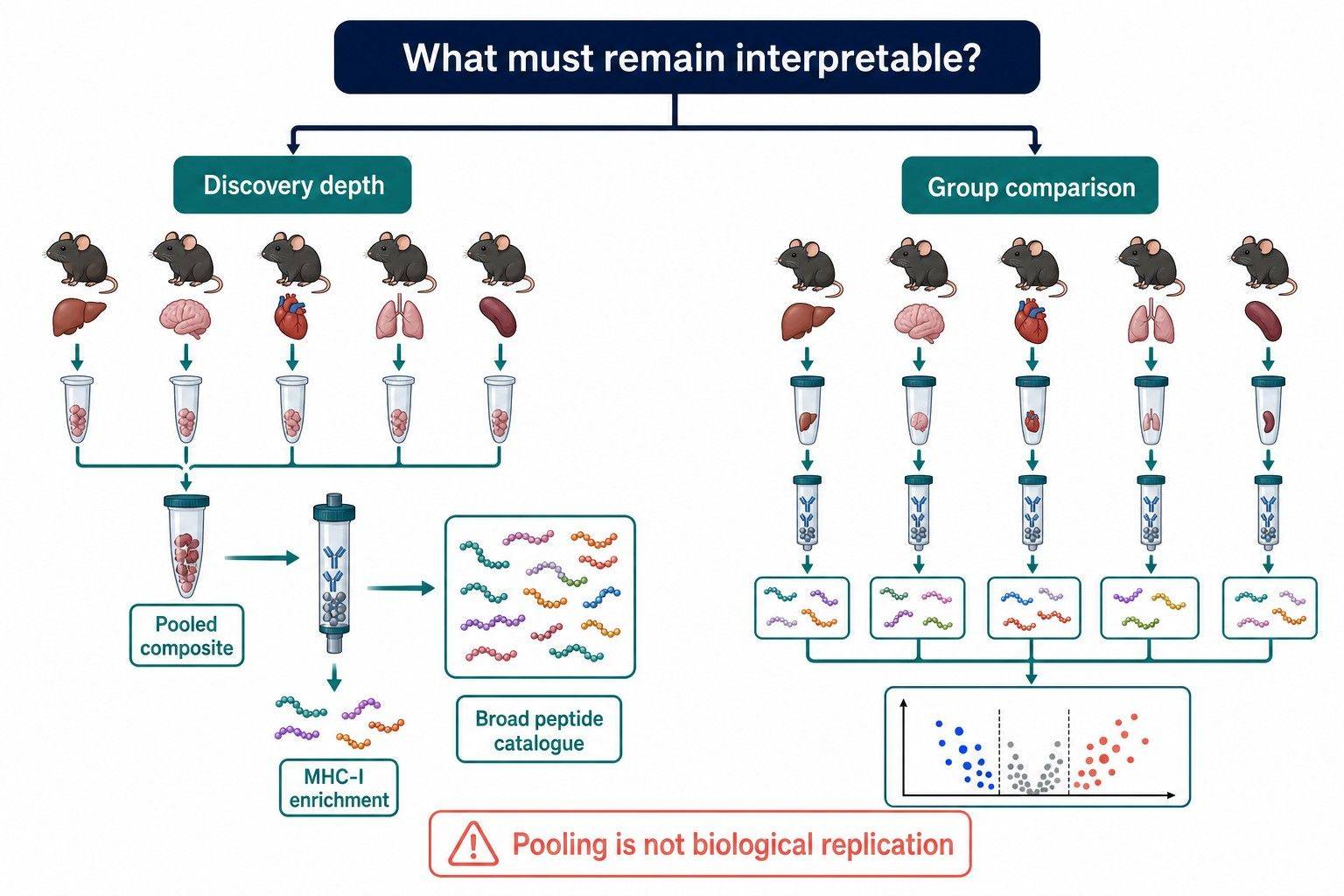
- Pooling is appropriate for a tissue-atlas or discovery objective when individual variation is not the endpoint; it is not a replacement for biological replication in group comparisons.
- Tissue mass alone is an unreliable input metric. A small immune-rich lesion, a cell pellet, and a low-MHC-I tissue can yield very different amounts of immunoaffinity-captured material.
- Pilot enrichment and peptide-level QC should establish feasibility before scarce samples are combined irreversibly.
Why H-2Kb and H-2Db Must Be Planned Separately in C57BL/6 Mice
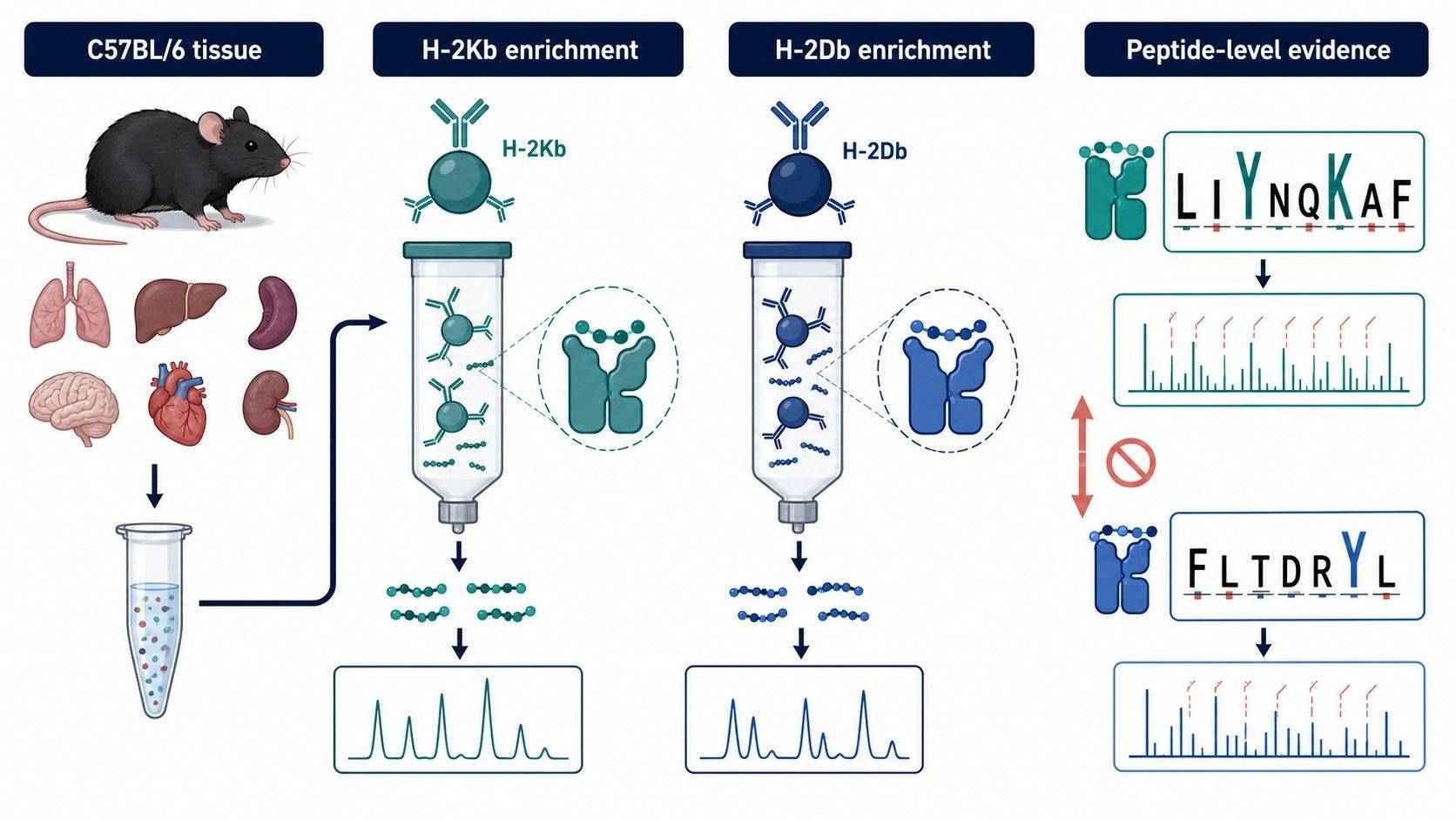
MHC-I immunopeptidomics identifies peptides naturally displayed by MHC class I molecules after immunoaffinity enrichment of MHC–peptide complexes and LC-MS/MS analysis. In the common C57BL/6 H-2b background, the classical H-2Kb and H-2Db molecules have different binding preferences. A pooled “mouse MHC-I” result can therefore contain two allele contexts that cannot be interpreted as one motif or one presentation rule.
Schuster and colleagues created a tissue-based C57BL/6 atlas by enriching H-2Db and H-2Kb independently from 19 normal tissues. The final atlas contained 28,448 high-confidence H-2Db/Kb-associated peptides and generated allele-specific spectral and assay libraries (Schuster et al., 2018). The study illustrates a key design point: separate enrichments preserve allele-level information even when the biological material is from the same strain and tissue.
This does not mean every study must separate alleles. The right design follows the biological claim. If the question is whether a treatment changes the total class-I-presented peptide landscape in one H-2b model, combined enrichment can be defensible. If the question is whether a candidate peptide is presented by H-2Kb or H-2Db, or whether allele-specific binding motifs change, separate enrichment is necessary.
Before sample collection, confirm:
- The strain, genetic background, and MHC haplotype of every experimental group.
- Whether the study requires total MHC-I coverage or H-2Kb/H-2Db-resolved results.
- Whether any engineered model changes MHC-I expression, beta-2-microglobulin dependence, or the source-cell composition of the tissue.
- Whether the reference proteome and predicted binding analysis match the species, strain, and allele context.
For studies that need direct evidence of presented ligands followed by computational ranking, an MHC-binding peptide identification workflow can align allele assignment, MS evidence, motif analysis, and source-protein annotation from the outset.
H-2Kb/H-2Db Enrichment: Antibody Specificity Determines What the MS Can See
The enrichment antibody is not a minor reagent choice. It defines which MHC–peptide complexes enter the MS workflow and therefore which peptide repertoire can be observed. A pan-MHC-I strategy may increase overall capture, but it does not necessarily capture all classical alleles equally or allow each peptide to be assigned to a specific allele.
Allele-specific enrichment for mechanistic questions
Allele-specific enrichment is preferred when a project asks about binding motifs, candidate antigen assignment, allele-selective presentation, or differences between H-2Kb and H-2Db. It supports a peptide list that can be evaluated against the appropriate allele model rather than inferring allele context from a mixed pool.
In the C57BL/6 tissue atlas, monoclonal antibodies B22-249.R1 and Y-3 were used to isolate H-2Db- and H-2Kb-associated peptides independently (Schuster et al., 2018). A separate evaluation of mouse MHC-I enrichment found that antibody choice materially affected H-2Kb and H-2Db recovery, reinforcing that a nominally broad MHC-I reagent should not be assumed to provide equivalent allele coverage (Nyayapathy et al., 2021).
Combined enrichment for total presentation questions
Combined enrichment can be useful when the biological endpoint is the total class-I peptide landscape and the final report will not make allele-specific claims. It can also be a sensible discovery option when input is limited and the team needs to determine whether a model yields a measurable repertoire at all.
The trade-off is interpretability. A peptide detected after combined H-2Kb/H-2Db capture may be predicted to bind one allele, both alleles, or neither under a chosen model. Such prediction is helpful for annotation but does not recreate direct allele-resolved capture. When the conclusion depends on allele identity, choose separate enrichments even if total peptide yield becomes lower.
Control logic for enrichment
The enrichment plan should include controls that can distinguish biological presentation from preparation background. At minimum, discuss a matrix-matched negative or isotype control, procedural blank, and a QC strategy suitable for the available input. The exact control set depends on whether material is tissue, a cell pellet, or a mixed cellular preparation.
The deliverable should retain the relationship between peptide, allele context, sample group, and confidence evidence. A source-protein name alone is not an immunopeptidomics result. The relevant evidence is the identified peptide sequence, its length and charge, MS/MS assignment quality, abundance pattern where quantification is intended, and the allele-enrichment context.
How Much Mouse Tissue Is Needed for MHC-I Immunopeptidomics?
There is no universal tissue-mass threshold for mouse MHC-I immunopeptidomics. Reporting a single number without knowing tissue type, strain, study objective, and MHC-I abundance would create false precision. A feasibility assessment should instead translate the available specimens into the amount of immunoaffinity-capturable MHC-I material and the number of independent biological samples required.
Factors that change usable input
MHC-I abundance. IFN-related activation, cell type, tissue physiology, and experimental perturbation can alter surface MHC-I abundance. More mass does not necessarily solve a biologically low MHC-I signal.
Cellularity and composition. Equal tissue weights can differ greatly in nucleated-cell content, extracellular matrix, necrosis, fat, or blood content. A dissociated cell preparation and an intact tissue specimen should not be planned as equivalent inputs solely by weight.
Target depth. Broad discovery needs more recoverable peptide material than confirmation of a small set of known ligands. The required input should be tied to the desired reportable output, not to an arbitrary extraction yield.
Allele resolution. Splitting one lysate into independent H-2Kb and H-2Db enrichments protects allele-specific interpretation but divides available input. This may motivate a pooling strategy for discovery, yet it should never be decided after the individual specimens have been discarded.
Sample handling. Delays, proteolysis, freeze–thaw cycles, and variable tissue processing can reduce the comparability of the recovered peptide repertoire. Consistent collection and rapid preservation are particularly important when pooling will combine material from multiple animals.
Use a staged feasibility pilot instead of assuming a fixed amount
A sensible pilot uses representative specimens to answer three questions. First, can the intended enrichment retrieve a detectable and biologically credible H-2b peptide repertoire? Second, does the available input support separate H-2Kb and H-2Db analysis or only a combined total-MHC-I result? Third, does the project need more material per analytical unit, or more independent biological units?
Published studies show why this is a study-specific decision. The tissue atlas pooled organs from five to six C57BL/6 mice for each tissue type, while its cell-line workflows used approximately 109 cells for MHC-I isolation (Schuster et al., 2018). Those values describe one large atlas-design choice, not a universal minimum. They are useful evidence that pooling can be necessary for deep tissue discovery, but they do not imply that every experimental comparison should pool five or six animals.
If specimen availability is limited, begin with a feasibility discussion through immunopeptidome profiling. State the tissue or cell type, strain, expected number of experimental groups, whether individual-level comparison is essential, and whether H-2Kb/H-2Db resolution is required. That information is more decision-useful than tissue mass alone.
When Should Mouse Tissues Be Pooled for MHC-I Peptide Enrichment?
Pooling is a sample-design decision, not a routine preparation step. It increases the total material available for enrichment but reduces the number of independently interpretable biological observations. The correct answer depends on whether the study’s endpoint is discovery depth or individual-level variation.
Pooling is appropriate when discovery depth is the endpoint
Pool specimens within a clearly defined biological unit when the aim is to create a qualitative tissue catalogue, build an allele-specific spectral library, or establish whether a model presents a candidate peptide class. In these cases, the pooled specimen represents the defined composite material; it should not be analyzed as if it were several independent biological replicates.
The C57BL/6 tissue atlas offers a concrete example: tissues from five to six mice were pooled per tissue type before separate H-2Db and H-2Kb isolation (Schuster et al., 2018). That design served a tissue-map objective by concentrating material for discovery. It did not test between-animal variability within a group.
Do not pool away a group-comparison endpoint
Avoid pooling all animals in each experimental group when the claim requires group-level statistics or assessment of biological heterogeneity. A single pooled control and a single pooled treated sample can produce an interesting peptide list, but it cannot estimate within-group variability. Multiple pooled biological units may be possible when individual samples are too small, provided each pool is created independently and the pooling plan is balanced across experimental variables.
For example, if the objective is to compare peptide presentation between two perturbation groups, preserve independent pools or individual samples at the experimental-unit level. Do not create one large pool after seeing which samples appear most abundant. Predefine which specimens enter each pool and keep their source metadata.
Pool only like with like
Pool only samples matched for strain, allele background, tissue or cell type, experimental condition, collection timing, and processing route. Pooling across different tissues or distinct cell compositions may increase detectability but creates a composite immunopeptidome that answers a different question.
If a project combines tissues because each is too small, state explicitly that the output represents the pooled composite rather than a tissue-specific presentation profile. The same discipline applies to sorting or enrichment of a cellular compartment before MHC-I capture: the biological unit must be defined before lysis.
QC Criteria That Make a Mouse MHC-I Peptide List Interpretable
An immunopeptidomics report should provide more than a peptide count. The following checks support interpretation of an H-2b result.
| QC domain | Questions to ask | Why it matters |
|---|---|---|
| Allele and strain | Are H-2Kb and H-2Db handled separately or intentionally combined? | Establishes the biological context for motif and binding annotation. |
| Enrichment evidence | Was the antibody selection fit for the stated allele objective? Were appropriate preparation controls included? | Helps separate a capture bias from a biological repertoire change. |
| Peptide properties | Do lengths and terminal motifs fit the enriched MHC-I context? | Flags inconsistent or low-confidence peptide populations. |
| Identification quality | Are peptide-level false-discovery controls and MS/MS evidence applied? | Protects against reporting unsupported sequences. |
| Quantitative completeness | For comparative projects, how often is each peptide observed across biological units and QC injections? | Distinguishes biological patterns from intermittent detectability. |
| Biological metadata | Are pool composition, tissue amount, cell counts where applicable, and collection conditions retained? | Makes the final result traceable and reproducible. |
Allele-specific binding prediction and motif analysis are useful secondary layers, but they are not substitutes for correct enrichment and MS evidence. A peptide predicted to bind H-2Kb is a hypothesis; a peptide observed after H-2Kb-specific enrichment carries a different level of contextual support.
For projects that will rank candidates for subsequent testing, peptidomics-based antigen discovery and prediction can connect experimentally observed ligands with sequence annotation and prioritization while retaining the experimental evidence layer.
A Practical Decision Framework for Mouse MHC-I Projects
Choose separate H-2Kb and H-2Db enrichment if the research question depends on allele-specific presentation, motif differences, or ligand assignment. Preserve separate eluates and report results at the peptide and allele level.
Choose combined MHC-I enrichment if the question concerns the total H-2b presentation landscape and material is insufficient for separate, meaningful enrichments. Do not make allele-specific claims from the combined result without independent supporting evidence.
Choose pooling for discovery if the purpose is a qualitative tissue map, spectral-library development, or initial feasibility assessment and individual variation is outside the study endpoint. Document the pool composition before processing.
Choose independent specimens or independent pools if the purpose is to compare experimental groups. Protect biological replication first, then decide how much material each analytical unit needs.
Creative Proteomics can help translate the strain, tissue availability, intended comparison, and desired allele resolution into an MHC-I enrichment and LC-MS/MS plan. A concise feasibility brief should include the model, H-2 haplotype, tissue or cell type, sample numbers, whether pooling is acceptable, and the intended level of peptide evidence.
FAQ: Mouse MHC-I Immunopeptidomics
Can H-2Kb and H-2Db peptides be analyzed in the same mouse study?
Yes. C57BL/6-derived material commonly contains both classical alleles. The key decision is whether they are enriched and reported separately for allele-resolved interpretation or intentionally combined for a total MHC-I presentation result.
How many mice should be pooled for a tissue immunopeptidomics experiment?
There is no fixed number that fits every tissue. Pooling should be based on expected MHC-I abundance, tissue cellularity, desired discovery depth, allele resolution, and whether the project needs independent biological comparisons. A pilot is the most reliable way to determine whether the available material supports the intended output.
Does pooling improve peptide identification?
Pooling can increase the total amount of captured MHC–peptide material and may improve discovery depth. It also removes individual-level variation within that pool, so it should be used for a defined discovery objective rather than as a substitute for biological replication.
Can a pan-MHC-I antibody replace H-2Kb and H-2Db-specific antibodies?
It may be useful for a total-MHC-I objective, but it should not be assumed to provide equal recovery or allele-level attribution for H-2Kb and H-2Db. If allele-specific conclusions are required, use a design that directly supports them.
What is the most important information to provide before requesting a quote?
Provide the mouse strain or MHC haplotype, specimen type, number of biological units, available tissue or cell quantity, experimental groups, desired allele resolution, and whether pooling is permitted. These details determine feasibility much more directly than a title such as “mouse immunopeptidomics.”
Can a tissue study be followed by targeted validation?
Yes. A discovery-stage enrichment can generate candidate peptide sequences, after which a focused immune peptide mass spectrometry analysis or targeted assay can be planned around a short, evidence-supported candidate list.
References
- Schuster H, Shao W, Weiss T, et al. A tissue-based draft map of the murine MHC class I immunopeptidome. Scientific Data. 2018;5:180157. doi: 10.1038/sdata.2018.157.
- Caron E, Kowalewski DJ, Chiek Koh C, Sturm T, Schuster H, Aebersold R. Analysis of Major Histocompatibility Complex (MHC) Immunopeptidomes Using Mass Spectrometry. Molecular & Cellular Proteomics. 2015;14(12):3105-3117. doi: 10.1074/mcp.O115.052431.
- Nyayapathy S, Jones R, Ford M, Pisano M, Rumble J. Immunopeptidome Analysis of Mouse MHC Class I. Poster presented at ASMS Conference on Mass Spectrometry and Allied Topics; 2021.
- Ghosh M, et al. Guidance Document: Validation of a High-Performance Liquid Chromatography-Tandem Mass Spectrometry Immunopeptidomics Assay for the Identification of HLA Class I Ligands Suitable for Pharmaceutical Therapies. Molecular & Cellular Proteomics. 2020;19(3):432-443. doi: 10.1074/mcp.C119.001652.
- Laumont CM, Vincent K, Hesnard L, et al. Noncoding regions are the main source of targetable tumor-specific antigens. Science Translational Medicine. 2018;10(470):eaau5516. doi: 10.1126/scitranslmed.aau5516.
Plan a Mouse MHC-I Immunopeptidomics Study with Defensible Input and Pooling Logic
Share the mouse strain, tissue, expected group structure, available input, and whether allele-resolved discovery or comparative biology is the priority. We can help align enrichment, pooling, QC, and peptide-level reporting with the research question.
Request a Quote