Top-down and bottom-up mass spectrometry do not provide interchangeable views of a protein. Bottom-up analysis measures proteolytic peptides and is highly effective for protein identification, sequence coverage, and localization of modifications within individual peptides. Top-down analysis measures intact protein ions and retains the linkage among sequence variation, terminal processing, and post-translational modifications (PTMs) present on the same molecular species. The appropriate workflow therefore depends on whether the analytical claim concerns local peptide evidence or the composition of an intact proteoform.
The distinction becomes critical when several PTMs may co-occur. If two modified peptides are detected after digestion, bottom-up data can establish that both sites occur somewhere in the protein population, but not necessarily that they coexist on the same molecule. An intact-mass feature can establish a combined mass shift, but mass alone may not identify which sites or modification types produce it. Top-down tandem MS can preserve connectivity and localize modifications, yet fragmentation coverage, precursor co-isolation, spectral complexity, and protein size may limit the final assignment.
The strongest strategy is often hierarchical. Bottom-up data define the base sequence and candidate modification sites with sensitive peptide-level evidence. Intact and top-down measurements establish proteoform mass, terminal state, and modification connectivity. Middle-down fragments can bridge the two scales when full intact-protein fragmentation is insufficient. Method selection should be expressed in terms of the evidence needed, not as a universal ranking of the platforms.
Analytical Summary
- Bottom-up answers peptide-level questions efficiently. It offers sensitive detection, mature chromatography and software, and strong localization when a modified peptide yields informative MS/MS.
- Digestion breaks long-range connectivity. PTMs observed on different peptides cannot automatically be assigned to the same protein molecule or proteoform.
- Top-down preserves the intact molecular context. Sequence variants, splice forms, truncations, terminal processing, and multiple PTMs remain linked in the precursor ion.
- Intact mass is not complete proteoform characterization. A mass difference can define total modification load while leaving modification type and site unresolved.
- Top-down localization depends on fragmentation coverage. Terminal fragments localize a modification only when cleavage positions bracket the site and the isolated precursor represents a sufficiently pure proteoform population.
- Middle-down is a practical connectivity bridge. Large peptides or protein subunits preserve more PTM relationships than bottom-up while remaining easier to separate and fragment than a full-length protein.
- Hybrid evidence is often decision-ready. Concordant intact mass, top-down or middle-down fragments, and site-specific bottom-up spectra provide stronger characterization than any one data layer alone.
Define the Required Level of Molecular Identity
A protein name, a peptide sequence, and a proteoform are different analytical entities. The project should define which one must be established before selecting the workflow.
| Analytical entity |
Required evidence |
Typical method |
Information not guaranteed |
| Protein identity |
Sequence-unique peptide or intact-protein evidence linked to a reference |
Bottom-up database search; targeted top-down identification |
Complete sequence, termini, or PTM configuration |
| Peptide sequence and site |
Product ions that establish residue order and localize the modification within a peptide |
Bottom-up LC-MS/MS |
Co-occurrence with sites on other peptides |
| Intact mass species |
Resolved charge-state envelope and deconvoluted neutral mass |
Intact LC-MS |
Base sequence, modification sites, or unique proteoform identity |
| Proteoform identification |
Base sequence plus intact mass and sufficient fragmentation evidence |
Top-down MS/MS |
Complete localization if fragmentation gaps remain |
| Proteoform characterization |
Base sequence, all relevant mass shifts, their identities and positions, and molecular connectivity |
Top-down supported by middle-/bottom-up evidence |
Biological function without separate functional data |
| Proteoform quantitation |
Selective and reproducible signal for each resolved molecular form |
Top-down, intact, or targeted hybrid assay |
Equal ion response among proteoforms unless demonstrated |
The term "proteoform" denotes a specific molecular form arising from a defined amino-acid sequence together with genetic variation, alternative splicing, proteolytic processing, and PTMs. Two molecules derived from the same gene but differing in one phosphorylation site, one terminal truncation, or one sequence variant are different proteoforms. A report that combines their peptide evidence into an "average protein" may be sufficient for some identification studies but does not describe their molecular connectivity.
Bottom-Up Protein Sequencing
In bottom-up analysis, the protein is denatured, reduced and alkylated where appropriate, digested into peptides, separated by liquid chromatography, and analyzed by MS/MS. Search algorithms match spectra to predicted peptides or infer sequences de novo. Accepted peptides are then mapped back to the protein reference.
Principal analytical strengths
Peptides generally separate efficiently by reversed-phase LC, ionize predictably, and fragment into interpretable b/y or c/z ion series. Bottom-up workflows can detect low-abundance species in complex samples and support high-throughput comparison across conditions. Multiple proteases can increase sequence coverage, move a target residue into a favorable peptide, and provide independent confirmation.
For a site-specific question, this scale is often ideal. A phosphopeptide spectrum can localize phosphorylation among neighboring residues; a glycopeptide can connect a glycan composition to one sequon; and a variant peptide can establish a substitution when diagnostic fragments bracket the site. Peptide mapping is therefore appropriate for primary-sequence confirmation and monitoring defined product quality attributes.
Protein inference and incomplete observation
Digestion increases the number of analytes and separates evidence from the intact molecule. Shared peptides may map to several isoforms or gene products. Hydrophobic, very short, very long, heavily modified, or poorly ionizing peptides may remain unobserved. Sequence coverage is calculated from detected residues, but it does not indicate that all molecules in the sample contain the same sequence or PTM configuration.
The absence of a modified peptide is also difficult to interpret without confirming that its unmodified counterpart, cleavage behavior, and analytical response are suitable. Enrichment can improve PTM detection but may distort relative representation and removes the direct denominator needed for site occupancy unless matched unenriched data are available.
Loss of long-range PTM connectivity
Suppose one peptide contains K9 acetylation and another contains K27 trimethylation. Bottom-up data demonstrate that the two modified peptides are present in the analyzed protein population. They do not establish whether K9ac and K27me3 occur on the same molecule, on different molecules, or in multiple combinations.
Connectivity can be retained only within the same peptide. Longer peptides from restricted digestion extend that window but eventually become a middle-down analysis. Computational reconstruction from peptide abundances may estimate compatible proteoform mixtures, yet the solution can be underdetermined when several combinations generate the same marginal site occupancies.
Top-Down Protein Sequencing
Top-down analysis introduces intact proteins into the mass spectrometer, resolves their charge-state distributions, isolates a proteoform ion, and fragments it without prior proteolysis. The precursor mass constrains the total molecular composition while product ions provide sequence and modification-localization evidence.
Connectivity retained in the precursor
Because the molecule remains intact, a precursor with an N-terminal acetylation, two phosphorylations, and a C-terminal truncation retains that combination as one analytical species. When chromatographic and mass resolution separate neighboring proteoforms, their relative signals can describe a proteoform distribution that bottom-up digestion would collapse into individual peptide measurements.
Top-down is particularly informative for histones, ribosomal proteins, proteolytically processed products, highly modified signaling proteins, small recombinant proteins, and intact subunits. It can distinguish splice or sequence forms that share most tryptic peptides and connect terminal processing with internal modifications.
Intact mass constrains but does not uniquely identify
The measured mass difference between an observed proteoform and a reference sequence indicates a net change. A +79.966 Da shift is consistent with phosphorylation, but an intact protein containing several possible phosphorylation sites may have many positional isomers. Multiple combinations of acetylation, methylation, adduction, sequence variation, and truncation can also yield identical or near-identical total mass.
Consequently, intact mass profiling should not be labeled top-down sequence characterization unless tandem spectra support the base sequence and modification assignment. An unidentified intact feature is a mass species, not necessarily a uniquely defined proteoform.
Fragmentation and the localization problem
Collision-based methods such as HCD or CID, electron-based methods such as ECD and ETD, hybrid EThcD, and UV photodissociation produce complementary intact-protein fragments. Terminal b/y or c/z ions localize a modification when the relevant cleavage sites bracket it. Electron-based methods are often valuable for retaining labile PTMs while cleaving the backbone.
Large proteins distribute signal across many charge states, isotopes, and fragments. Product-ion signal-to-noise falls, internal regions may have sparse cleavage, and deconvolution becomes more difficult. A high-confidence proteoform-spectrum match can therefore identify the base protein while leaving one or more PTM sites only partially localized. Internal-fragment analysis can fill central sequence gaps, but it also increases the candidate space and requires strict ambiguity control.
Practical mass range and sample complexity
Top-down performance does not have a universal molecular-weight cutoff. Many high-throughput studies are enriched for proteins below approximately 30 kDa because larger proteoforms are harder to separate, isotopically resolve, isolate, fragment, and identify. Specialized instrumentation and acquisition have extended robust observations into the 30–60 kDa range and beyond, but performance remains sample- and platform-dependent.
Complex lysates contain many proteoforms with overlapping charge envelopes. Offline fractionation, size-based separation, capillary electrophoresis, ion mobility, narrow isolation, and targeted acquisition reduce co-isolation. For a purified protein, top-down feasibility is usually greater than for the same proteoform at low abundance in a whole-cell extract.
 Figure 1. Bottom-up provides detailed peptide-level localization but breaks long-range connectivity, whereas top-down preserves intact proteoform combinations and requires sufficient fragmentation to localize them.
Middle-Down as a Connectivity Bridge
Middle-down workflows analyze large peptides or protein subunits, commonly produced by limited proteolysis or domain-specific cleavage. The analytes may range from several kilodaltons to tens of kilodaltons depending on the protein and method. They retain PTM relationships across a larger sequence segment than tryptic peptides while reducing the separation and fragmentation burden relative to the intact protein.
For monoclonal antibodies, analysis of light chains, heavy-chain subunits, Fc/Fab domains, or large proteolytic fragments can determine which mass shifts reside in a domain and connect modifications within that region. For histones, 50–60-residue N-terminal tails preserve combinations of regulatory marks that are separated across several bottom-up peptides.
Middle-down does not preserve connectivity across cleavage boundaries. A modification on an Fc fragment cannot be linked to a modification on the corresponding Fab molecule after dissociation unless the intact or another overlapping measurement supplies that relationship. It should therefore be described as extended regional connectivity, not complete proteoform resolution.
Co-Occurring PTMs and Proteoform Assignment
Site occupancy is not proteoform abundance
Bottom-up site occupancy estimates the fraction of observed peptide molecules modified at one site, subject to response and digestion assumptions. Proteoform abundance estimates the fraction of intact molecules carrying a specific combination of sequence and PTMs. These quantities are mathematically related but are not identical.
If phosphorylation occupancy is 40% at site A and 30% at site B, the abundance of the doubly phosphorylated proteoform is not determined. Depending on biological dependence, it could range from no co-occurrence to substantial overlap. Top-down or sufficiently long middle-down analytes are needed to measure that joint distribution directly.
Isobaric kindred proteoforms
Proteoforms can have the same base sequence and the same number and type of PTMs but at different sites. These positional isomers have identical intact mass. Chromatography or ion mobility may partially separate them, but unresolved co-isolation produces multiplexed MS/MS spectra in which shared fragments can be assigned to several candidates.
A site-localizing fragment is informative only if it can be attributed to a sufficiently pure precursor population. Narrow isolation windows, improved front-end separation, multiple charge states, and complementary fragmentation reduce ambiguity. The report should disclose whether one isolated precursor represented a single proteoform, a partially resolved family, or an unresolved envelope.
Modification type versus modification count
An intact mass shift may define the number of nominal modification equivalents while leaving chemistry uncertain. For example, acetylation and trimethylation differ by only about 0.036 Da. High resolving power can distinguish these modifications on favorable ions, but mass accuracy, isotopic resolution, charge-state assignment, and fragment localization all contribute.
Bottom-up spectra can identify the chemical modification on a peptide more readily, while top-down data establish which combination belongs to the intact molecule. This complementarity is a central reason to integrate both scales.
Sample Preparation, Separation, and Acquisition
Top-down sample preparation should preserve intact proteoforms while removing salts, detergents, polymers, and other components that suppress ionization or complicate charge envelopes. Bottom-up workflows tolerate more complex starting mixtures but introduce reduction, alkylation, digestion, desalting, and enrichment steps that can create artifacts or unequal peptide recovery.
| Method component |
Bottom-up considerations |
Top-down considerations |
| Extraction |
Maximize protein recovery; detergents may be removed before digestion |
Preserve intact forms and solubility; avoid irreversible precipitation and nonvolatile contaminants |
| Reduction/alkylation |
Improves peptide recovery but can create overalkylation or disulfide artifacts |
Optional depending on native/denaturing objective; changes intact mass and structural state |
| Digestion |
Protease specificity, missed cleavages, peptide length, PTM stability |
Not used for full top-down; limited cleavage may define middle-down workflow |
| Separation |
Reversed-phase peptide LC is mature and high capacity |
Intact-protein RP, ion exchange, GELFrEE, SEC, CZE, or ion mobility may be required |
| Precursor isolation |
Peptide isotope envelopes are comparatively simple |
Multiple charge states and kindred proteoforms increase co-isolation risk |
| Fragmentation |
HCD/CID commonly effective; ETD/EThcD useful for labile PTMs |
Complementary HCD, ETD/ECD, EThcD, UVPD, and internal fragments may be required |
| Quantitation |
Peptide response and digestion bias affect occupancy |
Charge-state summation, deconvolution, co-elution, and proteoform response affect abundance |
Method development should begin with a representative sample and a defined proteoform question. A clean purified 15 kDa protein and a 150 kDa glycoprotein in plasma do not present the same top-down experiment. Deglycosylation or subunit reduction may improve spectral interpretability but also changes the molecular entity being characterized; the resulting data should be described at the subunit or deglycosylated level.
Data Processing and Confidence Metrics
Bottom-up analysis generally reports peptide-spectrum matches, peptide and protein false discovery rates, sequence coverage, site-localization probability, and extracted-ion abundance. Top-down analysis reports deconvoluted precursor mass, proteoform-spectrum matches (PrSMs), fragment maps, proteoform-level false discovery rate, mass-shift assignment, and characterization or localization scores.
Deconvolution is part of the top-down evidence chain
Intact protein spectra contain many charge states and isotope distributions. Deconvolution converts them into neutral masses and consolidates fragment signals. Different deconvolution algorithms can disagree on precursor charge, monoisotopic mass, and fragment assignments. A comparative 2023 study found substantial workflow-specific proteoform identifications and inconsistent PTM detection among current pipelines.
For critical results, inspect the original m/z-domain spectrum, charge-state series, isotope fit, isolation window, and fragment evidence rather than accepting only the deconvoluted peak list. Report software versions and parameters because signal processing can change the candidate proteoform before database search begins.
Identification, characterization, and localization are separate levels
A PrSM may confidently identify a base protein sequence while leaving an unexplained mass shift. Matching the shift to a known PTM narrows the hypothesis. Localizing that PTM requires fragment ions that distinguish candidate sites. Complete characterization requires accounting for all material mass shifts and relevant sequence features.
The same separation applies to bottom-up data: a peptide may be identified, a modification composition assigned, and the site still ambiguous. Reports should not collapse these levels into a single "confidence score."
Workflow Selection by Analytical Question
| Analytical question |
Preferred primary workflow |
Complementary evidence |
| Confirm the expected amino-acid sequence of a known protein |
Multi-protease bottom-up LC-MS/MS |
Intact/subunit mass and terminal analysis |
| Localize one phosphorylation or glycosylation site |
Targeted bottom-up or middle-down MS/MS |
Intact mass for modification-load context |
| Determine whether two distant PTMs occur on the same molecule |
Top-down or a middle-down fragment spanning both sites |
Bottom-up spectra for site chemistry and localization |
| Resolve truncation plus internal PTM combinations |
Top-down intact precursor and MS/MS |
Terminal peptide mapping and orthogonal intact mass |
| Profile histone combinatorial marks |
Top-down or middle-down |
Bottom-up for sensitive site occupancy |
| Characterize a large heterogeneous antibody |
Bottom-up plus intact/subunit or middle-down analysis |
Targeted top-down on isolated chains/domains where feasible |
| Discover low-abundance proteins in a complex proteome |
Bottom-up discovery proteomics |
Targeted top-down after enrichment or fractionation |
| Quantify known intact proteoforms |
Targeted intact/top-down assay with resolved features |
Peptide-level attribute monitoring and reference standards |
For projects centered on intact molecular forms, top-down protein sequencing should be designed around precursor separation and fragment localization rather than intact mass alone. For extensive reference-free reconstruction, protein full-length sequencing may require overlapping bottom-up, middle-down, terminal, and intact evidence because neither a single digest nor one intact spectrum guarantees complete sequence definition.
Integrated Bottom-Up, Middle-Down, and Top-Down Strategy
A staged workflow uses each scale to constrain the next and avoids asking the most complex measurement to solve every problem simultaneously.
- Define the proteoform hypothesis. List expected sequence variants, termini, processing events, PTMs, disulfides, cofactors, and the specific connectivity question.
- Establish the base sequence by bottom-up analysis. Use complementary proteases where necessary and record residue-level coverage, modification sites, and unresolved regions.
- Profile intact or subunit masses. Determine how many mass species are present, their relative signals, and whether observed shifts agree with candidate modifications or processing.
- Reduce heterogeneity before top-down MS/MS. Fractionate by size, charge, hydrophobicity, capillary electrophoresis, or ion mobility; isolate individual chains or domains when scientifically appropriate.
- Acquire complementary fragmentation. Combine collision- and electron-based methods or UVPD to extend cleavage coverage and retain labile PTMs.
- Map peptide-level findings onto intact candidates. Use bottom-up sites to constrain top-down search space without assuming that every observed site occurs on every proteoform.
- Test connectivity explicitly. Require fragments or spanning middle-down peptides that distinguish competing PTM combinations.
- Quantify only resolved analytical species. Define charge-state integration, deconvolution, isotope handling, and response assumptions.
- Report remaining proteoform families. When positional isomers cannot be separated, state the compatible configurations rather than selecting one unsupported structure.
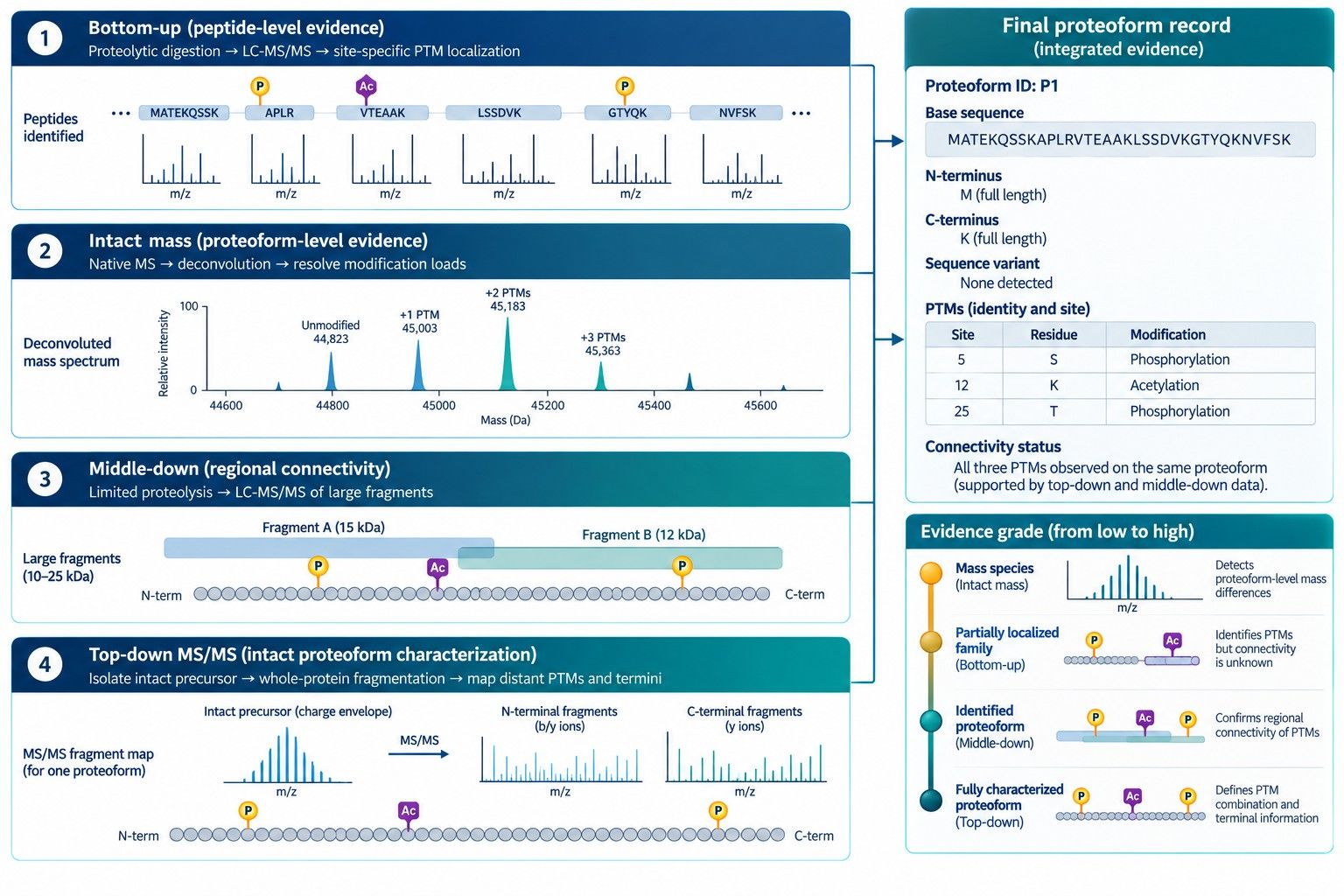 Figure 2. A hybrid evidence stack uses bottom-up site localization, intact mass, middle-down regional connectivity, and top-down fragmentation to define proteoform composition.
Data-Backed Literature Examples
Histone H3 combinatorial modification patterns
Top-down analysis of intact histone H3 from multiple-myeloma-derived cell lines identified proteoforms carrying coexisting K9, K27, and K36 methylation states. Trivalent hypermethylated forms associated with MMSET/NSD2 overexpression were not discovered in the corresponding bottom-up H3.1 peptide analysis. The case demonstrates the difference between measuring individual modified peptides and preserving disease-associated modification combinations on one protein molecule.
Integrated PTM-TBA analysis
A 2023 workflow combined top-down mass shifts, bottom-up data, and PTM annotations in SW480 cells. On average, top-down searches identified 2,000 mass shifts; 814.5 matched 11 common PTMs, 423 were localized, and 435 were verified using bottom-up evidence and annotations. The layered result shows that intact detection, PTM matching, site localization, and cross-platform verification are distinct analytical milestones.
Extending the top-down mass range
High-field 21 T FT-ICR analysis of a human colorectal-cancer-cell lysate identified 3,238 proteoforms at 1% false discovery rate, including 372 above 30 kDa detected at isotopic resolution. A separate quadrupole-Orbitrap study captured 439 proteoforms in the 30–60 kDa range using tailored isolation and acquisition. These studies demonstrate that 30 kDa is not a physical ceiling while confirming that larger-proteoform performance depends on specialized separation, resolution, and acquisition.
Complementary fragmentation for intact proteins
A 2026 CZE-MS/MS study integrated HCD, ETD, EThcD, and UVPD across standard proteins of approximately 10–70 kDa. Combining the modes produced 67% sequence coverage for 30 kDa carbonic anhydrase and 98% for 12 kDa thioredoxin; adding internal fragments further increased reported coverage. The numerical results belong to the tested proteins and platform, but they illustrate why complementary ion chemistry can fill different intact-protein fragmentation gaps.
Decision-Ready Reporting
A proteoform report should make clear which molecular relationships were measured and which were inferred. Include:
- sample origin, extraction conditions, enrichment, fractionation, reduction, alkylation, digestion, and PTM-preservation controls;
- reference sequence, isoform, residue-numbering convention, expected termini, and database version;
- bottom-up peptide list with uniqueness, sequence coverage, PTM identity, site-localization evidence, and occupancy calculation where used;
- intact or subunit charge-state spectra, deconvoluted masses, mass errors, observed relative signals, and unresolved envelopes;
- top-down precursor isolation window, charge state, co-isolation assessment, fragmentation modes, and annotated terminal/internal fragments;
- explicit proteoform definition including base sequence, terminal state, sequence variants, PTM types, sites, and connectivity status;
- evidence distinguishing positional isomers and kindred proteoforms;
- software, deconvolution engine, database, search parameters, FDR method, and manual-review rules;
- concordance and disagreement among bottom-up, middle-down, intact, and top-down data;
- quantitative integration rules, response assumptions, replicate precision, and method capability; and
- final classification as identified proteoform, fully characterized proteoform, partially localized proteoform family, intact mass species, or unresolved feature.
The conclusion should not state that bottom-up data proved long-range PTM co-occurrence, or that intact mass alone defined a proteoform. A transparent partial characterization is more useful than a complete-looking structure assembled from disconnected evidence.
Technical FAQ
What is the main difference between top-down and bottom-up protein sequencing?
Bottom-up measures peptides produced by digestion and is optimized for peptide sequence and local PTM evidence. Top-down measures intact protein ions and preserves the connection among sequence, termini, and multiple modifications on the same molecular species.
Can bottom-up LC-MS/MS identify proteoforms?
It can identify proteoform-specific peptides when a unique splice junction, sequence variant, terminus, or local PTM combination is observed. It generally cannot reconstruct the complete intact PTM configuration when distinguishing features occur on separate peptides.
Does intact mass analysis count as top-down sequencing?
Intact mass profiling is an important top-down-level measurement, but sequencing or full proteoform characterization requires MS/MS or other evidence that establishes the base sequence and localizes relevant mass shifts. A deconvoluted mass peak alone may have several structural explanations.
Why is top-down analysis more difficult for large proteins?
Signal is distributed across many charge states, isotopes, and fragments. Large proteoforms are harder to separate and isotopically resolve, and their interior sequences may receive sparse terminal-fragment coverage. Heterogeneity further increases co-isolation and deconvolution complexity.
What is middle-down protein sequencing?
Middle-down analyzes large peptides, domains, chains, or subunits rather than short tryptic peptides or the entire protein. It preserves PTM connectivity within a larger region while reducing the analytical burden of full top-down analysis.
Which approach is better for PTM localization?
Bottom-up often provides denser local fragmentation and higher sensitivity for an individual modified peptide. Top-down is required when the objective is to connect distant PTMs to the same intact molecule. A hybrid workflow can provide both site localization and molecular connectivity.
Can top-down distinguish proteoforms with the same intact mass?
Only if separation or fragment evidence distinguishes them. Positional PTM isomers can share an identical precursor mass. Chromatography, ion mobility, narrow isolation, complementary fragmentation, and diagnostic product ions may resolve them; otherwise they should be reported as a proteoform family.
How are proteoforms quantified?
Relative signal can be estimated across deconvoluted intact features or selected charge states, but ionization and transmission may differ among forms. Quantitation requires selective feature definition, consistent charge-state handling, replicate performance, and standards or response studies when accurate molar abundance is needed.
When should both top-down and bottom-up be used?
Use both when the project needs confident base sequence and site localization together with termini, truncations, isoform identity, or long-range PTM co-occurrence. Bottom-up constrains candidate sites; top-down tests which combinations occur on intact molecules.
References
- Smith LM, Kelleher NL; Consortium for Top Down Proteomics. Proteoform: A Single Term Describing Protein Complexity. Nat Methods. 2013;10(3):186-187. doi:10.1038/nmeth.2369.
- Toby TK, Fornelli L, Kelleher NL. Progress in Top-Down Proteomics and the Analysis of Proteoforms. Annu Rev Anal Chem. 2016;9:499-519. doi:10.1146/annurev-anchem-071015-041550.
- Ntai I, Fornelli L, DeHart CJ, et al. Precise Characterization of KRAS4b Proteoforms in Human Colorectal Cells and Tumors Reveals Mutation/Modification Cross-Talk. Proc Natl Acad Sci USA. 2018;115(16):4140-4145. doi:10.1073/pnas.1716122115.
- Melani RD, Skinner OS, Fornelli L, et al. Mapping Proteoforms and Protein Complexes from King Cobra Venom Using Both Denaturing and Native Top-Down Proteomics. Mol Cell Proteomics. 2016;15(7):2423-2434. doi:10.1074/mcp.M115.056523.
- Chen W, Ding Z, Zang Y, Liu X. Characterization of Proteoform Post-Translational Modifications by Top-Down and Bottom-Up Mass Spectrometry in Conjunction with Annotations. J Proteome Res. 2023;22(10):3178-3189. doi:10.1021/acs.jproteome.3c00207.
- Pesavento JJ, Bullock CR, LeDuc RD, Mizzen CA, Kelleher NL. Combinatorial Modification of Human Histone H4 Quantitated by Two-Dimensional Liquid Chromatography Coupled with Top Down Mass Spectrometry. J Biol Chem. 2008;283(22):14927-14937. doi:10.1074/jbc.M709796200.
- Zheng Y, Fornelli L, Compton PD, et al. Unabridged Analysis of Human Histone H3 by Differential Top-Down Mass Spectrometry Reveals Hypermethylated Proteoforms from MMSET/NSD2 Overexpression. Mol Cell Proteomics. 2016;15(3):776-790. doi:10.1074/mcp.M115.053819.
- Anderson LC, DeHart CJ, Kaiser NK, et al. Identification and Characterization of Human Proteoforms by Top-Down LC-21 Tesla FT-ICR Mass Spectrometry. J Proteome Res. 2017;16(2):1087-1096. doi:10.1021/acs.jproteome.6b00696.
- Schaffer LV, Millikin RJ, Shortreed MR, et al. Identification and Quantification of Proteoforms by Mass Spectrometry. Proteomics. 2019;19(10):e1800361. doi:10.1002/pmic.201800361.
- Schmitt ND, Berger JM, Conway JB, Agar JN. Increasing Top-Down Mass Spectrometry Sequence Coverage by an Order of Magnitude through Optimized Internal Fragment Generation and Assignment. Anal Chem. 2021;93(16):6355-6362. doi:10.1021/acs.analchem.0c04670.
- Donnelly DP, Rawlins CM, DeHart CJ, et al. Best Practices and Benchmarks for Intact Protein Analysis for Top-Down Mass Spectrometry. Nat Methods. 2019;16(7):587-594. doi:10.1038/s41592-019-0457-0.
- McCool EN, Lubeckyj RA, Shen X, et al. Deep Top-Down Proteomics Using Capillary Zone Electrophoresis-Tandem Mass Spectrometry: Identification of 5,700 Proteoforms from the Escherichia coli Proteome. Anal Chem. 2018;90(9):5529-5533. doi:10.1021/acs.analchem.8b00693.
- Wang Q, Wang Q, Melani RD, Liu Q, Nurmi P, Sun L. Extensive Backbone Cleavage Coverage of Intact Proteoforms in a Mass Range of 10–70 kDa by Integrating Electron, Collision, and Photon-Based Fragmentation Techniques during an Electrophoretic Time Scale. J Am Soc Mass Spectrom. 2026;37(2):505-513. doi:10.1021/jasms.5c00384.
For research use only, not intended for any clinical use.


