A single-amino-acid substitution can represent an intended mutation, a cell-line sequence variant, translational misincorporation, a sample-specific polymorphism, an undocumented construct difference, or an incorrect database assignment. LC-MS/MS can provide residue-level evidence, but a software-reported variant peptide is not automatically a confirmed substitution. The analytical conclusion depends on whether the precursor mass shift is chemically distinctive, whether fragment ions localize the difference, whether alternative modifications and sequences have been excluded, and whether the result is reproducible at the required abundance.
The first decision is whether the study is targeted or discovery-based. In a targeted confirmation study, the expected wild-type and variant sequences are known and acquisition can be optimized for the diagnostic peptide. In an untargeted sequence-variant analysis, many possible substitutions, modifications, and cleavage products compete in a much larger search space. The latter demands stronger false-positive controls and usually a separate confirmation stage.
For biopharmaceutical development, confirmation and quantitation are also distinct claims. A high-quality product-ion spectrum may identify the variant sequence, while relative abundance requires chromatographic integration, response assessment, replicate precision, and a defined calculation model. Biological impact is a third question requiring functional and structural characterization. A defensible workflow keeps these claims separate and reports the evidence supporting each one.
Analytical Summary
- Precursor mass difference is a screening clue, not complete proof. Many substitutions have characteristic exact-mass shifts, but the same shift may also arise from a post-translational modification, chemical artifact, alternate peptide, or monoisotopic assignment error.
- Site localization requires product ions on both sides of the changed residue. Variant-specific fragment ions should distinguish the proposed site from every plausible alternative position within the peptide.
- The wild-type peptide is an analytical control. Its retention time, fragmentation pattern, isotope distribution, and abundance help distinguish a true variant from a modified or misassigned peptide.
- Discovery and confirmation should be separated. Error-tolerant, open, or broad substitution searches generate candidates; targeted reprocessing or reacquisition tests the candidate under a constrained hypothesis.
- Leu/Ile remains an exceptional case. An L↔I substitution produces no precursor or conventional backbone-fragment mass change and requires side-chain diagnostic fragmentation or orthogonal evidence.
- Relative abundance is method-dependent. Variant-to-wild-type peak-area ratios can be biased by digestion, ionization, chromatography, and fragmentation and should not be presented as absolute protein-level abundance without qualification.
Define the Substitution Question Before Method Development
The phrase "confirm this mutation" can describe several different analytical objectives. Each objective changes the database, sample design, acquisition method, review burden, and strength of conclusion.
| Study objective |
Primary question |
Appropriate result |
Principal risk |
| Expected substitution confirmation |
Does the sample contain the predefined residue change? |
Variant peptide identity and localized residue, with wild-type status |
Confirmation bias and failure to test alternative explanations |
| Reference sequence confirmation |
Does the protein match the supplied construct across a defined region? |
Coverage map plus residue-level MS/MS evidence |
Coverage reported without direct support at the target site |
| Low-level sequence-variant screening |
Are unintended substitutions present above the method capability? |
Verified variant list with estimated relative abundance |
Large search space and false-positive candidates |
| Clone or process comparison |
Does variant abundance differ among clones, batches, or conditions? |
Comparable variant/wild-type ratios and trend statistics |
Run-order, carryover, digestion, or response bias |
| Unknown protein or proteogenomic analysis |
Does a peptide support a sequence absent from the standard database? |
Variant peptide plus genomic, transcriptomic, or homology context |
Alternate protein mappings and non-unique peptides |
| Impact assessment |
Could the substitution alter structure, binding, potency, or safety? |
Integrated structural and functional risk assessment |
Treating analytical detection as evidence of biological effect |
For an intended construct change, peptide mapping can be designed around the expected wild-type and variant peptides. For an undocumented sequence, the initial task may instead require de novo protein and peptide sequencing or a proteogenomic search before targeted confirmation becomes possible.
Targeted confirmation versus untargeted discovery
A targeted search contains the two candidate peptide sequences and a controlled set of expected modifications. This improves interpretability and permits inclusion-list acquisition, optimized collision energy, or parallel reaction monitoring. However, it can only test hypotheses included in the method.
An untargeted sequence-variant search may allow every plausible residue substitution, nonspecific cleavage, or wide precursor mass differences. It increases discovery scope but also multiplies candidate explanations. Search-engine score and global false discovery rate do not eliminate every site-specific false positive, especially for a single rare event. Broad search results should therefore be treated as candidate generation rather than final confirmation.
Predict the Analytical Consequence of the Substitution
Before data review, calculate the theoretical mass change between the original and substituted residues. The expected variant peptide mass equals the wild-type peptide mass plus the residue mass difference, provided that cleavage and modification states are otherwise identical.
The same substitution can change more than mass. Replacing Lys or Arg can create or remove a tryptic cleavage site. Substitution of a hydrophobic residue with a charged residue can substantially shift reversed-phase retention. A change adjacent to a protease site can alter digestion kinetics. The method should predict precursor m/z values for relevant charge states, expected peptide boundaries, missed-cleavage forms, fragment-ion mass shifts, retention behavior, and possible modification mimics.
Unique, near-isobaric, and isobaric mass changes
Some residue pairs have readily distinguished exact masses. Others differ by only a few hundredths of a dalton and require adequate resolving power and mass accuracy. Leucine and isoleucine are constitutional isomers with identical residue mass and cannot be differentiated by precursor mass or ordinary b/y ions.
Peptide mass alone may uniquely suggest a substitution when the changed residue occurs only once and the mass difference has no plausible competitor. Even then, MS/MS is needed to establish sequence and site. A mass shift that could be placed at several residues, explained by a PTM, or reproduced by a different peptide composition is not residue-level evidence.
Substitution-versus-modification conflicts
A true amino-acid replacement changes the covalent primary sequence, whereas oxidation, deamidation, dehydration, alkylation, metal adduction, and sample-preparation reactions modify an existing residue. Search algorithms can interchange these explanations when their mass shifts are identical or close within tolerance.
Examples include deamidation-related shifts near +0.984 Da, oxidation at +15.995 Da, and multiple residue substitutions with similar net masses. The correct comparison is not only "variant versus reference" but "variant versus every chemically plausible modified or alternatively cleaved form." Diagnostic fragments should place the mass difference specifically at the substituted residue and, when possible, include ions that exclude the competing PTM site.
Minimum LC-MS/MS Evidence for Confirmation
A substitution is most convincing when precursor, chromatographic, product-ion, comparative, and reproducibility evidence converge. No single score substitutes for this evidence stack.
Precursor identity and isotope pattern
The selected precursor should have the expected monoisotopic m/z, charge state, and isotope envelope. Review whether the acquisition software selected the monoisotopic peak rather than the M+1 or M+2 isotope, whether an overlapping precursor was co-isolated, and whether the observed mass error is consistent across the chromatographic peak.
For low-abundance variants near a high-abundance wild-type or modified peptide, extracted-ion chromatograms should be inspected at appropriate mass tolerance. Carryover, in-source fragments, column bleed, and adduct clusters can produce signals at the expected m/z. Blank injections and process controls are particularly important when the claimed abundance is near the reporting threshold.
Fragment ions that localize the residue
The product-ion spectrum should contain a sequence-informative ion on each side of the proposed substitution. For a b/y spectrum, this means a pair of ions whose mass difference spans the target residue and whose assignments change as predicted between wild-type and variant sequences. Ideally, multiple independent ions support each side, including complementary b and y series when observable.
Variant-specific ions should be defined before manual review. A fragment shared by both candidate sequences confirms peptide context but not the substitution. A fragment whose mass includes the changed residue but also several possible modification sites may be insufficient to localize the event. The annotated spectrum should show both supporting and unexplained high-intensity peaks, not only selected matches.
Comparative evidence from the wild-type peptide
The expected wild-type peptide provides a reference for digestion, charge state, fragmentation, and retention. Directly compare theoretical and observed masses, isotope distributions, product ions, retention times, and chromatographic peak shapes. In a mixture, the variant and wild-type signals should behave independently but consistently across replicate preparations and dilution levels.
Absence of the wild-type peptide can be meaningful for a high-abundance intended mutation, but only if the method is capable of detecting it and the peptide is not lost through altered cleavage or modification. For trace variant analysis, co-detection of the abundant wild-type peptide supports peptide context but can also increase co-isolation and ion suppression.
Reproducibility and targeted reacquisition
A candidate found in one data-dependent MS/MS event should be confirmed in an independent preparation or targeted injection. Inclusion lists, parallel reaction monitoring, or data-independent acquisition can improve repeated observation of the variant precursor and diagnostic fragments. Replication should address sample preparation as well as instrument injection because digestion artifacts and carryover can reproduce within a single prepared vial.
 Figure 1. Confirmation requires agreement among precursor, chromatographic, site-localizing fragment-ion, wild-type comparator, and replicate evidence.
Search and Data-Review Strategies
Custom FASTA and targeted candidate search
For a known substitution, create a database containing the expected sequence, the variant sequence, relevant processing forms, host-cell proteins, and common contaminants. Keep modification settings chemically justified. The search should permit direct comparison of candidate scores and site-determining ions without an unnecessarily large hypothesis space.
When the substitution creates or removes a protease cleavage site, include both predicted peptide products and plausible missed-cleavage forms. A negative result for only the original peptide boundary is not evidence that the variant is absent.
Error-tolerant and open searches
Error-tolerant searches test substitutions or modifications not present in the initial database. Open searches evaluate a broad range of precursor mass shifts and can reveal unexpected chemistry. These methods are valuable for discovery but may assign a mass difference to the wrong residue, choose a substitution instead of a PTM, or match an unrelated sequence with similar fragments.
Candidates should be re-searched in a constrained database, reviewed manually, and reacquired when material permits. The variant peptide should also be checked against comprehensive protein databases to determine whether it maps exactly to a contaminant, homolog, isoform, or other expected protein.
Proteogenomic confirmation
Genomic or transcriptomic variants can be translated into sample-specific protein sequences and added to the search database. Detection of the corresponding peptide supports translation of the nucleotide change, provided the peptide is unique and the spectrum localizes the altered residue. Conversely, a protein-level variant with no DNA change may indicate transcriptional error, translational misincorporation, mixed clones, or an analytical false positive.
Nucleotide evidence is orthogonal but not interchangeable with protein evidence. DNA or RNA establishes the encoded candidate; LC-MS/MS establishes whether the variant peptide is present in the analyzed material.
Excluding False-Positive Variant Assignments
Co-isolated and chimeric spectra
A wide isolation window can fragment two peptides simultaneously. The search engine may assemble fragments from both precursors into a plausible variant match. Review isolation purity, precursor traces, fragment co-elution, and whether major unmatched ions correspond to another peptide. Targeted reacquisition with a narrower isolation window can clarify the result.
Incorrect monoisotopic peak selection
Selecting an isotope peak as the monoisotopic precursor creates an apparent mass error near integer multiples of 1.00335 Da divided by charge. Search algorithms may explain that error with a substitution or modification. Inspect the full isotope envelope and chromatographic consistency rather than relying on the acquisition-reported precursor mass.
Sample-preparation artifacts
Oxidation, deamidation, disulfide scrambling, incomplete alkylation, overalkylation, and nonspecific cleavage can arise during denaturation, reduction, alkylation, digestion, or storage. Their abundance may change with preparation time, pH, temperature, reagent age, or light exposure. A suspected substitution that tracks preparation stress rather than biological sample level is more likely to be artifactual.
Process blanks, time-course controls, alternative digestion conditions, and an orthogonal protease help distinguish preparation chemistry from primary-sequence change. A second protease is especially useful when the initial peptide contains multiple plausible modification sites or unfavorable fragmentation.
Alternate peptide and protein mappings
The proposed variant peptide may be an exact peptide from a host-cell protein, contaminant, isoform, or homolog. Search the sequence against the complete relevant proteome and contaminant database. Protein-level context, additional unique peptides, expected molecular mass, and sample purification history should support the mapping.
Isobaric sequences and sequence permutations
Different residue combinations can have the same or nearly the same mass. Isobaric dipeptide compositions and rearrangement products can produce misleading fragment matches. A 2025 study of therapeutic proteins and recombinant vaccine antigens documented commercial-software misassignments involving isobaric or near-isobaric sequence alternatives and modification compensation. Manual site-level spectrum review remains necessary even when the software workflow itself is validated.
Leucine/isoleucine substitutions
An L↔I change is invisible to precursor mass and routine b/y series. Direct confirmation requires diagnostic side-chain fragments, such as suitable d- or w-type ions from high-energy or electron-based fragmentation, a validated multistage immonium-ion method, or orthogonal evidence. A database letter or retention-time difference without qualified standards should not be presented as direct proof.
Sample Preparation and Acquisition Design
The target peptide should be analytically favorable: unique to the protein, neither extremely short nor excessively long, compatible with the LC gradient, and capable of generating informative charge states and fragments. If trypsin produces a poor peptide, Lys-C, Glu-C, chymotrypsin, Asp-N, or another justified cleavage strategy can reposition the site.
| Design factor |
Risk to substitution confirmation |
Mitigation |
| Variant changes a protease site |
Expected peptide absent or new semi-specific products formed |
Predict both cleavage states and use a complementary protease |
| Multiple modification-prone residues |
Variant mass shift can be localized incorrectly |
Shorten peptide, change protease, and acquire complementary fragmentation |
| Very low variant abundance |
Stochastic precursor selection and weak MS/MS |
Targeted inclusion, PRM, enrichment, longer fill time, or fractionation |
| Co-elution with wild-type or another peptide |
Ion suppression and chimeric spectra |
Optimize chromatography and isolation width; compare fragment co-elution |
| Poor fragmentation near the site |
Sequence identified but site not localized |
Adjust collision energy or use complementary HCD/ETD fragmentation |
| Carryover from a high-level control |
False low-level detection in subsequent samples |
Blank injections, randomized order, wash assessment, and acceptance criteria |
| Labile PTM near the site |
Neutral loss or modification relocation |
Use complementary activation and explicit modified-candidate searches |
For broader primary-structure work, mass spectrometry-based protein sequencing can integrate intact or subunit mass information with peptide-level localization. Intact mass is valuable when the substitution produces a measurable global shift at high abundance, but heterogeneous glycosylation, terminal processing, and other modifications often prevent a unique intact-mass assignment.
Quantitation of a Confirmed Sequence Variant
After identity and site localization are established, relative abundance is commonly estimated from extracted-ion chromatogram areas of the variant peptide and its wild-type counterpart:
Relative variant response (%) = Area_variant / (Area_variant + Area_wild-type) × 100
The calculation is a response ratio, not automatically a molar protein percentage. The two peptides can differ in ionization efficiency, charge-state distribution, digestion yield, recovery, retention, and susceptibility to modification. The assumption of equal response should be tested when the number will support clone selection, process control, specification, or safety assessment.
Elements of a quantitative method
- integrate all justified charge states and isotope peaks consistently;
- define peak boundaries using retention time and fragment-ion co-elution;
- assess selectivity against wild-type, PTM, blank, and carryover signals;
- evaluate repeatability across independent digestions and injections;
- establish linearity or dilutional behavior using a representative mixture;
- determine method-specific detection, quantitation, and reporting limits;
- assess stability during preparation, autosampler storage, and reinjection;
- use stable-isotope-labeled or structurally matched standards when absolute accuracy is required; and
- state whether the result is peptide response, estimated molecule percentage, or calibrated concentration.
Low-level sequence-variant studies have used dilution series to distinguish true sample-dependent signals from false positives and to test linear response below 0.1% in specific systems. Such literature performance is not a universal platform claim; each peptide, matrix, instrument, and processing pipeline requires its own demonstrated capability.
Evidence Requirements by Project Scenario
| Scenario |
Minimum confirmation evidence |
Additional evidence for decision use |
| Intended mutation in a purified recombinant protein |
Correct variant precursor plus site-localizing MS/MS; expected peptide boundaries reviewed |
Wild-type negative/positive controls, complementary protease, construct sequence |
| Unexpected high-level variant |
Variant and wild-type comparison, localization, alternative-explanation review |
Intact/subunit mass, cell-line DNA/RNA, clone analysis |
| Trace biopharmaceutical sequence variant |
Independent preparation, targeted reacquisition, blanks, isotope and co-isolation review |
Calibrated quantitation, process trend, orthogonal genetic investigation |
| Biosimilar comparative assessment |
Matched method and sample handling; verified variant identities in both products |
Lot-to-lot distribution, method capability, structural/functional risk assessment |
| Proteogenomic single-amino-acid variant |
Unique variant peptide, site-determining ions, reference-proteome exclusion |
DNA/RNA support, multiple PSMs, independent peptide or protease |
| Unknown protein sequence difference |
De novo or homology-supported candidate plus local MS/MS |
Overlapping peptides, alternative fragmentation, nucleotide or Edman evidence |
Data-Backed Literature Examples
Sequence-variant screening in recombinant antibodies
Yang and colleagues combined tryptic and chymotryptic peptide mapping with an error-tolerant search. Model mixtures covered 0.5–5.0%, and case studies identified M83R at approximately 5%, P274T at 42%, and L413Q at 0.3% relative to the expected antibody sequence. The study demonstrates both the value of complementary proteases and the different review burden for high- and low-level variants.
Cell-line heterogeneity detected through Y376Q
An early recombinant-antibody study recovered tryptic peptides containing either the expected Tyr or Gln at heavy-chain position 376. The Y376Q level varied from 27% to 1% with cell age, and subsequent PCR and subcloning linked the protein variant to genetic heterogeneity in the production cell line. Protein and genetic evidence together established origin more convincingly than either measurement alone.
Sub-percent Phe-to-Tyr misincorporation
An optimized differential peptide-mapping workflow detected sequence variants at sub-percentage abundance. One Thr-to-Asn event correlated with a DNA change, whereas a Phe-to-Tyr substitution correlated with amino-acid limitation in fermentation medium. These cases illustrate why an identical protein-level observation can arise from different biological mechanisms and therefore requires process-context investigation.
Targeted SRM for process control
A 2021 monoclonal-antibody case study used LC-MS/MS for variant identification and developed a selected-reaction-monitoring method for quantitation across purification. The reported process reduced the sequence variant to below 0.05% in the final drug product. The numerical result is specific to that product, peptide, purification process, and validated SRM method; it should not be generalized as an LC-MS/MS detection guarantee.
Decision-Ready Reporting
A report should make it possible for another scientist to reconstruct why the substitution was accepted, rejected, or retained as ambiguous. At minimum, include:
- sample, lot, clone, process stage, preparation replicate, and control identities;
- reference sequence version and residue-numbering convention;
- wild-type and variant peptide sequences, protease, missed-cleavage state, modifications, and theoretical masses;
- precursor m/z, charge state, observed mass, mass error, isotope pattern, retention time, and isolation-purity review;
- annotated MS/MS spectra showing ions on both sides of the site and a table of variant-specific fragments;
- comparison with the wild-type spectrum and explicit evaluation of PTM or alternate-sequence explanations;
- search software, version, database, search space, tolerances, scoring outputs, and false-discovery strategy;
- independent preparation or targeted reacquisition evidence;
- quantitative calculation, integration boundaries, response assumptions, precision, and method capability when abundance is reported;
- orthogonal DNA, RNA, intact-mass, synthetic-standard, Edman, or functional evidence where applicable; and
- a conclusion classified as confirmed substitution, reference-supported assignment, candidate variant, not detected above method capability, or unresolved.
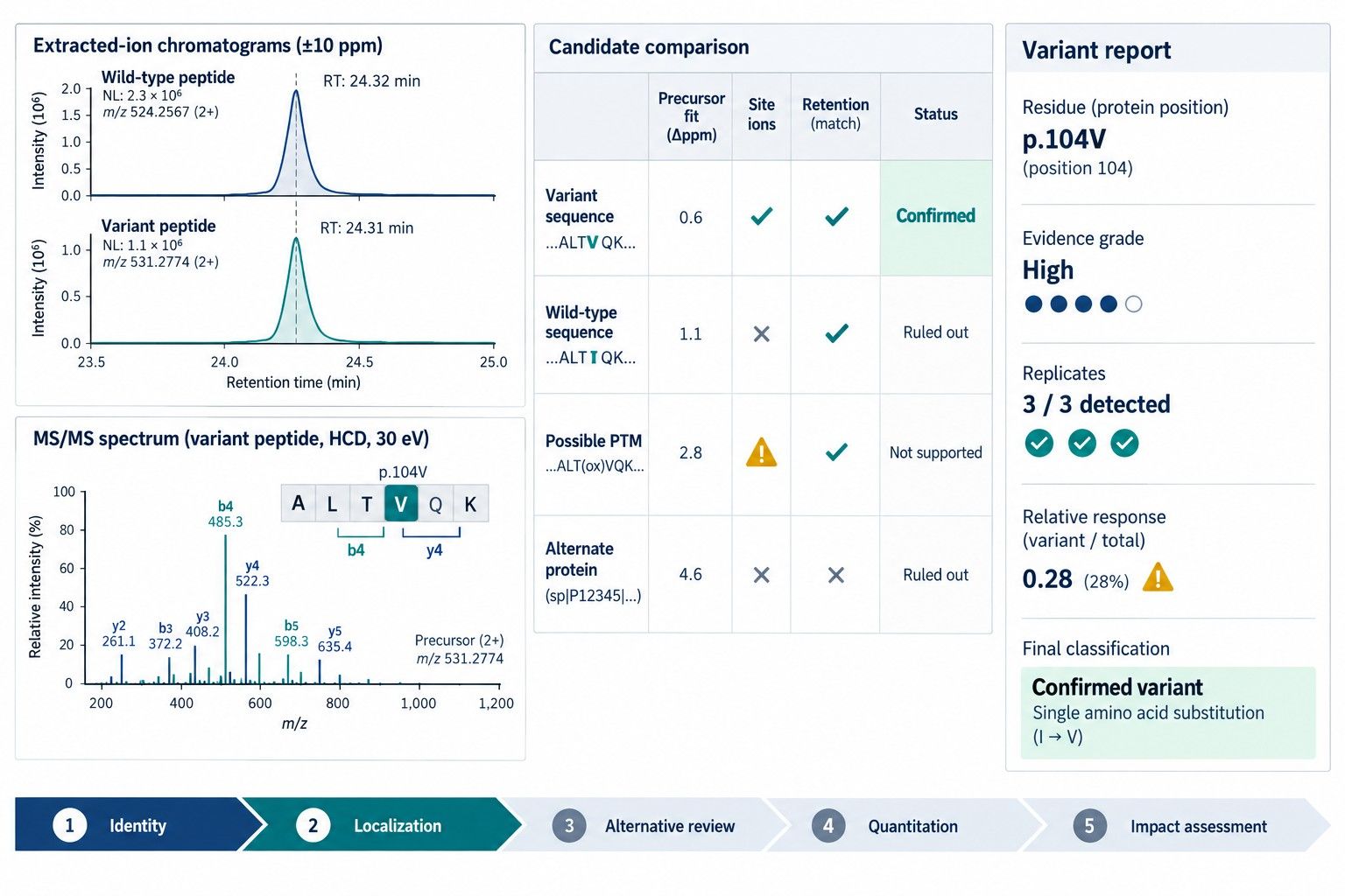 Figure 2. A decision-ready sequence-variant report links the wild-type and variant chromatograms to site-localizing MS/MS evidence, alternative-explanation review, and qualified abundance.
Technical FAQ
Is an accurate precursor mass sufficient to confirm an amino-acid substitution?
No. The mass shift may identify a candidate composition, but it does not necessarily establish peptide sequence or residue position. Product ions must localize the difference, and chemically plausible PTMs, isotope errors, and alternative peptides must be evaluated.
How many fragment ions are required around the substitution site?
There is no universal number. The essential requirement is that the observed ions distinguish the proposed site and sequence from credible alternatives. Multiple ions from both sides and complementary series strengthen the assignment, especially for low-abundance or regulatory-relevant results.
Can peptide coverage confirm a substitution?
Coverage shows that a peptide overlaps the residue, not that the substitution itself is distinguished. The target site must be supported by sequence-determining MS/MS ions or suitable orthogonal evidence. A high overall coverage percentage does not compensate for missing site-level evidence.
What if the variant peptide is detected only once?
A single high-quality spectrum can generate a credible candidate, but independent preparation or targeted reacquisition is preferable for confirmation. Rare data-dependent selection, chimeric spectra, and preparation artifacts are difficult to exclude from one event.
Can an open search discover all possible substitutions?
Open searching expands candidate detection but cannot guarantee discovery of every substitution. Low abundance, unsuitable peptide formation, poor ionization, missing MS/MS, isobaric events, and search-scoring limitations remain. Open-search candidates still require constrained verification.
How can a substitution be distinguished from a PTM with the same mass shift?
Use fragment ions that localize the change, compare modified and substituted candidate spectra, evaluate retention behavior, change peptide boundaries, and use complementary fragmentation or synthetic standards. Preparation-condition dependence also helps identify artifacts.
Does DNA sequencing confirm the protein substitution?
DNA identifies the encoded residue and can explain a genetically derived variant. It does not by itself prove the composition or abundance of the protein present in the sample. Protein LC-MS/MS and nucleotide analysis are complementary.
How are low-level sequence variants quantified?
They are commonly estimated from variant and wild-type extracted-ion chromatogram areas, sometimes with targeted SRM or PRM. Because peptide responses can differ, the ratio should be validated or described as a relative response unless calibrated standards establish molar accuracy.
What does "not detected" mean in a sequence-variant report?
It means no acceptable signal was observed under the stated method and sample conditions. It should be qualified by sequence coverage, target-peptide detectability, interference, and the method-specific reporting limit; it is not proof of absolute absence.
References
- Yang Y, Strahan A, Li C, et al. Detecting Low Level Sequence Variants in Recombinant Monoclonal Antibodies. MAbs. 2010;2(3):285-298. doi:10.4161/mabs.2.3.11718.
- Tanaka K, Takenaka S, Tsuyama S, Wada Y. Determination of Unique Amino Acid Substitutions in Protein Variants by Peptide Mass Mapping with FT-ICR MS. J Am Soc Mass Spectrom. 2006;17(4):508-513. doi:10.1016/j.jasms.2005.12.006.
- Harris RJ, Murnane AA, Utter SL, et al. Assessing Genetic Heterogeneity in Production Cell Lines: Detection by Peptide Mapping of a Low Level Tyr to Gln Sequence Variant in a Recombinant Antibody. Biotechnology (N Y). 1993;11(11):1293-1297. doi:10.1038/nbt1193-1293.
- Zeck A, Regula JT, Larraillet V, et al. Low Level Sequence Variant Analysis of Recombinant Proteins: An Optimized Approach. PLoS One. 2012;7(7):e40328. doi:10.1371/journal.pone.0040328.
- Ren D, Zhang J, Pritchett R, et al. An Optimized Approach to the Rapid Assessment and Detection of Sequence Variants in Recombinant Protein Products. MAbs. 2015;7(3):560-572. doi:10.1080/19420862.2015.1016692.
- Xu X, Huang Y, Pan H, et al. A General Evidence-Based Sequence Variant Control Limit for Recombinant Therapeutic Protein Development. MAbs. 2020;12(1):1791399. doi:10.1080/19420862.2020.1791399.
- Sissolak B, Lingg N, Sommeregger W, et al. Identification, Characterization and Control of a Sequence Variant in Monoclonal Antibody Drug Product: A Case Study. Sci Rep. 2021;11:11882. doi:10.1038/s41598-021-90780-3.
- Lund A, Ren D, Rogers RS, Rouse JC, Yu XC, Valliere-Douglass JF. Scientific Best Practices for Primary Sequence Confirmation and Sequence Variant Analysis in the Development of Therapeutic Proteins. J Pharm Sci. 2021;110(2):619-626. doi:10.1016/j.xphs.2020.11.007.
- Dobrowolski M, Urbaniak M, Pietrucha T. Peptide Mapping for Sequence Confirmation of Therapeutic Proteins and Recombinant Vaccine Antigens by High-Resolution Mass Spectrometry: Software Limitations, Pitfalls, and Lessons Learned. Int J Mol Sci. 2025;26(20):9962. doi:10.3390/ijms26209962.
- International Council for Harmonisation. ICH Q6B: Specifications—Test Procedures and Acceptance Criteria for Biotechnological/Biological Products. 1999.
- United States Pharmacopeia. General Chapter <1055> Biotechnology-Derived Articles—Peptide Mapping. Official December 1, 2024.
For research use only, not intended for any clinical use.


