Sequence coverage is routinely reported in LC-MS/MS peptide-mapping datasets, but it is frequently interpreted as direct evidence of complete primary sequence confirmation. The two terms describe related but different analytical attributes. Sequence coverage measures the proportion of a reference sequence represented by accepted peptide identifications; primary sequence confirmation evaluates whether the combined precursor- and product-ion evidence supports the expected amino-acid sequence at the resolution required for the study.
This distinction is material in recombinant construct verification, biotherapeutic primary-structure characterization, comparability studies, sequence-variant analysis, mutation confirmation, and investigations of unexpected molecular forms. A high global coverage value can coexist with an unobserved terminal peptide, an unsupported engineered residue, insufficient localization of a modification, or a peptide-spectrum match (PSM) that does not distinguish a credible alternative assignment.
The appropriate interpretation is therefore fit for purpose. The analytical team must define the sequence feature that controls the project decision, determine what LC-MS/MS evidence is required for that feature, and document any residual ambiguity separately from the global coverage percentage.
Analytical Summary
- Coverage is a breadth metric. It is usually calculated as the number of reference residues represented by accepted peptide identifications divided by the total sequence length.
- Confirmation is an evidence claim. It depends on peptide specificity, MS/MS fragment support, localization, search assumptions, and the scientific question.
- High coverage can coexist with an important unresolved region. A short missing terminus, mutation site, processing junction, or highly modified peptide may be more consequential than the overall percentage suggests.
- Precursor mass alone is not residue-level evidence. Fragment ions, retention behavior, complementary peptides, and expert review help distinguish plausible assignments from demonstrated ones.
- Multiple proteases are useful when they close a defined gap. The goal is complementary evidence, not a larger number for its own sake.
Definition and Calculation of Protein Sequence Coverage
In a bottom-up workflow, a protein is digested into peptides, the peptides are separated by liquid chromatography, and precursor and fragment-ion spectra are acquired. Identified peptides are mapped back to a supplied or inferred protein sequence. A common calculation is:
Sequence coverage (%) = covered amino-acid positions / total amino-acid positions × 100
If accepted peptides collectively represent 360 positions in a 400-residue protein, the reported coverage is 90%. Overlapping peptides can strengthen evidence for a region, but a residue is normally counted once in the coverage percentage. The result is therefore a map of observed sequence breadth—not a direct score of confidence at every residue.
Coverage also depends on analysis rules. Enzyme specificity, allowed missed cleavages, variable modifications, peptide confidence thresholds, false-discovery controls, minimum peptide length, and whether an assignment requires confirmatory MS/MS can change the reported percentage. The denominator may also differ when signal peptides, affinity tags, propeptides, or unresolved sequence regions are included or excluded. A coverage value is not fully interpretable unless the reference sequence and calculation rules are stated.
Detected, identified, covered, and confirmed are not interchangeable
A peptide feature may be detected at the MS1 level, assigned to a sequence by database searching, and supported by an MS/MS spectrum. These are related but distinct stages. For residue-level confirmation, the decisive question is whether the fragment-ion series distinguishes the expected sequence from credible alternatives and localizes any difference.
| Term |
What it answers |
What it does not establish by itself |
| Detected feature |
Was an ion observed at a given mass, charge, and retention time? |
Peptide identity or residue order |
| Peptide identification |
Is a peptide-spectrum match consistent with a candidate sequence under the search model? |
Unambiguous localization of every residue or modification |
| Sequence coverage |
What fraction of the reference is represented by accepted peptides? |
Equal confidence across the entire protein |
| Sequence confirmation |
Does the combined evidence support the expected sequence at the required resolution? |
Absence of every low-level variant in a heterogeneous sample |
| Sequence-variant analysis |
Is there evidence for an unintended amino-acid substitution or related variant? |
Complete characterization without suitable controls and targeted review |
Sequence coverage and fragment-ion coverage answer different questions
Protein sequence coverage maps accepted peptides onto the full-length reference. Fragment-ion coverage evaluates the peptide bonds represented by observed product ions within an MS/MS spectrum. These metrics operate at different levels. A peptide may extend across ten residues and contribute all ten positions to protein sequence coverage even when its spectrum contains only a few sequence-informative product ions. In that case, the peptide supports coverage of the region, but individual internal residues may not be independently localized.
Software packages also use different terminology. "MS/MS-confirmed coverage" may refer to residues contained in peptides with an accepted MS/MS identification, not to observation of product ions at every backbone cleavage. "Fragment coverage" may refer to the proportion of theoretical b/y or c/z ions matched in one spectrum. The report should define the metric rather than relying on the label alone.
Four evidence levels should be reported separately
| Evidence level |
Typical basis |
Appropriate interpretation |
| Accurate-mass peptide evidence |
Precursor mass, isotope pattern, charge state, retention behavior |
Consistent with a candidate peptide under the specified search space |
| PSM-supported sequence coverage |
Accepted database-search assignment with confirmatory MS/MS |
The peptide sequence is supported at the confidence level and search conditions applied |
| Residue- or site-specific confirmation |
Sequence-diagnostic product ions bracketing the residue, junction, or modification site |
The local assignment is supported against defined alternatives |
| Orthogonal primary-structure confirmation |
Complementary digest, terminal method, intact/subunit mass, or top-down/middle-down evidence |
Independent evidence addresses limitations of the original peptide map |
These levels are cumulative only when the underlying observations are compatible. A high-confidence PSM does not automatically localize every modification within a peptide, and an intact mass agreement does not compensate for a missing sequence-specific peptide at a critical region.
Sources of Incomplete or Ambiguous Sequence Evidence
The total percentage compresses a position-specific map into one number. That is useful for orientation, but it can hide where the missing or weak evidence occurs.
Global coverage can mask a critical sequence gap
A protein with 98% coverage may still lack the N-terminus, C-terminus, a cleavage junction, an engineered mutation, or a binding-site residue. For construct identity, processing verification, or mutation confirmation, that two-percent gap can be the only region that matters.
Conversely, a lower global percentage may be sufficient for a focused question if the target region is represented by specific peptides with strong fragment support. The interpretation should therefore begin with the study objective and residue map, not an arbitrary universal cutoff.
A peptide assignment may not provide residue-level localization
A peptide can match the expected precursor mass while its MS/MS spectrum contains too few informative product ions to support every internal position. Long peptides, low-abundance precursors, co-isolated ions, labile modifications, and inefficient backbone fragmentation can all produce discontinuous ion series. For a candidate amino-acid substitution or processing junction, the decisive evidence is generally a set of sequence-diagnostic product ions that bracket the position of interest rather than the precursor mass or aggregate PSM score alone.
Near-isobaric or isomeric possibilities require special care. Leucine and isoleucine have the same nominal and exact residue mass, so routine collision-based spectra generally cannot distinguish them from precursor mass alone. Some short sequence permutations or modification-versus-substitution combinations can also produce plausible automated assignments. Recent peptide-mapping work documents software-generated false assignments and emphasizes manual review of decisive spectra.
Shared peptide sequences may not identify a unique molecular source
When a peptide sequence is shared by isoforms, homologs, antibody chains, or contaminating proteins, its identification may support the presence of a protein family but not uniquely prove the intended construct. Unique peptides, junction-spanning peptides, or orthogonal intact/subunit evidence may be needed to resolve the source.
Sample heterogeneity limits proteoform-level inference
Bottom-up data combine peptides released from all molecular forms in the sample. The expected peptide set can confirm that the reference sequence derived from the expression construct is present while a low-level sequence variant, clipped form, or modified proteoform also exists. A sequence-confirmation claim should not be interpreted as proof that every molecule in the sample is identical.
Sample-Preparation, Acquisition, and Processing Effects on Coverage
An uncovered or weakly supported region does not have one universal cause. The limitation may originate during protein denaturation, reduction and alkylation, proteolysis, chromatographic recovery, ionization, precursor selection, fragmentation, or data processing. Repeating the same LC-MS/MS method without identifying the limiting step may reproduce the same evidence gap.
| Stage |
Mechanism affecting the result |
Observable consequence |
Appropriate diagnostic response |
| Reduction and alkylation |
Incomplete reduction, under-alkylation, over-alkylation, or disulfide scrambling |
Missing cysteine-containing peptides, multiple unexpected forms, or incorrect disulfide interpretation |
Review reagent ratios and reaction conditions; compare reduced and non-reduced strategies where relevant |
| Proteolysis |
Missed cleavage, nonspecific cleavage, autolysis, steric restriction, or modification of a cleavage residue |
Peptides outside the useful mass range, semitryptic products, and poor reproducibility |
Evaluate digestion efficiency and select a complementary cleavage specificity |
| Sample handling |
Digestion-induced deamidation, oxidation, carbamylation, or other preparation artifacts |
Artificial heterogeneity and competing modified peptide assignments |
Minimize exposure time, temperature, pH, and reactive reagents; include appropriate controls |
| LC recovery |
Very hydrophilic, highly hydrophobic, very short, or aggregation-prone peptides |
Peptide absent despite a favorable theoretical cleavage pattern |
Modify chromatography or redesign peptide boundaries with another protease |
| Electrospray ionization |
Ion suppression, unfavorable charge-state distribution, or low analyte abundance |
Weak precursor signal and stochastic MS/MS sampling |
Improve separation, enrichment, loading strategy, or targeted acquisition |
| Data-dependent acquisition |
Dynamic exclusion, duty-cycle limitations, or co-isolation |
Missing MS/MS, chimeric spectra, or inconsistent selection across replicates |
Use inclusion lists, targeted MS/MS, or acquisition settings matched to the critical peptide |
| Data processing |
Incorrect monoisotopic peak selection, overly broad modification search, permissive mass tolerance, or database incompleteness |
High-scoring false assignments, unexplained mass compensation, or missed variants |
Constrain the initial search, inspect decisive spectra, then expand hypotheses deliberately |
Sample preparation is particularly important in biotherapeutic peptide mapping because conditions that improve denaturation and digestion can also promote artificial oxidation or deamidation. The NISTmAb method-development study treated digestion efficiency and artifact control as linked optimization objectives rather than independent checks.
Evidence Requirements Beyond Global Sequence Coverage
A decision-ready review separates breadth, depth, specificity, localization, and orthogonality.
- Map accepted peptides to the reference sequence. Show covered and uncovered positions and distinguish peptides supported by MS/MS from features supported only at precursor level.
- Mark critical regions before interpreting the total. These may include termini, signal-peptide boundaries, tag-cleavage sites, disulfide-linked regions, engineered substitutions, active-site residues, and known PTM sites.
- Inspect peptide uniqueness. Determine whether each decisive peptide maps uniquely to the intended sequence under the relevant database and whether homologous proteins or construct variants contain the same subsequence.
- Review product-ion support. Confirm that sequence-diagnostic ions bracket the residue, junction, or modification whose identity must be established. Record the ion series and mass error used for the assignment.
- Evaluate competing explanations. Consider isobaric residues, near-isobaric sequence permutations, chemical modifications, incorrect isotope selection, and unmodeled construct-derived sequence.
- Seek independent support where the risk warrants it. An overlapping peptide from a complementary digest, a terminal method, or an intact/subunit mass result can test an assignment through a different measurement principle.
- Record ambiguity explicitly. Separate "detected," "consistent with," "PSM-supported," "site-localized," and "not distinguished under the applied method" rather than forcing every observation into a binary confirmed/not-confirmed label.
For studies that need a reference-aware peptide map, peptide mapping can provide the coverage and modification framework. When the question extends to residue-level interpretation or an unexpected sequence, mass spectrometry-based protein sequencing adds targeted MS/MS and sequence analysis.
 Figure 1. Relationship among peptide-level sequence coverage, product-ion evidence, and local residue confirmation.
Complementary Proteolysis for Sequence-Coverage Gap Closure
Trypsin is widely used because it often produces peptides with favorable length, charge, chromatographic behavior, and predictable C-terminal basic residues. It does not, however, create an informative peptide for every sequence region. A region may contain too many or too few tryptic cleavage sites, generate a peptide outside the useful mass range, or yield a peptide with poor recovery or fragmentation.
Complementary proteases cut at different residues and redraw the peptide boundaries. Published studies show that combining data from multiple proteases can increase sequence coverage and reach regions inaccessible to a single digest. The benefit is protein- and question-dependent: an appropriate complementary protease produces an observable, sequence-specific peptide across the unresolved region and, ideally, places the critical residue within a product-ion-rich portion of the peptide rather than at an uninformative terminus.
| Protease |
Typical cleavage preference |
When it may help |
Planning caution |
| Trypsin |
C-terminal to Lys or Arg |
General mapping and database searching |
Can generate peptides that are too short in Lys/Arg-rich regions or too long in sparse regions |
| Lys-C |
C-terminal to Lys |
Complementary boundaries and robust digestion under some denaturing conditions |
May still leave long peptides in Lys-poor regions |
| Glu-C |
Commonly C-terminal to Glu; behavior depends on conditions |
Regions poorly represented by basic-residue cleavage |
Specificity can depend on buffer and method conditions |
| Asp-N |
Commonly N-terminal to Asp |
Orthogonal peptide orientation around a target region |
Peptide recovery and fragmentation still require evaluation |
| Chymotrypsin |
Prefers aromatic and selected hydrophobic residues |
Hydrophobic or trypsin-resistant regions |
Broader specificity can increase interpretation complexity |
Run complementary digests separately when possible so each peptide can be traced to a defined cleavage scheme. Combining data after acquisition preserves interpretability while allowing overlapping evidence to be assembled into a more complete map. A parallel-digest design also avoids overdigestion that can occur when several enzymes act in the same reaction and produce peptides too short for confident assignment.
More coverage is not automatically more confirmation. If a second digest adds peptides only to noncritical regions but still leaves the target residue without sequence-diagnostic product ions, the analytical gap remains. Enzyme selection should therefore be guided by an in-silico digest, expected peptide physicochemical properties, potential PTMs, and the position of the missing evidence.
Data-Backed Examples of Coverage and Confirmation
NISTmAb: critical-region coverage and artifact-controlled digestion
The NISTmAb LC-MS/MS peptide-mapping protocol was optimized around three linked stages: enzymatic digestion, peptide separation, and peptide detection. The authors emphasized that a high overall sequence coverage value was not sufficient if product-specific complementarity-determining regions remained unobserved. Digestion conditions were also evaluated for artificially induced deamidation and oxidation because these changes can complicate primary-structure interpretation even when coverage is high.
The transferable principle is that coverage targets should be sequence-aware. For an antibody, the CDRs may control identity and function; for another recombinant protein, the decisive regions may be a signal-peptide junction, catalytic residue, engineered substitution, fusion linker, or terminal processing site.
Multi-protease yeast study: complementary cleavage outperformed repeat acquisition
Swaney, Wenger, and Coon compared trypsin, Lys-C, Arg-C, Asp-N, and Glu-C in a large-scale yeast proteomics dataset. Moving from one to five proteases increased sequence coverage by 172%, and the added coverage was more effective than repeated analysis of a single-protease digest. This result demonstrates the value of orthogonal cleavage specificity, but it should not be converted into a performance promise for an individual purified protein. The magnitude of improvement depends on sequence composition, abundance, digestion chemistry, LC-MS method, and acceptance criteria.
CAP256 antibody clipping: coverage alone did not establish the molecular event
In a study of the CAP256 broadly neutralizing antibody, reducing capillary electrophoresis detected clipping ranging from 1% to 31%, and a sample containing approximately 17% clipped material was selected for detailed characterization. Intact, reduced, middle-up, tryptic, chymotryptic, and disulfide-mapping measurements were combined. Separate trypsin and chymotrypsin digests improved coverage and helped localize the cleavage region, while subunit mass and orthogonal observations established that the evidence belonged to a clipped molecular form.
This example illustrates a central limitation of global coverage: bottom-up peptide identifications can localize a candidate region, but intact or subunit evidence may still be required to connect the peptide observation to the molecular species detected in the sample.
Orthogonal Strategies for Unresolved Sequence Regions
The analytical claim should determine the minimum evidence package. Protein identification, construct verification, primary sequence confirmation, and low-level sequence-variant analysis are not interchangeable objectives.
| Study objective |
Minimum useful evidence |
Additional evidence commonly required |
| Protein identification |
Multiple sequence-unique peptides with accepted PSMs |
Intact mass or another identity measurement when homologous proteins are plausible |
| Recombinant construct verification |
Coverage of engineered residues, processing junctions, tags, and termini relevant to the construct |
Sequence-diagnostic MS/MS ions and targeted terminal analysis for unresolved boundaries |
| Primary sequence confirmation |
Extensive reference-sequence coverage with MS/MS support across product-specific regions |
Complementary digestion, intact/subunit mass, and appropriate terminal or disulfide evidence |
| Single-amino-acid substitution confirmation |
Variant peptide plus product ions bracketing the candidate residue |
Wild-type reference comparison, complementary peptide, and evaluation of isobaric alternatives |
| Low-level sequence-variant analysis |
Variant-specific detection strategy, suitable controls, and defined search space |
Targeted extraction, manual spectrum review, quantitative evaluation, and orthogonal confirmation where necessary |
| Unknown-sequence determination |
High-quality MS/MS data with de novo sequence tags and overlapping peptides |
Multiple fragmentation modes, complementary digests, homology search, and terminal information |
After the objective is defined, the next method should address the specific source of uncertainty.
| Evidence gap |
Useful next step |
What the added evidence can resolve |
| Missing internal region after tryptic mapping |
Complementary protease digest and targeted LC-MS/MS |
New peptide boundaries and fragment ions across the gap |
| Unconfirmed N-terminus or processing site |
N-terminal sequencing or targeted terminal peptide analysis |
Actual starting residue, clipping, blockage, or signal-peptide processing |
| Unconfirmed C-terminus |
C-terminal sequencing or targeted terminal strategy |
Terminal residue, clipping, extension, or incomplete processing |
| Expected and observed intact masses disagree |
Intact/subunit mass plus peptide-level localization |
Whether a global molecular difference is present and where it maps |
| Candidate sequence is absent or unreliable |
De novo sequencing with complementary peptide overlaps |
Sequence evidence without requiring the correct answer in the initial database |
| Co-occurring PTMs or proteoform connectivity matters |
Top-down or middle-down analysis where suitable |
Whether modifications and sequence features coexist on the same molecular form |
| Leu/Ile or another isomeric ambiguity controls the decision |
Specialized fragmentation, targeted chemical derivatization, or orthogonal sequence evidence |
Evidence beyond routine precursor mass and standard collision-induced product-ion series |
If the objective is to reconstruct or verify an extensive primary sequence, full-length protein sequencing can combine complementary peptide information and targeted gap closing. If preserving the connection between multiple modifications and a single molecular form is central, top-down protein sequencing may provide complementary proteoform-level information.
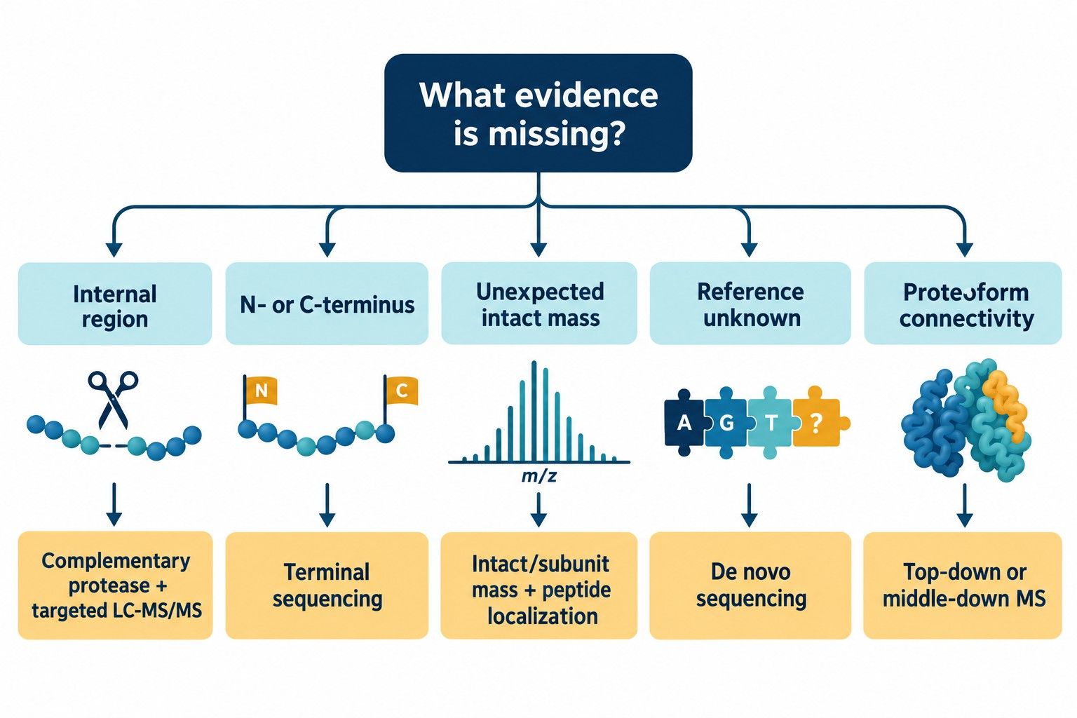 Figure 2. Method-selection framework based on the location and analytical type of unresolved sequence evidence.
Reporting Requirements for Primary Sequence Confirmation
A useful report makes the analytical claim auditable. It should allow the reader to see not only the final percentage, but also how that percentage was obtained and where uncertainty remains.
- The exact reference sequence, including tags, leaders, linkers, expected processing, and known engineered changes
- Sample preparation and digestion conditions, including the protease and reduction/alkylation strategy
- LC-MS/MS acquisition and database-search settings relevant to interpretation
- A peptide coverage map with accepted peptides and uncovered positions
- Peptide-level mass error, charge state, score or confidence measure, and modification assignment where relevant
- Annotated MS/MS spectra for peptides supporting critical residues, termini, junctions, or substitutions
- Distinction between unique and shared peptides
- A list of ambiguities, unsupported positions, and plausible alternative explanations
- The rationale for any complementary digest or orthogonal method
- A conclusion written at the level supported by the evidence
For example, "95% sequence coverage was obtained" is a factual breadth statement. "The expected sequence was confirmed at the engineered substitution and both processing junctions; residues 184–190 were not observed" is more informative because it states what the data prove and what remains unresolved.
Example of a decision-oriented result summary
| Report field |
Example result statement |
Interpretation |
| Reference definition |
Mature recombinant sequence, affinity tag removed; residues numbered according to the processed construct |
Establishes the denominator and residue-numbering convention |
| Global sequence coverage |
95% of reference positions represented by accepted peptides |
Describes breadth but does not assign equal confidence to every position |
| MS/MS-supported coverage |
91% of positions represented by peptides meeting the defined MS/MS acceptance criteria |
Separates precursor-consistent features from product-ion-supported assignments |
| Critical-region status |
Engineered residue 128 supported by diagnostic product ions on both sides; processing junction covered by two overlapping peptides |
Supports the project-specific confirmation claim |
| Uncovered region |
Residues 184–190 not observed after tryptic mapping |
Defines the remaining analytical gap |
| Ambiguity |
Leu/Ile assignment at residue 242 not distinguishable under the applied HCD method |
Prevents an isomeric assignment from being overstated |
| Orthogonal evidence |
Intact mass agrees with the expected processed construct within the stated mass tolerance |
Supports overall molecular consistency without replacing local sequence evidence |
| Recommended action |
Glu-C digest with targeted MS/MS across residues 180–195 |
Connects the unresolved region to a justified follow-up experiment |
The numerical values above are illustrative reporting examples, not universal acceptance criteria. Actual thresholds should be defined from method performance, sample type, project phase, reference material, and the analytical decision being supported.
Fit-for-Purpose Interpretation and Study-Specific Acceptance Criteria
Coverage can be sufficient when the research question is limited and the decisive regions have suitable evidence. Examples include confirming the identity of a purified target with multiple unique peptides, verifying a known engineered residue with diagnostic fragments, or comparing whether the same expected peptide map is present across exploratory samples.
Deeper confirmation is warranted when:
- the missing region contains the study's critical residue or processing boundary;
- the expected reference may be wrong or incomplete;
- a single-amino-acid substitution must be localized;
- a terminal truncation or extension is suspected;
- isoforms or homologous proteins share most observed peptides;
- intact mass indicates an unexplained molecular form;
- co-occurring modifications must be assigned to the same proteoform; or
- automated identification conflicts with the raw spectrum or orthogonal data.
The most defensible target is therefore not a universal coverage percentage. It is an evidence package matched to the decision the researcher needs to make.
Technical FAQ
Is 100% protein sequence coverage the same as complete sequence confirmation?
Not necessarily. A 100% map indicates that accepted peptides collectively span every reference position. Complete residue-level confirmation additionally requires sufficient MS/MS evidence, peptide specificity, and review of ambiguous assignments at the resolution relevant to the study.
What is a good protein sequence coverage percentage?
There is no universal percentage that fits every protein and purpose. Protein size, sequence composition, modifications, digestion behavior, sample complexity, and the location of critical residues all affect what is achievable and what is scientifically adequate.
Can MS1 peptide mass confirm an amino-acid sequence?
MS1 mass can support a peptide assignment, but different sequences or modification combinations can share or closely approximate the same precursor mass. MS/MS fragment ions are generally required to localize sequence information within the peptide.
Why is my N- or C-terminus missing from the coverage map?
Terminal peptides may be too short, too long, highly hydrophilic or hydrophobic, modified, blocked, or outside the acquisition method's effective range. A complementary protease or a dedicated terminal method may be more informative than repeating the same digest.
Does using more proteases always improve sequence confirmation?
No. Multiple proteases are valuable when their cleavage patterns generate informative peptides across a defined gap. They also add sample preparation and interpretation complexity. An in-silico digest and region-specific plan should guide enzyme selection.
Can bottom-up LC-MS/MS prove that all modifications occur on the same protein molecule?
Usually not. Bottom-up digestion separates the original protein into peptides, so connectivity between distant features can be lost. Intact, top-down, or middle-down measurements may be needed when proteoform-level linkage is the question.
How should an ambiguous residue be reported?
Report the observed peptide, the evidence supporting the assignment, the unresolved alternatives, and the method needed to distinguish them. Terms such as "consistent with," "localized," and "not distinguishable under the applied method" are more useful than overstating certainty.
Creative Proteomics supports research-use protein sequence characterization with study-specific digestion, LC-MS/MS, sequence interpretation, and orthogonal strategies. A project discussion should begin with the exact sequence question and the residues or molecular forms that control the next decision.
References
- International Council for Harmonisation. Q6B: Specifications—Test Procedures and Acceptance Criteria for Biotechnological/Biological Products. 1999.
- Lund A, Ren D, Rogers RS, Rouse JC, Yu XC, Valliere-Douglass JF. Scientific Best Practices for Primary Sequence Confirmation and Sequence Variant Analysis in the Development of Therapeutic Proteins. Journal of Pharmaceutical Sciences. 2021;110(2):619–626. doi:10.1016/j.xphs.2020.11.007.
- Mouchahoir T, Schiel JE. Development of an LC-MS/MS Peptide Mapping Protocol for the NISTmAb. Analytical and Bioanalytical Chemistry. 2018;410:2111–2126. doi:10.1007/s00216-018-0848-6.
- Dobrowolski M, Urbaniak M, Pietrucha T. Peptide Mapping for Sequence Confirmation of Therapeutic Proteins and Recombinant Vaccine Antigens by High-Resolution Mass Spectrometry: Software Limitations, Pitfalls, and Lessons Learned. International Journal of Molecular Sciences. 2025;26(20):9962. doi:10.3390/ijms26209962.
- Swaney DL, Wenger CD, Coon JJ. The Value of Using Multiple Proteases for Large-Scale Mass Spectrometry-Based Proteomics. Journal of Proteome Research. 2010;9(3):1323–1329. doi:10.1021/pr900863u.
- Biringer RG, Amato H, Harrington MG, Fonteh AN, Riggins JN, Hühmer AFR. Enhanced Sequence Coverage of Proteins in Human Cerebrospinal Fluid Using Multiple Enzymatic Digestion and Linear Ion Trap LC-MS/MS. Briefings in Functional Genomics and Proteomics. 2006;5(2):144–153. doi:10.1093/bfgp/ell026.
- Zhou M, Gucinski AC, Boyne MT II. Performance Metrics for Evaluating System Suitability in Liquid Chromatography–Mass Spectrometry Peptide Mass Mapping of Protein Therapeutics and Monoclonal Antibodies. mAbs. 2015;7(6):1104–1117. doi:10.1080/19420862.2015.1074364.
- Ivleva VB, Schneck NA, Gollapudi D, Arnold F, Cooper JW, Lei QP. Investigation of Sequence Clipping and Structural Heterogeneity of an HIV Broadly Neutralizing Antibody by a Comprehensive LC-MS Analysis. Journal of the American Society for Mass Spectrometry. 2018;29(7):1512–1523. doi:10.1007/s13361-018-1968-0.
Author: CAIMEI LI, Senior Scientist
For research use only, not intended for any clinical use.


