Leucine (Leu, L) and isoleucine (Ile, I) are constitutional isomers with the same elemental composition and monoisotopic residue mass. Consequently, a Leu-to-Ile substitution does not shift the precursor mass of an otherwise identical peptide, and the ordinary backbone fragments generated by routine low-energy collision-induced dissociation (CID) or higher-energy collisional dissociation (HCD) remain isobaric. This is a chemical limitation of the measurement, not a software-scoring problem.
The distinction nevertheless matters. A single Leu/Ile assignment can define an antibody complementarity-determining region, differentiate a recombinant construct from a homolog, establish the sequence of a natural peptide, or identify an isomeric impurity in a synthetic peptide product. In these situations, a conventional de novo sequence containing an unresolved L/I position is not equivalent to a residue-specific assignment.
Leu/Ile differentiation therefore requires an evidence strategy designed around the position of interest. Specialized fragmentation can produce side-chain-derived d- or w-type ions whose losses differ between the branched side chains. Reference sequences, nucleotide data, Edman degradation, synthetic comparators, or stable-isotope labeling can provide complementary evidence, but each supports a different analytical claim. A defensible report must state whether the final residue identity was directly distinguished by mass spectrometry, inferred from an external reference, or retained as an unresolved isomeric ambiguity.
Analytical Summary
- Routine precursor mass does not distinguish Leu from Ile. Both residues contribute the same exact mass to a peptide, so mass accuracy alone cannot resolve the assignment.
- Conventional b- and y-ion series usually localize an isobaric residue but do not identify which isomer is present. More backbone coverage can narrow the ambiguous position without changing the underlying L/I equivalence.
- Side-chain fragmentation provides direct mass-spectrometric discrimination. Diagnostic d- and w-type ions can reflect different radical losses from leucine and isoleucine side chains when suitable activation and ion chemistry are achieved.
- Method suitability is peptide- and position-dependent. Charge state, sequence context, residue position, precursor abundance, fragmentation efficiency, co-isolation, and instrument architecture affect whether the required diagnostic ions are observable.
- External sequence agreement is valuable but should be labeled as inference. A database, homolog, germline sequence, cDNA, or construct record can resolve a biological hypothesis without proving that the analyzed molecule contains that residue.
- Unresolved positions should remain explicit. Reporting L/I, Xle, or another clearly defined ambiguity is more scientifically accurate than selecting the higher-scoring candidate without isomer-specific evidence.
Molecular Basis of the Leu/Ile Isomeric Ambiguity
Leucine and isoleucine both have the molecular formula C6H13NO2 as free amino acids. Their difference is connectivity: leucine contains an isobutyl side chain, whereas isoleucine contains a sec-butyl side chain and an additional stereogenic center. After incorporation into a peptide, both have the same monoisotopic residue mass, approximately 113.0841 Da. No resolving power can separate two peptide ions that have the same elemental composition, charge state, and isotopic composition solely on the basis of mass-to-charge ratio.
This distinguishes the L/I problem from near-isobaric assignments. Lysine and glutamine, for example, differ by approximately 0.036 Da as residues and may be distinguishable with sufficient mass accuracy. Leucine and isoleucine have no such mass defect. Higher resolution improves separation from interfering peaks and accuracy of fragment assignment, but it does not create a mass difference between the two isomers.
What a conventional spectrum can still establish
Routine LC-MS/MS can often determine that a residue of mass 113.0841 Da occupies a specific sequence position. If b- and y-type ions bracket the residue, the sequence interval can be localized and the rest of the peptide may be strongly supported. The correct conclusion is then a position-specific Leu/Ile ambiguity, not an unlocalized sequence gap.
The spectrum may also exclude other candidate residues, confirm peptide termini, support neighboring substitutions, and link the peptide to an overlapping sequence. These are valuable results. They simply do not distinguish the two side-chain connectivities.
Why the common immonium ion is insufficient
Leu- and Ile-containing peptides can both generate an immonium ion near m/z 86.0964. Detection of that ion supports the presence of an Xle-type residue but ordinarily does not identify its isomer. Any interpretation based only on the presence or intensity of the shared m/z 86 ion is vulnerable to sequence context, collision energy, co-fragmentation, and other contributors in the low-mass region.
Recent multistage approaches have fragmented the Xle immonium ion further and evaluated lower-mass products or their intensity ratios. Such a strategy can become discriminating when it is experimentally validated on the relevant instrument and peptide classes, but the initial m/z 86 signal alone remains non-specific.
Evidence Available from Routine LC-MS/MS
Most bottom-up and de novo sequencing experiments use collision-based fragmentation because it provides robust peptide identification and sequence coverage. The resulting evidence should be separated into localization, contextual inference, and isomer-specific discrimination.
| Evidence type |
What it can establish |
What it cannot establish alone |
| Accurate precursor mass |
Peptide elemental composition within measurement uncertainty; consistency with an L- or I-containing candidate |
Leu versus Ile when all other residues are identical |
| Consecutive b/y ions bracketing the site |
Position of a 113.0841 Da residue and neighboring sequence order |
Side-chain connectivity of the residue |
| Shared Xle immonium ion |
Presence of a Leu/Ile-like residue in a sufficiently clean spectrum |
Which Xle isomer occupies the localized position |
| Database or germline match |
Best sequence explanation within the supplied reference space |
Direct observation of the isomer in the sample |
| Retention-time agreement |
Consistency with a comparator under controlled chromatography |
Identity without a qualified reference and sufficiently selective method |
| Diagnostic d/w side-chain ions |
Direct MS evidence for Leu or Ile at a defined position when correctly assigned |
Universal discrimination if ions are absent, interfered with, or produced outside a validated context |
Database search can conceal the ambiguity
Many protein-search workflows treat I and L as equivalent because the theoretical precursor and conventional product-ion masses are identical. A peptide-spectrum match can therefore be correct at the mass-sequence level while the reported letter follows the FASTA entry rather than direct isomer-specific evidence. Protein identification remains valid when other sequence-unique evidence is sufficient, but the individual L/I residue has been reference-assigned.
This distinction is particularly important in unknown protein sequence analysis. A search result may nominate a homolog and fill every L/I position from that homolog, whereas a reference-free interpretation should retain positions not directly resolved. The report should disclose whether sequence letters were propagated from the database, inferred by homology, or confirmed experimentally.
More coverage does not automatically resolve the isomer
Complementary proteases are essential for full sequence reconstruction because they create overlapping peptides, move difficult residues away from peptide termini, and produce alternative charge states. However, ten conventional HCD spectra containing the same Leu/Ile site do not become isomer-specific merely through replication. They increase confidence in localization and surrounding sequence, but direct differentiation still requires a diagnostic measurement or an orthogonal source of residue identity.
Specialized Mass-Spectrometric Strategies
Leu/Ile differentiation by MS relies on fragment pathways that retain information about side-chain structure. The most established approaches generate radical fragments or high-energy side-chain cleavages, producing d-, v-, or w-type ions in addition to ordinary backbone ions.
ECD, ETD, and EThcD-derived w ions
Electron capture dissociation (ECD) and electron transfer dissociation (ETD) cleave the peptide N–Cα bond and commonly form c- and z-type fragments. Radical z ions can undergo secondary side-chain losses. For an appropriately positioned Xle residue, loss of an isopropyl radical from leucine or an ethyl radical from isoleucine produces w-type ions with different masses. In practical notation, the diagnostic ion must be related to the correct z fragment and residue position; a low-mass peak without that structural relationship is not sufficient.
EThcD combines electron-transfer fragmentation with supplemental collisional activation. It can increase product-ion richness and help generate or reveal diagnostic w ions. A published Orbitrap-based study used ETD or ETcaD to form z ions followed by broad-band HCD and correctly assigned 83 of 93 Leu/Ile residues across a set of tryptic and non-tryptic peptides. That result demonstrates feasibility, not a universal 89% performance specification: the success rate belongs to the tested sequences, instrument, acquisition settings, and interpretation rules.
Radical migration is a central interpretive risk. If the radical relocates before side-chain loss, a w-like product may no longer report the intended residue position. Neighboring Leu/Ile residues, short terminal fragments, low product abundance, and competing fragmentation can also complicate assignment. Diagnostic-ion interpretation should therefore consider the complete precursor-to-primary-fragment-to-secondary-fragment relationship and, where possible, both complementary evidence and reference peptides.
High-energy collision and multistage HCD/CID
High-energy CID can produce charge-remote side-chain fragments, including d- and w-type ions. Modern instrument implementations may use targeted MS3 experiments: an informative primary fragment or shared immonium ion is isolated and subjected to a second activation event. Xiao and colleagues integrated HCD-MSⁿ and ETD-HCD MS3 on an Orbitrap Fusion to assign Leu/Ile residues in peptides and monoclonal-antibody variable regions. The method was designed around predefined decision rules rather than a conventional untargeted HCD spectrum.
A 2026 high-resolution HCD/CID study generated the shared Xle immonium ion and then fragmented it to lower-mass diagnostic products at m/z 44.0 and 69.0. The relative I44/I69 response was used to distinguish Leu and Ile on the tested Orbitrap Eclipse platform. Because intensity ratios depend on instrument configuration, collision settings, isolation purity, and matrix, they should be calibrated with suitable standards before being used as a general identity rule.
Hot ECD, UV photodissociation, CTD, and other radical methods
Hot ECD and hybrid ECD methods can increase the abundance of side-chain fragments compared with conventional electron-driven fragmentation. Ultraviolet photodissociation (UVPD) can produce multiple backbone and side-chain ion types, including w ions. Charge transfer dissociation (CTD) has also generated d, w, and v ions from singly and doubly charged peptides; in one six-peptide evaluation, reliable Xle-associated d or w ions were observed in more than 80% of tested instances.
These techniques expand the available evidence but differ in precursor-charge requirements, fragmentation pathways, duty cycle, hardware availability, spectral complexity, and software support. A published success rate for one technique cannot be transferred to another instrument or sample type. Method selection should be based on whether the platform can generate and accurately measure the diagnostic ions for the actual peptide containing the site of interest.
 Figure 1. Evidence pathway from an isobaric L/I site in routine HCD to targeted electron- or high-energy fragmentation and side-chain diagnostic-ion assessment.
Orthogonal Strategies for Residue Assignment
Specialized MS/MS is not the only way to resolve a Leu/Ile position. An orthogonal method may be preferable when the peptide has unsuitable charge state, the site lies at an unfavorable terminus, the diagnostic fragment is too weak, or a biological reference can answer the project question more directly.
Nucleotide or verified construct sequence
A high-quality cDNA, genomic, or expression-construct sequence distinguishes the codons encoding leucine and isoleucine. This may provide the strongest biological explanation for a recombinant product or expressed antibody sequence. However, nucleotide evidence describes the encoded sequence, whereas protein MS measures the material present. Translation errors, sample swaps, unrecorded construct changes, sequence variants, or chemical synthesis errors are not excluded by agreement with a design file.
When the analytical claim is "consistent with the supplied construct," reference-based evidence may be sufficient. When the claim is "the protein sample directly contains Ile at position 52," an isomer-specific protein measurement or qualified comparator is required.
Edman degradation
Sequential Edman degradation converts the accessible N-terminal residue into a phenylthiohydantoin derivative that can be identified chromatographically. PTH-Leu and PTH-Ile are chemically distinct and can be separated under suitable conditions. Edman sequencing is therefore a useful orthogonal option when the ambiguous residue lies within the readable N-terminal cycles of an unblocked, sufficiently pure peptide or protein.
Its applicability is constrained by N-terminal accessibility, sample amount and purity, cycle yield, heterogeneous termini, and the location of the ambiguous site. An internal residue may require isolation of a proteolytic peptide with that site repositioned near its N-terminus. Protein N-terminal sequencing should be planned as a targeted orthogonal experiment, not assumed to resolve every internal L/I position in an intact protein.
Synthetic reference peptides and chromatographic comparison
Synthetic peptides containing the alternative Leu and Ile sequences can support assignment through co-analysis. The most informative design includes both isomeric standards, identical sample preparation, and matched LC-MS conditions. Retention behavior, targeted product-ion patterns, and co-injection can then be assessed.
Chromatographic selectivity is sequence- and method-dependent. Co-elution is not proof of identity when the method does not resolve the isomers, whereas a reproducible retention-time match plus discriminating MSn behavior can be compelling. Reference-peptide purity and sequence identity must themselves be qualified because a mislabeled or isomerically contaminated standard transfers its uncertainty to the sample conclusion.
Stable-isotope labeling and biological incorporation
In a system where metabolic labeling is possible, isotopically labeled leucine can create a residue-specific mass shift while isoleucine remains unshifted. A bank-vole protein study used deuterated leucine incorporation and fragment-ion doublets to resolve L/I positions during de novo sequencing. This is direct and elegant for controlled biological systems, but it is generally unavailable for an existing purified product, clinical specimen, or synthetic peptide.
Targeted Workflow for Resolving a Defined L/I Site
The most efficient strategy begins with a site-level analytical question rather than an untargeted request for "higher coverage." A suitable workflow preserves the routine sequence evidence, isolates the unresolved position, and adds only the experiments needed to change the residue-level conclusion.
- Define the claim and candidate sequences. Specify the protein or peptide, residue numbering system, expected Leu and Ile alternatives, neighboring sequence, and whether direct protein-level proof is required.
- Map existing evidence. Identify all peptides that contain the site, their charge states, retention times, precursor intensities, isolation purities, fragmentation modes, and product ions bracketing the residue.
- Redesign peptide boundaries where necessary. Use complementary proteases or controlled cleavage to move the site away from an uninformative terminus, reduce multiple Xle residues in one peptide, and generate a charge state compatible with electron-based fragmentation.
- Acquire a routine reference spectrum. Confirm precursor identity, localize the 113.0841 Da residue, evaluate co-isolation, and establish the surrounding backbone sequence before interpreting side-chain fragments.
- Select an isomer-specific activation route. Apply EThcD, ETD-HCD MS3, high-energy MSn, UVPD, hot ECD, CTD, or a platform-appropriate alternative with settings qualified for diagnostic d/w-ion formation.
- Assign diagnostic ions structurally. Require mass accuracy, expected charge state, correct relationship to the primary a- or z-type ion, site specificity, adequate signal-to-noise, and absence of plausible interferences.
- Test both candidate explanations. Evaluate the ions predicted for Leu and Ile rather than searching only for support of the expected sequence. Absence of one ion is not affirmative evidence for the other unless the method has validated sensitivity.
- Add orthogonal confirmation when the site is critical. Compare synthetic isomers, verify the encoding nucleotide sequence, perform Edman analysis on an appropriate terminus, or analyze an independently generated peptide.
- Classify the result. Report direct assignment, orthogonally supported inference, reference-based assignment, or unresolved L/I according to the evidence actually obtained.
Workflow Selection by Project Scenario
| Project scenario |
Primary analytical risk |
Preferred first-line evidence |
Escalation when unresolved |
| De novo antibody sequencing |
Incorrect L/I in a CDR or framework position; germline inference may conceal a true difference |
Multi-protease sequence assembly plus targeted EThcD or MS3 at non-germline-resolved sites |
Synthetic isomer comparison and nucleotide evidence when available |
| Recombinant protein sequence confirmation |
Design file may not reflect the expressed material |
Routine peptide mapping to localize the site and confirm surrounding sequence |
Targeted diagnostic fragmentation for sample-level residue proof |
| Unknown natural peptide |
No definitive database or encoding sequence |
De novo backbone sequencing with electron/high-energy side-chain fragments |
Edman sequencing, derivatization, or orthogonal purified-peptide analysis |
| Synthetic peptide identity or impurity investigation |
Leu/Ile isomeric substitution produces no precursor-mass shift |
Paired Leu and Ile reference peptides under qualified LC-MSn conditions |
Orthogonal chromatography or targeted immonium/side-chain fragmentation |
| Single residue in an inaccessible peptide |
Poor charge state, terminal location, multiple Xle residues, or low abundance |
Alternative protease or chemical cleavage to generate a more favorable peptide |
Enrichment, targeted acquisition, isolated peptide Edman analysis, or nucleotide evidence |
| Broad discovery proteomics |
L/I identity may not affect protein-level identification |
Preserve I/L equivalence in search and report sequence tags appropriately |
Target only sites that affect biological interpretation |
For projects requiring broad sequence reconstruction, de novo protein and peptide sequencing should incorporate L/I resolution as a targeted module rather than promise universal assignment from every discovery spectrum. For known proteins, mass spectrometry-based protein sequencing can combine reference-guided mapping with targeted review of positions that alter the final claim.
Common Failure Modes and Misinterpretations
Reporting the database letter as an observed residue
A search engine may display L because the FASTA sequence contains L, even though an I-containing peptide would have the same theoretical precursor and conventional b/y fragments. The correct report distinguishes database-supported sequence identity from direct isomer discrimination.
Treating higher mass resolution as a solution
Resolution separates ions with different m/z values. Leu- and Ile-substituted versions of an otherwise identical peptide have the same elemental composition and therefore the same exact mass. Higher resolution improves spectral cleanliness and diagnostic-ion accuracy but does not separate the intact isomers by mass alone.
Using the shared m/z 86 ion as a binary marker
The common Xle immonium ion does not by itself distinguish the residues. Diagnostic use requires a validated secondary fragmentation pattern or another isomer-selective response, not merely observation of m/z 86.
Interpreting absence as proof
A missing Leu-associated or Ile-associated diagnostic ion can result from low abundance, poor precursor selection, interference, unfavorable residue position, insufficient activation, excessive secondary fragmentation, or radical migration. Unless detectability has been established for the method and sequence context, non-observation is an unresolved result.
Ignoring peptide position and multiple Xle residues
Diagnostic fragments must isolate the relevant side chain. A peptide containing several Leu/Ile residues can generate overlapping candidate fragments, and a terminal Xle site may not yield the same evidence as an internal position. Peptide redesign is often more informative than simply increasing injection time on an unsuitable precursor.
Calling a homology-consistent sequence "complete de novo sequencing"
Homologs are valuable for assembly and error detection, but sequence letters copied from a homolog are not de novo observations. A complete sequence claim should identify every position supported only by homology, especially isomeric residues and regions without overlapping peptide evidence.
Data-Backed Literature Examples
Targeted EThcD discrimination across 93 residues
Artemenko and colleagues evaluated an EThcD-based workflow in which ETD/ETcaD-derived z ions were subjected to HCD and the resulting w ions were used for L/I discrimination. Across 43 tryptic peptides plus three non-tryptic peptides, 83 of 93 Leu/Ile residues were correctly assigned. The unresolved or doubtful cases illustrate why diagnostic-ion generation and radical-site behavior must be assessed per peptide rather than represented as an automatic capability.
Integrated LC-MSn for monoclonal-antibody variable regions
Xiao, Vecchi, and Wen combined HCD-MSⁿ with ETD-HCD MS3 on an Orbitrap Fusion. The study assigned L/I residues in peptides containing multiple Xle sites and addressed monoclonal-antibody variable-region positions that could not be determined from germline alignment. The result supports targeted multistage acquisition for critical antibody positions; it does not imply that routine HCD-only peptide mapping directly differentiates every L/I site.
EThcD-supported sequencing of plasma antibodies
A 2024 de novo antibody study used EThcD and manual assessment of residue-specific w ions to resolve L/I ambiguities in antibody sequences recovered from human plasma. This shows how L/I analysis functions within a broader evidence stack: multiple proteases and sequence assembly establish the antibody sequence, while targeted side-chain fragments address the isomeric positions that ordinary backbone ions leave unresolved.
CTD evaluation on singly and doubly charged peptides
Edwards and co-workers evaluated four model peptides and two wild-type sequences using CTD. The experiment produced d, w, and v ions and reported reliable Xle-associated d or w ions in more than 80% of the tested cases. Applicability to singly charged precursors is analytically interesting, but the limited sequence set and modified instrumentation require project-specific feasibility assessment.
Evidence Grading and Decision-Ready Reporting
A residue-level result should be graded according to the origin and specificity of the evidence. Peptide-level confidence scores, protein-level coverage, and L/I identity are related but not interchangeable metrics.
| Evidence grade |
Minimum basis |
Appropriate wording |
| Direct MS assignment |
Site-specific diagnostic d/w or validated MSn ion pattern with correct precursor and fragment relationships, adequate signal, and interference review |
"Ile was directly assigned at position X by diagnostic side-chain fragmentation." |
| Orthogonally confirmed |
Two independent discriminating methods, such as targeted MSn plus synthetic comparator or Edman sequence |
"Ile at position X was confirmed by targeted MSn and orthogonal reference analysis." |
| Reference-supported inference |
Conventional peptide evidence localizes Xle and a verified construct, cDNA, database, or homolog specifies one isomer |
"The site is mass-spectrometrically localized as L/I and assigned as Ile from the supplied construct sequence." |
| Unresolved isomer |
Routine spectrum localizes the residue but no valid isomer-specific evidence is observed |
"Position X remains Leu/Ile (Xle); the current data do not distinguish the isomers." |
| Unsupported |
Letter chosen from software output, expectation, or non-specific ion without adequate site evidence |
Do not report a residue-specific conclusion |
A decision-ready result package should include:
- sample identity, preparation history, and peptide-generation method;
- sequence and residue numbering convention, including mature-chain versus precursor numbering;
- precursor m/z, charge, retention time, mass error, and isolation-purity assessment;
- annotated routine spectrum demonstrating site localization and neighboring sequence;
- fragmentation mode, activation settings, MS stage, and instrument configuration used for discrimination;
- table of diagnostic ions with theoretical and observed m/z, charge, mass error, intensity or signal-to-noise, and structural assignment;
- explicit evaluation of the alternative Leu and Ile candidates;
- reference standard, nucleotide, Edman, or other orthogonal evidence where used;
- status of each Xle position across the sequence: direct, orthogonally confirmed, reference-inferred, or unresolved; and
- a conclusion whose wording matches the achieved evidence grade.
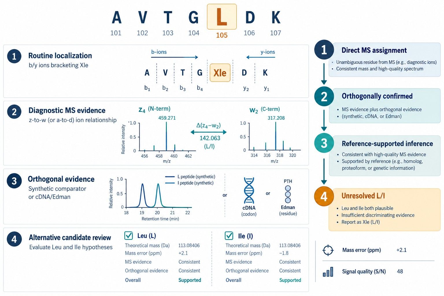 Figure 2. Residue-level reporting separates routine localization, diagnostic side-chain evidence, orthogonal confirmation, and explicitly unresolved Leu/Ile positions.
Technical FAQ
Can standard HCD distinguish leucine from isoleucine?
Routine HCD dominated by b- and y-type backbone ions generally cannot distinguish the residues because their precursor and backbone-fragment masses are identical. High-energy or multistage HCD strategies may become discriminating when they generate validated side-chain or secondary immonium-ion products; this is a different evidence basis from a standard HCD peptide-spectrum match.
Does high-resolution accurate-mass MS solve the L/I problem?
No. High resolution can separate interferences and improve product-ion assignment, but L- and I-containing versions of an otherwise identical peptide have the same elemental composition and exact mass. The distinction requires structural fragmentation, selective chromatography with qualified references, or another orthogonal method.
What are w ions, and why are they useful?
w ions arise from side-chain loss associated with radical z-type fragments. Leucine and isoleucine can undergo different characteristic radical losses because their side-chain connectivity differs. When the precursor, primary z ion, and secondary w ion are correctly related to one residue position, the mass difference can provide direct isomer-specific evidence.
Is EThcD always sufficient for every Leu/Ile position?
No. Diagnostic-ion formation depends on peptide sequence, charge, residue position, abundance, activation conditions, and instrument performance. C-terminal or adjacent Xle residues and radical migration can be problematic. Alternative peptide boundaries, MS3, another radical method, or orthogonal analysis may be required.
Can a database search distinguish L from I?
It can report the letter present in a candidate database sequence, but routine mass matching does not independently observe that difference. The assignment should be described as reference-supported unless diagnostic fragmentation or other isomer-specific evidence confirms the residue in the analyzed protein.
Should de novo software output L, I, or J?
Software conventions vary. Some workflows use I/L equivalence, Xle, or J to represent an unresolved leucine/isoleucine position. The report should define the notation explicitly and avoid converting the ambiguity into a specific residue without supporting evidence.
Can complementary proteases resolve Leu/Ile by themselves?
They do not change the equal residue masses, but they can create a peptide with better charge state, cleaner isolation, fewer Xle residues, or more favorable position for diagnostic fragmentation. They are therefore an important method-development step even though the cleavage pattern alone is not necessarily isomer-specific.
When is Edman degradation useful?
Edman analysis is useful when the ambiguous position is within the readable cycles of a pure peptide or protein with an accessible N-terminus. It may directly distinguish PTH-Leu and PTH-Ile chromatographically. Internal sites may require prior proteolysis and isolation of a suitable peptide.
How should an unresolved position appear in the final sequence?
It should remain visibly ambiguous, for example as L/I or Xle, with a defined notation and residue-level evidence status. A separate table should state whether each position was directly measured, inferred from a reference, or not resolved.
References
- Xiao Y, Vecchi MM, Wen D. Distinguishing between Leucine and Isoleucine by Integrated LC-MS Analysis Using an Orbitrap Fusion Mass Spectrometer. Anal Chem. 2016;88(21):10757-10766. doi:10.1021/acs.analchem.6b03409.
- Artemenko KA, Zubarev AR, Samgina TY, Lebedev AT, Savitski MM, Zubarev RA. An EThcD-Based Method for Discrimination of Leucine and Isoleucine Residues in Tryptic Peptides. J Am Soc Mass Spectrom. 2017;28(8):1655-1662. doi:10.1007/s13361-017-1672-7.
- Gupta K, Kumar M, Chandrashekara K, Krishnan KS, Balaram P. Combined Electron Transfer Dissociation-Collision-Induced Dissociation Fragmentation in the Mass Spectrometric Distinction of Leucine, Isoleucine, and Hydroxyproline Residues in Peptide Natural Products. J Proteome Res. 2012;11(2):515-522. doi:10.1021/pr200091v.
- Edwards HM, Wu HT, Julian RR, Jackson GP. Differentiation of Leucine and Isoleucine Residues in Peptides Using Charge Transfer Dissociation Mass Spectrometry. Rapid Commun Mass Spectrom. 2022;36(5):e9246. doi:10.1002/rcm.9246.
- Wu P, Zhang T, Wang G, et al. Direct Characterization of Leucine and Isoleucine Residues in Peptides Using High-Resolution Tandem Mass Spectrometry. Anal Chem. 2026;98(28):20912-20925. doi:10.1021/acs.analchem.6c02059.
- Wee S, O'Hair RAJ, McFadyen WD. Side-Chain Radical Losses from Radical Cations Allows Distinction of Leucine and Isoleucine Residues in the Isomeric Peptides Gly-Xxx-Arg. Rapid Commun Mass Spectrom. 2002;16(9):884-890. doi:10.1002/rcm.658.
- Muth T, Hartkopf F, Vaudel M, Renard BY. A Potential Golden Age to Come—Current Tools, Recent Use Cases, and Future Avenues for De Novo Sequencing in Proteomics. Proteomics. 2018;18(18):e1700150. doi:10.1002/pmic.201700150.
- Le Bihan T, Nunez de Villavicencio Diaz T, Reitzel C, et al. De Novo Protein Sequencing of Antibodies for Identification of Neutralizing Antibodies in Human Plasma Post SARS-CoV-2 Vaccination. Nat Commun. 2024;15:8790. doi:10.1038/s41467-024-53105-8.
- Ramsay SL, Steinborner ST, Waugh RJ, Dua S, Bowie JH. A Simple Method for Differentiating Leu and Ile in Peptides: Negative-Ion Mass Spectra of PTH-Leu and PTH-Ile. Rapid Commun Mass Spectrom. 1995;9(13):1241-1243. doi:10.1002/rcm.1290091305.
- Shaw JB, Malhan N, Vasil'ev YV, et al. Sequencing Grade Tandem Mass Spectrometry for Top-Down Proteomics Using Hybrid Electron Capture Dissociation Methods in a Benchtop Orbitrap Mass Spectrometer. Anal Chem. 2018;90(18):10819-10827. doi:10.1021/acs.analchem.8b01901.
For research use only, not intended for any clinical use.


